O R I G I N A L R E S E A R C H A R T I C L E
Open Access
Flexible bronchoscopy may decrease respiratory
muscle strength: premedicational midazolam
in focus
Baykal Tulek
*, Fikret Kanat, Sule Tol and Mecit Suerdem
Abstract
Background:Flexible bronchoscopy (FB) is a procedure accepted to be safe in general, with low complication rates reported.On the other hand, it is known that patients with pre-existing respiratory failure have developed
hypoventilation following FB. In this study the effects of FB on respiratory muscle strength were investigated by measuring maximum respiratory pressures.
Methods:One hundred and forty patients, aged between 25 and 90 years, who had undergone diagnostic bronchoscopy between February 2012 and May 2012, were recruited to the study. Pre- and post-procedure maximal inspiratory pressure (MIP) and maximal expiratory pressure (MEP) were measured. A correlation between the MIP and MEP changes and patient characteristics and FB variables were investigated.
Results:Significant decreases in both MIP and MEP values were observed following FB (p < 0.001 for both). Decreases were attributed to the midazolam used for sedation. Significant decreases in respiratory muscle strengths were observed especially in the high-dose midazolam group, compared to both low-dose and non-midazolam groups.
Conclusions:It was determined that respiratory muscle weakness may arise post-procedure in patients who have undergone FB, and this is constitutively related to midazolam premedication. Respiratory muscle weakness might play a role in potential hypoventilation in critical patients who undergo FB.
Keywords:Bronchoscopy, Midazolam, Respiratory muscle strength
Background
Flexible bronchoscopy (FB) is a procedure that is widely used in the diagnosis and management of airway and lung diseases, and is accepted to be safe in general [1]. Data regarding bronchoscopy complications and low complication rates are mostly obtained from retro-spective studies. However, the increase in therapeutic bronchoscopy applications, which are becoming more and more complex, and the bronchoscopy applications in risky patient groups cause an increase in complication rates [2].
Hypoventilation has been shown to develop during and after the procedure in patients who undergo bron-choscopy [3,4]. This has mostly been linked to sedative
agents in relevant studies. However, to our knowledge, the effects of bronchoscopy itself and/or sedative agents on respiratory muscle strength have yet to be studied. The aim of this study was to evaluate the effects of bronchoscopy on respiratory muscle strength, which as an important role in respiratory function and induction of cough reflex [5,6].
Methods Patients
One hundred and forty patients, aged between 25 and 90 years, who had undergone diagnostic bronchoscopy between February 2012 and May 2012 were recruited to the study. Patients with hemodynamic instability (heart rate below 50 bpm or above 120 bpm, systolic arterial blood pressure below 90 mmHg or above 180 mmHg), basal oxygen saturation <90%, PaCO2> 45 mmHg, liver * Correspondence:[email protected]
Selcuk University, Selcuklu Faculty of Medicine, Department of Chest Diseases, Selçuklu 42075, Konya, Turkey
or kidney dysfunction, those who were diagnosed with a neuromuscular disease, had cancer with cachexia, medium (FEV1between 50 and 80%), severe (FEV1 be-tween 30 and 50%) or very severe (FEV1<30%) COPD, and those who could not cooperate with respiratory muscle strength test or with benzodiazepine allergy have not been included into the study. Study protocol was approved by Selcuk University Selcuklu Medical Faculty Ethics Committee. Written informed consent was obtained from all participants.
Bronchoscopy
All bronchoscopy procedures were performed by the same experienced investigator in accordance with the international guidelines [7]. In the absence of any con-traindications, IM atropine (0.01 mg/kg) was given to the patients half an hour prior to the procedure. 2% lido-caine was applied topically to the inside of the mouth, pharynx, upper respiratory airways and endobronchially. As a routine application at our clinic, IV midazolam was offered to all patients by the bronchoscopist prior to the procedure and 1 mg IV midazolam was given to those who accepted the offer 3 minutes prior to the procedure. While no additional doses were given to patients who had adequate sedation during the procedure, additional doses of 1 mg were given in 2-minute intervals to patients considered to be in need by the bronchoscopist. At the end of the study, patients were retrospectively separated into three groups: no midazolam (Group A), low-dose midazolam (Group B), and high-dose midazo-lam (Group C).
2–4 ml/min oxygen supplementation was performed
in all patients, and oxygen flow was increased in case of hypoxemia. Flumazenil was kept available for suspected cases of respiratory depression. An IV vascular access was prepared, and electrocardiogram (ECG), oxygen sa-turation and non-invasive blood pressure monitoring were performed throughout the procedure. Total du-ration of bronchoscopy, midazolam dose and adverse events during the endoscopic procedure were recorded. Cardiopulmonary complications observed during the flexible bronchoscopy were defined as the following: hypotension (systolic blood pressure (BP) <90 mmHg or mean arterial blood pressure <60 mmHg), hypertension (systolic BP >180 mmHg or diastolic BP >90 mmHg), tachycardia (HR >100/min and/or a variation of >20% from baseline value), bradycardia (HR <50/min), and oxygen desaturation (SaO2 decrease <90% for >30s). Monitoring was continued for 2 hours following the pro-cedure. 0–100 mm visual analogue scale (VAS) was used two hours after the flexible bronchoscopy to investigate the comfort of the procedure. A higher score meant better satisfaction or less discomfort (0: worst imaginable health state, 100: best imaginable health state).
Maximum respiratory pressure measurements
A hand-held respiratory pressure meter (Micro RPM; Micro Medical, Chatham, UK) was used to measure respiratory muscle strength [8]. Maximal inspiratory pressure (MIP) was acquired from residual volume (RV) and the maximal expiratory pressure (MEP) was acquired from total lung capacity (TLC). Measurements were taken prior to bronchoscopy before premedication and after 30 minutes following the procedure by the same investigator without any knowledge of the clinical condi-tions of the patients. The operator explained the proced-ure before beginning and showed the correct manoeuvre. MIP was measured first, followed by MEP. Patients per-formed 5 maneuvers in intervals of 1 minute for each measurement and the highest values (<20% variance) were recorded [9].
Statistical analyses
Descriptive statistics are shown as mean (±SD) or count (percentage). T-test was used to compare respiratory muscle strengths before and after bronchoscopy for all patients.T-test or One-Way ANOVA were used to com-pare each variable for the mean MIP and MEP changes. One-Way ANOVA and Chi-square tests were utilized in the comparison of continuous and categorical variables between various midazolam dose groups, respectively. The post-hoc comparisons of significant difference between the groups were performed using the Tukey test. The statistical significance level was set to 0.05.
Results
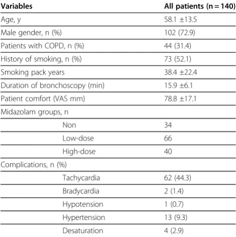
Bronchoscopies were successfully performed in all of the 140 patients included in the study. As to the midazolam dosages: 34 patients did not want midazolam adminis-tration (Group A), 66 were administered 1 mg of mida-zolam prior to the procedure, and 40 received high doses (mean, 0.05 ± 0.03 mg/kg) of midazolam (Group C). Patient characteristics and complications developed during the bronchoscopy are presented in Table 1. The study population mostly comprised middle- and old-aged males. One third of the patients had mild COPD and the most common complication observed was tachycardia. On the contrary, hypertension, bradycardia, hypotension and desaturation were rarely encountered complications.
When the overall pre- and post-procedure MIP and
MEP values were compared (73.9 ±24.4 cmH20 vs
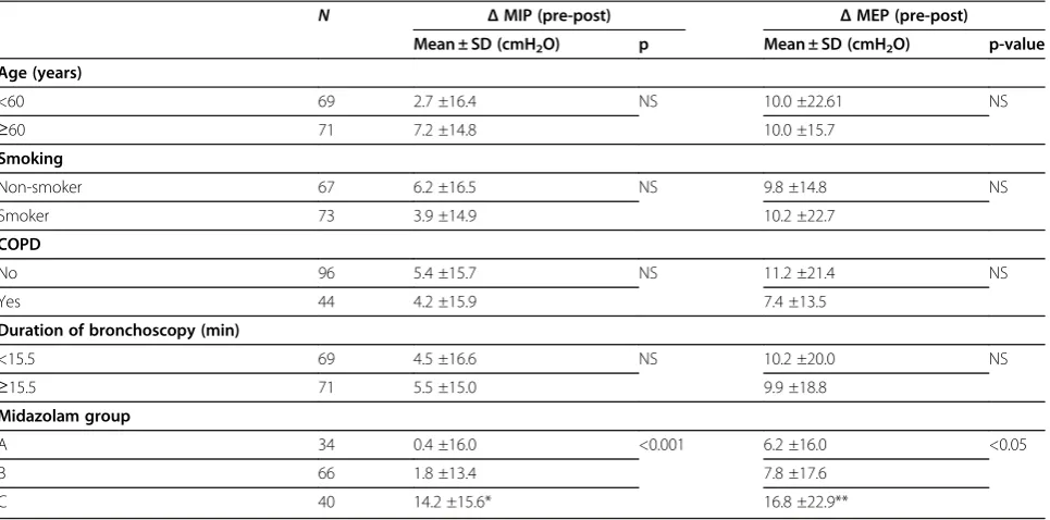
addition, the pre- and post-procedure MIP and MEP values were investigated between the different midazo-lam dose groups as well. Among these variables, a sig-nificant difference was determined only in different
midazolam dose groups in terms ofΔ MIP andΔ MEP
(Table 2).
The post-hoc analysis in the midazolam groups revealed no significant difference between the Groups A and B in terms of Δ MIP (p > 0.05) while Δ MIP was higher in Group C when compared to Group A (p < 0.001) and Group B (p < 0.001). There was no sig-nificant difference between Group A and Group B in
terms of Δ MEP (p > 0.05). Δ MEP was higher in
Group C when compared to Groups A (p = 0.04) and B (p = 0.05).
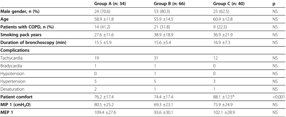
The comparison of pre- and post-procedure MIP and MEP values among different midazolam dose groups using T-test revealed no significant difference in terms of MIP measurements in the Groups A and B; however, there was a significant decrease in post-procedure MEP values in both groups. Significant decreases in both the post-procedure MIP and MEP values were determined in Group C (Table 3).
No significant differences were determined in terms of age, sex, smoking status, ratio of patients with COPD, duration of bronchoscopy, and basal MIP/MEP mea-surements in different midazolam groups. However, there was a significant difference between the groups in terms of patient comfort assessed using VAS (p < 0.001), and while there was no significant difference between the Groups A and B in thepost-hoc test, the VAS scores
of Group C was determined to be higher than the patients in Group A and Group B (p < 0.001 for both comparisons; Table 4).
Discussion
In this study, where -to our knowledge- the effects of FB and premedication on respiratory muscle strengths were investigated for the first time, it was revealed that mida-zolam, administered for sedation purposes in bronchos-copy, might affect negatively the respiratory muscle strengths, as shown with MIP and MEP measurements.
Flexible bronchoscopy is generally regarded as a safe procedure [1]. Hypoventilation is one of the most common complications and many causes of hypoven-tilation related to FB have been defined. These include ventilation-perfusion mismatch related to the procedure itself, upper airway obstruction, sedation-related central respiratory depression, and increased resistance due to introduction of bronchoscope into the trachea [3,10]. Our findings show that sedation-related respiratory muscle weakness can be included among these mechanisms.
Pulse oxymetry is utilized in many centers for moni-toring hypoventilation during bronchoscopy. On the other hand, patients are routinely given oxygen during FB in many centers. Bronchoscopy guidelines recom-mend oxygen supplementation to maintain oxygen sa-turation at a minimum of 90%, because it will reduce the risk of arrhythmia during and after bronchoscopy [7]. However, even though severe CO2retention may occur during the procedure, oxyhemoglobin desaturation may not be observed. Chhajed et al.[3] have performed cuta-neous carbon dioxide tension (PcCO2) measurements in addition to the oxymetry in patients that underwent FB in which sedation was achieved with intermittent intra-venous midazolam and 5 mg of hydrocodone, and deter-mined an increase in PcCO2in all but one patient. The highest PcCO2 value was significantly associated to the
Table 1 Demographics and clinical and bronchoscopic characteristics of patients
Variables All patients (n = 140)
Age, y 58.1 ±13.5
Male gender, n (%) 102 (72.9)
Patients with COPD, n (%) 44 (31.4)
History of smoking, n (%) 73 (52.1)
Smoking pack years 38.4 ±22.4
Duration of bronchoscopy (min) 15.9 ±6.1
Patient comfort (VAS mm) 78.8 ±17.1
Midazolam groups, n
Non 34
Low-dose 66
High-dose 40
Complications, n (%)
Tachycardia 62 (44.3)
Bradycardia 2 (1.4)
Hypotension 1 (0.7)
Hypertension 13 (9.3)
Desaturation 4 (2.9)
baseline PcCO2(p < 0.0001) and lowest SpO2(p = 0.016). Dreher et al. [4] have utilized PcCO2 measurements in order to evaluate alveolar hypoventilation in patients with pre-existing respiratory failure, and determine a sig-nificant increase in PcCO2during the procedure. In their study, while no significant difference was determined in
terms of PcCO2 during FB between the groups where
the patients were sedated with either midazolam alone or midazolam plus alfentanil, PcCO2was higher compared to baseline in the midazolam alone group 120 minutes after the procedure than the midazolam plus alfentanil group. The cause for prolonged hypoventilation was con-sidered to be midazolam due to the fact that the midazo-lam alone group received twice the amount of midazomidazo-lam than the midazolam plus alfentanil group (4 mg vs. 2 mg). We believe that the decrease in both the inspiratory and expiratory respiratory muscle strengths, which was deter-mined in the high-dose (0.05 ± 0.03 mg/kg) midazolam group in our study, might play an important role in hypoventilation that has been determined in the two studies mentioned above. However, because we did not
measure PcCO2 in our study, we do not know whether
there was an increase in the CO2values or not, and thus, we do not know whether the determined muscle weak-ness has any clinical importance or not.
The effects of midazolam on respiratory muscle strength have been previously shown in experimental and clinical studies. Fujii et al. [11] have investigated the effects of midazolam and propofol on diaphragm con-tractility in dogs. In that study, they have induced dia-phragmatic fatigue with intermittent supramaximal bilateral electrophrenic stimulation at low or high fre-quency (20 and 100 Hz, respectively), and after the induction of fatigue, in order to assess the diaphragm contractility, transdiaphragmatic pressure (Pdi) and inte-grated electrical activity of the crural (Edi-cru) and costal (Edi-cost) parts of the diaphragm were measured. Their findings showed that midazolam caused a decrease in Pdi at both frequencies when compared to fatigued values (p < 0.05), and that Edi-cru and Edi-cost at 100 Hz stimulation during midazolam administration were below the baseline values (p < 0.05). They have also reported a lower Pdi value in the midazolam group than in the pro-pofol group (p < 0.05).
Table 2 Comparison of the differences in pre- and post-procedure maximum respiratory pressures (Δ: pre-post) between different patient groups
N ΔMIP (pre-post) ΔMEP (pre-post)
Mean ± SD (cmH2O) p Mean ± SD (cmH2O) p-value
Age (years)
<60 69 2.7 ±16.4 NS 10.0 ±22.61 NS
≥60 71 7.2 ±14.8 10.0 ±15.7
Smoking
Non-smoker 67 6.2 ±16.5 NS 9.8 ±14.8 NS
Smoker 73 3.9 ±14.9 10.2 ±22.7
COPD
No 96 5.4 ±15.7 NS 11.2 ±21.4 NS
Yes 44 4.2 ±15.9 7.4 ±13.5
Duration of bronchoscopy (min)
<15.5 69 4.5 ±16.6 NS 10.2 ±20.0 NS
≥15.5 71 5.5 ±15.0 9.9 ±18.8
Midazolam group
A 34 0.4 ±16.0 <0.001 6.2 ±16.0 <0.05
B 66 1.8 ±13.4 7.8 ±17.6
C 40 14.2 ±15.6* 16.8 ±22.9**
*Viapost-hocanalysis, pair-wised comparison of Group C with both Group A and B (p < 0.001 for both). ** Viapost-hocanalysis, pair-wised comparison of Group C with Group A (p = 0.04) and Group B (p = 0.05).
Table 3 Comparison of pre- and post-flexible bronchoscopy MIP and MEP values in different midazolam dose groups
MIP (cm H2O) p MEP (cm H2O) P
Pre Post Pre Post
Group A 80.5 ±25.2 80.0 ±24.7 NS 109.4 ±27.6 103.2 ±21 =0.03
Group B 69.3 ±23.1 67.6 ±22.8 NS 93.6 ±30.3 85.8 ±29.0 =0.001
In another study, the effects of sedative (0.1 mg/kg/h) and anesthetic (0.5 mg/kg/h) dosages of midazolam on the decrease in diaphragm contractility, fatigue (detail fatigue rating [DFR]), have been evaluated in dogs [12]. They showed that an infusion of midazolam has caused a decrease from baseline values (p < 0.05) in Pdi at 20 and 100 Hz stimulations, and that%Edi-cru and % Edi-cost values at 100 Hz were below baseline (p < 0.05) in both sedative and anesthetic groups. They have also demon-strated that the Pdi and % Edi decrease was greater in the anesthetic dose group than in the sedative dose group (p < 0.05). Their findings show that contractility of fatigued diaphragm dose-dependently decreases with midazolam.
Molliex et al.[13] have studied the effects of midazo-lam, with a dosage of 0.1 mg/kg, on total pulmonary resistance and diaphragmatic, intercostal and abdominal muscle patterns in 9 healthy volunteers. Changes in gas-tric pressure (ΔPga) and pleural pressure (ΔPpl) were measured in all participants, and the reduction in dia-phragm contractility was evaluated with ΔPga/ ΔPpl. Midazolam was determined to increase total pulmonary resistance in sedative doses, and associatively, an increase in intercostal muscle activity was also determined; however, the diaphragmatic contribution to respiratory process was found to be decreased. This was explained as a shift from an abdominal breathing to predominantly rib cage breathing rather than a decrease in diaphragm contractility. Even though upper airway measurements were not taken in the current study, the increase in pul-monary resistance is thought to be associated with upper airway occlusion. Diazepam causing a decrease in the activity of the genioglossus muscle, which has an
important role in maintaining the patency of the upper airway, supports this hypothesis [14].
It is known that bronchoscopy deleteriously affects pulmonary mechanics and lung volumes [10,15]. This may arise a question regarding whether the significant reductions in maximal pressures are related to lung volumes rather than muscle weakness or respiratory muscle strength. MIP is measured at or close to RV and MEP at or close to TLC. Sometimes these measurements were performed at functional residual capacity. Although the latter may be more accurate for some studies, in that case the lung volumes should be specifically stated [16]. In patients with abnormally high lung volumes, a low MIP may partly reflect the shortened inspiratory muscle fiber length associated with increased lung volume at RV rather than reduced inspiratory muscle strength; how-ever, in our study all moderate to very severe COPD patients were not taken into the study and also the groups were not different regarding the number of mild COPD patients. Therefore it may be considered that the reductions in maximal pressures are probably not related to lung volumes.
Although there is limited falls in MEP values in patients who were not sedated during bronchoscopy,
when Δ MEP values were compared between groups,
thepost-hocanalysis revealed that the difference was sta-tistically significant only with group A and C and the decline in MEP values was higher in the high-dose mida-zolam group. When pre and post MEP values within each group were compared, post MEP values in group B and C were significantly lower than group A. It is notable that although post MIP values were significantly lower only in Group C, there were remarkable falls in post
Table 4 Comparison regarding patient demographics, bronchoscopic characteristics and pre-procedure maximum respiratory pressures among different midazolam dose groups
Group A (n: 34) Group B (n: 66) Group C (n: 40) p
Male gender, n (%) 24 (70.6) 53 (80.3) 25 (62.5) NS
Age 58.9 ±11.8 55.9 ±14.5 60.9 ±12.8 NS
Patients with COPD, n (%) 14 (41.2) 21 (31.8) 9 (22.5) NS
Smoking pack years 27.6 ±11.6 38.9 ±18.9 36.9 ±21.9 NS
Duration of bronchoscopy (min) 15.5 ±5.9 15.6 ±5.4 16.9 ±7.3 NS
Complications
Tachycardia 19 31 12 NS
Bradycardia 1 1 0 NS
Hypotension 0 1 0 NS
Hypertension 5 5 3 NS
Desaturation 2 1 1 NS
Patient comfort 76.2 ±17.4 74.4 ±17.4 88.1 ±12.5* <0.001
MIP 1 (cmH2O) 80.5 ±25.2 69.3 ±23.1 75.9 ±24.9 NS
MEP 1 109.4 ±27.6 93.6 ±30.1 102.1 ±28.9 NS
MEP values in all groups. The fall in post MEP values even in group A, i.e. in groups who were not sedated with midazolam might be due to exhaustion of the patient after repeated maneuvers for correct MIP measurements.
Even though the overall bronchoscopy comfort scores were quite high in our study, the fact of significantly higher VAS scores of the patients in the high-dose rather than in the low-dose midazolam group, and of the simi-larity of the patient comfort levels in the low-dose and no midazolam groups can be explained by the bronch-oscopist’s tendency to administer low doses. It is known that objective techniques, such as electroencephalogram-based bispectral index [17] or Ramsay sedation score [18], allow a more effective titration of the sedatives. In addition, it is also shown that both inter-individual and intra-individual variations are seen in the online sedation monitoring in healthy volunteers who were sedated with midazolam [19]. This condition may explain why some patients needed high doses and some low doses of mida-zolam in our study although the bronchoscopist decided the appropriate sedation level in patients subjectively. In humans, midazolam is mostly metabolized by CYP3A4⁄5 isozymes to one major metabolite, 1-hydroxymidazolam and to some extent to 4-hydroxymidazolam and 1,4-dihydroxymidazolam [20,21]. It is well known that wide inter-individual variations in hepatic and intestinal CYP3A activity are seen in the human population [22].
Conclusion
In conclusion, midazolam for premedication purposes has been determined to increase comfort in patients undergoing flexible bronchoscopy, however, causing sig-nificant decrease in respiratory muscle strength. This might increase the probability of possible complications following bronchoscopy in critical patients who have advanced COPD with limited respiratory reserves or respiratory insufficiency. Certainly there is a special need for further studies with risk patient groups and the use of high-dose midazolam.
Competing interests
The authors of the paper”Flexible bronchoscopy may decrease respiratory muscle strength: premedicational midazolam in focus”declare to have no competing interests that might be related to the contents of the manuscript.
Authors’contribution
BT helped design the study, conduct of the study, collection of data, analysis of data and prepare the manuscript. FK helped conduct of the study, collection of data, analysis of data and prepare the manuscript. ST helped conduct of the study, collection of data. MS helped conduct of the study, collection of data, and prepare the manuscript. All authors read and approved the final manuscript.
Received: 12 July 2012 Accepted: 7 September 2012 Published: 25 September 2012
References
1. Facciolongo N, Patelli M, Gasparini S, Lazzari Agli L, Salio M, Simonassi C, Del Prato B, Zanoni P:Incidence of complications in bronchoscopy.
Multicentre prospective study of 20,986 bronchoscopies.Monaldi Arch Chest Dis2009,71(1):8–14.
2. Jin F, Mu D, Chu D, Fu E, Xie Y, Liu T:Severe complications of bronchoscopy.Respiration2008,76(4):429–433.
3. Chhajed PN, Rajasekaran R, Kaegi B, Chhajed TP, Pflimlin E, Leuppi J, Tamm M:
Measurement of combined oximetry and cutaneous capnography during flexible bronchoscopy.Eur Respir J2006,28(2):386–390.
4. Dreher M, Ekkernkamp E, Storre JH, Kabitz HJ, Windisch W:Sedation during flexible bronchoscopy in patients with pre-existing respiratory failure: Midazolam versus Midazolam plus Alfentanil.Respiration2010,
79(4):307–314.
5. Begin P, Grassino A:Inspiratory muscle dysfunction and chronic hypercapnia in chronic obstructive pulmonary disease.Am Rev Respir Dis 1991,143(5 Pt 1):905–912.
6. Park JH, Kang SW, Lee SC, Choi WA, Kim DH:How respiratory muscle strength correlates with cough capacity in patients with respiratory muscle weakness.Yonsei Med J2010,51(3):392–397.
7. British Thoracic Society guidelines on diagnostic flexible bronchoscopy.
Thorax2001,56(1):1–21.
8. Dimitriadis Z, Kapreli E, Konstantinidou I, Oldham J, Strimpakos N:
Test/retest reliability of maximum mouth pressure measurements with the MicroRPM in healthy volunteers.Respir Care2011,56(6):776–782. 9. Evans JA, Whitelaw WA:The assessment of maximal respiratory mouth
pressures in adults.Respir Care2009,54(10):1348–1359. 10. Matsushima Y, Jones RL, King EG, Moysa G, Alton JD:Alterations in
pulmonary mechanics and gas exchange during routine fiberoptic bronchoscopy.Chest1984,86(2):184–188.
11. Fujii Y, Toyooka H:Midazolam versus propofol for reducing contractility of fatigued canine diaphragm.Br J Anaesth2001,86(6):879–881. 12. Fujii Y, Uemura A, Toyooka H:Midazolam-induced muscle dysfunction
and its recovery in fatigued diaphragm in dogs.Anesth Analg2003,
97(3):755–758.
13. Molliex S, Dureuil B, Montravers P, Desmonts JM:Effects of midazolam on respiratory muscles in humans.Anesth Analg1993,77(3):592–597. 14. Leiter JC, Knuth SL, Krol RC, Bartlett D Jr:The effect of diazepam on
genioglossal muscle activity in normal human subjects.Am Rev Respir Dis 1985,132(2):216–219.
15. Peacock AJ, Benson-Mitchell R, Godfrey R:Effect of fibreoptic bronchoscopy on pulmonary function.Thorax1990,45(1):38–41. 16. ATS/ERS Statement on respiratory muscle testing.Am J Respir Crit Care
Med2002,166(4):518–624.
17. Bower AL, Ripepi A, Dilger J, Boparai N, Brody FJ, Ponsky JL:Bispectral index monitoring of sedation during endoscopy.Gastrointest Endosc2000,
52(2):192–196.
18. Ramsay MA, Savege TM, Simpson BR, Goodwin R:Controlled sedation with alphaxalone-alphadolone.Br Med J1974,2(5920):656–659.
19. Haenggi M, Ypparila-Wolters H, Hauser K, Caviezel C, Takala J, Korhonen I, Jakob SM:Intra- and inter-individual variation of BIS-index and Entropy during controlled sedation with midazolam/remifentanil and dexmedetomidine/remifentanil in healthy volunteers: an interventional study.Crit Care2009,13(1):R20.
20. Kronbach T, Mathys D, Umeno M, Gonzalez FJ, Meyer UA:Oxidation of midazolam and triazolam by human liver cytochrome P450IIIA4.
Mol Pharmacol1989,36(1):89–96.
21. Wandel C, Bocker R, Bohrer H, Browne A, Rugheimer E, Martin E:Midazolam is metabolized by at least three different cytochrome P450 enzymes.
Br J Anaesth1994,73(5):658–661.
22. Wandel C, Witte JS, Hall JM, Stein CM, Wood AJ, Wilkinson GR:CYP3A activity in African American and European American men: population differences and functional effect of the CYP3A4*1B5'-promoter region polymorphism.Clin Pharmacol Ther2000,68(1):82–91.
doi:10.1186/2049-6958-7-31
Cite this article as:Tuleket al.:Flexible bronchoscopy may decrease respiratory muscle strength: premedicational midazolam