ISSN: 2250-1177 [412] CODEN (USA): JDDTAO Available online on 15.06.2019 at http://jddtonline.info
Journal of Drug Delivery and Therapeutics
Open Access to Pharmaceutical and Medical Research© 2011-18, publisher and licensee JDDT, This is an Open Access article which permits unrestricted non-commercial use, provided the original work is properly cited
Open Access
Research Article
Protective effects of some Generally Recognized As Safe (GRAS) grade food
preservatives against experimentally induced renal dysfunction
Barsha Dassarma
1, Saptadip Samanta
2, Dilip K Nandi
1*, Somnath Gangopadhyay
3* 1 Department of Physiology, Raja N.L. Khan Women’s College, Midnapore, 721102, West Bengal, India. 2 Department of Physiology, Midnapore College, Midnapore, 721101, West Bengal, India.3 Department of Physiology, Occupational Ergonomics Laboratory, University College of Science and Technology, University of Calcutta, 92 APC Road, Kolkata, 700009, West Bengal, India.
ABSTRACT
Drug induced nephrotoxicity is the current concern of research due to its awful worldwide occurrences. Generally recognized as safe (GRAS) grade food preservatives e.g. butylated hydroxyanisole (BHA), butylated hydroxytoluene (BHT), L-ascorbic acid (Vit.C) and gamma-tocopherol (Vit. E) exhibits potent antioxidant, anti-inflammatory properties against severe oxidative stress. The aim of this study was to evaluate the efficacy of food preservatives on carbon tetrachloride (CCl4)-induced (230 mg/ kg b wt/ rat/day) nephritic damage in rats. Nephritic markers like serum urea, blood urea nitrogen, serum creatinine; antioxidant markers such as GSH, SOD, CAT, GPx, and lipid peroxidation end product, MDA were measured to establish anti-oxidant properties of said food preservatives and vitamins. The results had shown an elevated level of serum urea (387.30%), blood urea nitrogen (376%), serum creatinine (646.82%) and marked decreased activity of antioxidant markers like SOD (81.03%), CAT (72.24%), GSH (63.04%), GPx (50.34%) as well. CCl4 induced nephrotoxicity also caused 48.14% and 59.47% increase in sodium and potassium concentration. Histological studies also confirmed that antioxidant status in renal cells was restored as BHA, BHT, L-ascorbic acid, and gamma-tocopherol successfully ameliorated certain degenerative changes caused due to CCl4 intoxication. Therefore, it can be concluded that supplementation of certain food preservatives like BHA, BHT and like Vitamins L-ascorbic acid, gamma-tocopherol may be potentially beneficial to the community affected by severe renal dysfunction.
Keywords: butylated hydroxyanisole, butylated hydroxytoluene, L-ascorbic acid, gamma-tocopherol, CCl4 intoxication.
Article Info:Received 03 May 2019; Review Completed 01 June 2019; Accepted 06 June 2019; Available online 15 June 2019
Cite this article as:
Dassarma B, Samanta S, Nandi DK, Gangopadhyay S,Protective effects of some Generally Recognized As Safe (GRAS) grade
food preservatives against experimentally induced renal dysfunction, Journal of Drug Delivery and Therapeutics. 2019;
9(3-s):412-419 http://dx.doi.org/10.22270/jddt.v9i3-s.2871
*Address for Correspondence:
Dilip K Nandi, Department of Physiology, Raja N L K W College, Midnapore, 721102, West Bengal, India. And
Somnath Gangopadhyay, Department of Physiology, Occupational Ergonomics Laboratory, University College of Science and Technology,
University of Calcutta, 92 APC Road, Kolkata, 700009, West Bengal, India.
1.
INTRODUCTION
In recent times, large-scale urbanization has effects on several risk factors for the development of several communicable diseases (NCD). The occurrence of non-communicable, noninfectious diseases becomes the major cause of change in mortality and morbidity worldwide. Now
days, drug-induced nephrotoxicity is a widespread clinical
problem which is creating a grave threat to public health in developing countries due to its high cost combating measures1. Therefore, the present article is able to introduce
some low cost preventive measures against the said disorder.
Carbon tetrachloride (CCl4), a colorless, odorant solvent
which is uses as metal degreasing, dry cleaning, fabric-spotting, grain fumigant, to make refrigerants, propellant for
aerosols, as a major component in pesticide2. Exposure on
CCl4 may cause Headache, dizziness, vomiting, stomach pain,
lightheadedness, tiredness, weakness and blurred vision. The principal target organs of carbon tetrachloride in humans are the lungs, liver, and kidney. Carbon
tetrachloride (CCl4) is able to absorb swiftly by several route
of exposure (through nasal cavity, epidermis, orally) in humans and animals and distributed among the tissues, especially those which possess a high lipid percentage in
their structure, specifically PUFA. Acute exposure to CCl4
the generation of free radicals (trichloromethyl radical). Later molecule is able to bind with membrane lipid molecules directly and thus increases the rate of lipid
peroxidation3. Rapid absorption of CCl4 occurs through skin
which reaches peak concentrations in <1–6 µg/ hours and is
metabolized mainly by the liver3. Antioxidants are known to
diminish several ROS depended cyto-toxicity. Several
antioxidative compounds which include ginkgo sp, black tea
extracts and melatonin have been reported to ameliorate
CCl4 -induced nephrotoxicity4,5.
Butylated hydroxyanisole (BHA), Butylated hydroxytoluene (BHT) are lipophilic, phenol derivative synthetic antioxidants used as a food preservative for the purpose of food products last longer. This synthetic antioxidant mostly found in several food products like cooking oils, fat-containing foods, pulses, cereals, processed rice, butter, snacks, baked goods, meat, chewing gum as well as some
canned and packaged foods as a preservative6,7. These waxy
solids are reported as synthetic analog of vitamin E as well as a chain-breaking antioxidant and is able to act as a terminating agent which suppresses auto-oxidation thus
prevents food from becoming rancid8. BHA, BHT are
reported as potent molecule responsible for lowering lipid peroxidation (LPO) activity and controlling oxidative stress
(OS) in an experimental model9. Vitamin C (Vit C), also
known as ascorbic acid or L- ascorbic acid an organic, hydrophilic, antioxidant as well as micronutrient which also acts as a food preservative10. The acidity of ascorbic acid
protects food from becoming spoilt by neutralization of oxygen through inhibition in oxidation accelerating enzyme
phenolase11. Vitamin E is a lipophilic, organic micronutrient
consist of tocopherols and tocotrienols12. Vitamin E is reported as an efficient antioxidant which may involve protecting cell membrane and helps to maintain cellular integrity13. This chain breaking antioxidant had been using as a food preservative in a broad range of products such as food and beverages, pharmaceutical drugs, biological samples, cosmetics since many years and prevents products from becoming decomposed. This chemical preservative help to reduce the risk of foodborne infections in cheese, butter, mayonnaise, sunflower oil, wheat-gram oil, chips, wine, baked goods by preventing auto-oxidation and lipid-
peroxyl radical formation14. These can naturally be found
within broccoli, spinach, cauliflower, fish, oyster, grape seed,
wheat-germ, almond, peanut, cashew nut, avocado etc14.
These synthetic antioxidants as well as vitamins are classified under generally recognized as safe (GRAS) grade of food preservatives based on rat and mice model studies in
National Cancer Institute15.
This study had been performed to compare nephro-protective activity among synthetic and natural food
preservatives against CCl4 induced nephrotoxicity in Wistar
strain male albino rats.
2.
MATERIAL AND METHODS
2.1. Chemicals:
Carbon tetrachloride, Butylated hydroxianisole, Butylated hydroxytoluene, Vitamin C (Ascorbic acid), Vitamin E (α- tocopherol), Methanol, Alcohol, Chloroform, Sodium chloride (NaCl), Ethylene diamine tetra acetate (EDTA), Tris buffer, Triton-X 100, Trichloro acetic acid (TCA), Thiobarbituric acid (TBA), Potassium hydroxide (KOH), Potassium
dihydrogen phosphate (KH2PO4), Dipotassium hydrogen
phosphate (K2HPO4), Sodium hydroxide (NaOH), were
procured from Merck Ltd., SRL Pvt. Ltd., India. Standard
reduced glutathione (GSH), 5, 5-dithio (bis)-2-
nitrobenzoic acid (DTNB) were procured from Sigma (USA).
2.2. Animal care and Selection of animals:
All the experimental animal care was provided according to
the guidelines for the Care and Use of Animals16. Thirty six
(36) healthy Wistar strain male albino rats supplied by CPSEA, Govt. of India registered firm with 100 ±15 g body weight were taken for the study and were acclimatized in laboratory condition prior to the commencement of the experiment for two weeks. They were housed (three rats/cage) in a room with temperature of 22 ±2ºC, 12–12 h
dark–light cycles, 50 ±10% humidity and water ad libitum.
2.2.1. Experimental Design:
To carry out the study, rats were distributed into six equal groups (n= 6/gr) as follows: Group I- control group: feed
normal diet + water ad libitum.
Group II- CCl4 treated group: normal diet and water ad
libitum + subcutaneous injection of CCl4 at a dose of 230
mg/kg body wt/rat/day diluted in Olive oil17.
Group III- pre-treated group of BHA: BHA pre-treatment with a dose of 0.5 mg/kg with normal diet + subcutaneous
injection of CCl4 at a dose of 230 mg/kg body wt/rat/day
diluted in olive oil17,18.
Group IV- pre-treated group of BHT: BHT pre-treatment with a dose of 0.8 mg/kg with normal diet + subcutaneous
injection of CCl4 at a dose of 230 mg/kg body wt/rat/day
diluted in olive oil17,18.
Group V- treated group of vitamin C: Vit. C pre-treatment with a dose of 100 mg/kg with normal diet +
subcutaneous injection of CCl4 at a dose of 230 mg/kg body
wt/rat/day diluted in olive oil17, 19.
Group VI- treated group of vitamin E: Vit. E pre-treatment with a dose of 50 mg/kg with normal diet +
subcutaneous injection of CCl4 at a dose of 230 mg/kg body
wt/rat/day diluted in Olive oil17, 20 .
2.3. Sacrifice of animals and collection of blood and tissues:
The experimental schedule was continued for 28 days and later the animals were sacrificed afterward blood as well as organ (kidney) was collected to perform different biochemical and histological studies. The tissues were stored into -80ºC prior to the preparation of tissue homogenates. For histological examination, kidney was preserved in 10%
formaldehyde solution until further process18.
a. Separation of serum and homogenization of liver and kidney:
Kidney (1.5 g) tissue was washed with 0.9% saline prior to
prepare tissue homogenates. Then immediate
homogenization in the ice-cold PBS buffer (pH 7.4) was done
and after centrifugation (600×g, 10 min at 4ºC) the
supernatant was stored (-80°C) for further assessments.
Serum was separated from the collected blood by
centrifugation (1500×g, 15 min) and was preserved (-80ºC)
for further use17.
b. Biochemical determinations:
i. Biochemical markers of nephrotoxicity:
Nephrotoxicity markers like serum urea21, serum blood urea
nitrogen21, serum creatinine22 concentration were measured
by using assay kits Sigma (USA).
ISSN: 2250-1177 [414] CODEN (USA): JDDTAO Concentrations of major electrolytes such as sodium and
potassium ion in serum were measured by Electrolyte analyzer.
iii. Oxidative stress profile:
Lipid peroxidation (LPO) level by estimation of MDA content in renal tissue homogenate was measured to evaluate the degree of intracellular damage. Renal tissue homogenate was mixed with 1.34% TBA (1.5 ml) and 20% TCA (1.5 ml) allowed to boiled for 30 minute and cooled after addition of 2.5 ml butanol. Later the mixture was centrifuged (5 min in
2000×g)andsupernatant was collected. The optical density
of the supernatant was measured at 535 nm and calculated
by using the molar extinction coefficient 1.43×10⁻³M-1Cm-1
and expressed as nmol of MDA formed/mg protein 23.
iv. Antioxidant Enzyme Profile:
Activities of superoxide dismutase (SOD), catalase (CAT), reduced glutathione (GSH), glutathione peroxidase (GPx) were measure from renal tissue homogenate for estimation of intracellular antioxidant enzyme status.
SOD activity of renal homogenate was measured through its capability to inhibit the auto-oxidation of pyrogallol by the
method of Mestro and McDonald 198624. SOD activity was
expressed as unit/mg protein as the reaction mixtures were
measured at 420 nm at 25C for 3 min.
CAT activity of renal tissue homogenate was assessed by the
method of Luck, 196325, using the molar extinction
coefficient of 43.6 M−1cm−1for H2O2 and the values were
expressed as unit/mg protein.
The value of GSH estimated from renal tissue homogenate was by the modified method of Ellman (1959). The tissue homogenate was mixed with 25% of TCA and then after
centrifugation (2,000×g, 15 min) the supernatant was
diluted to 1 mL with the help of 0.2 M sodium phosphate buffer (pH 8.0) followed by an addition of 2 mL DTNB (0.6 mM) and incubated for 10 minutes at room temperature. The optical density of the yellow-colored complex was measured at 405 nm which was formed due to the reaction of GSH and DTNB (Ellman’s reagent). The values of GSH
were expressed as μg of GSH/mg protein26.
The activity of GPx from renal tissue homogenate was
evaluated by the method of Paglia and Valentine 196727 in
which the absorbance of reaction mixtures were recorded at 340 nm for 5 min and values were expressed in nmol of NADPH oxidized to NADP per min/mg protein by using the
extinction coefficient of 6.2× 10³M−1cm−1.
c. Histological Study:
Histological analysis of liver tissue for every single experimental Group was performed by the method of
Iranloye and Bolarinwa 200928. Kidney tissues which were
kept into formalin (10%) were proceeding for dehydration with ascending grade of alcohol (70%-100%). Then those tissues were kept in xylene overnight for the purpose of remove of alcohol and then followed by embedding block was prepared. Afterward histological sections were made
with a thickness of 5 μm by using a microtome and then the
sectioned tissues were placed on glass slides and stained with haematoxylin-eosin, also mounted with DPX medium. Prepared slides were then assessed for histopathological alterations under microscope (400X).
d. Data Analysis:
The data were calculated and statistical analyses were done by using a statistical package, Origin 6.1, Northampton, Mass, USA. The statistically calculated data were expressed
as mean ± SEM, n= 6. Comparisons were done between the
means of control and CCl4 administered group, by one way
ANOVA, P<0.05, level of significance.
3.
RESULT
i. Biochemical markers of nephrotoxicity:
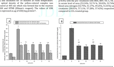
Fig.1 shows subcutaneous administration of CCl4 caused a
significant (p<0.001, p<0.05) increase in serum level of urea, blood urea nitrogen and creatinine 387.30%, 376% and
646.82% respectively in CCl4 treated Group compared to
control which was then ameliorated significantly (p<0.001, p<0.05) with the pre- treatment with BHA, BHT, Vit. C, Vit. E in serum level of urea (53.24%, 52.74 %, 50.02%, 52.74%), blood urea nitrogen (52.75%, 51.17%, 49.25%, 51.01%) and creatinine (58.01%, 57.11%, 57.28%, 57.42%) respectively
compared to CCl4 treated group.
I II III IV V VI
0 10 20 30 40 50 60 70 80 90 *# **# *# *# *# *# *# *# ** ** C ON C E N T R A T IO N O F S E R U M U R E A & B L OO D U R E A N ITRO GE N (m g /d l) GROUPS CONCENTRATION OF SERUM UREA CONCENTRATION OF BLOOD UREA NITROGEN
I II III IV V VI
0 1 2 3 4 5 *# *# ** ## ** ## *
CONCENTRATION OF SERUM CRETININE
C ON C E N T R A T IO N O F S E R U M C R E A T IN IN E (m g /d l) GROUPS
A
B
ISSN: 2250-1177 [415] CODEN (USA): JDDTAO
ii. Electrolyte profile:
Concentrations of major electrolytes such as sodium and potassium ion in serum were measured. CCl4 induced nephrotoxicity
showed significant (p<0.001, p<0.05) increase in serum sodium and potassium concentration by 48.14%, 59.47% compared to control which was significantly (p<0.001, p<0.05) reduced by the pre- treatment with BHA (19.79%, 19.637%), BHT (19.47%,
19.06%), Vit. C (19.36%, 19.59%), Vit. E (19.34%, 19.65%) respectively compared to CCl4 treated group (Fig.2).
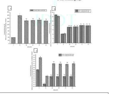
iii. Oxidative stress and Antioxidant enzyme profile:
In vivo production of malondyaldehyde (MDA) determines
rate of lipid peroxidation. Intoxication of CCl4 leads to an
abrupt (p<0.001) elevation in tissue MDA level within kidney (360.54%) cells compared to control (Fig.3). However, pretreatment with BHA (18.54%), BHT (17.98%), Vit. C (17.65%) and Vit. E (18.35%) significantly (p<0.001, p<0.05) ameliorated high level of tissue MDA content in both liver
and kidney cells compared to CCl4 treated group.
Subcutaneous intoxication of CCl4 leads to decreased
concentrations of intracellular antioxidant compounds (SOD,
CAT, GPx, GSH)which helps to maintain normal cellular homeostasis. Concentrations of SOD, CAT, GPx, and GSHin renal tissue (81.03%, 72.24%, 50.34%, 63.04%) homogenate had been decreased significantly (p<0.001, p<0.05) due
toCCl4intoxication compared to control (Fig.3). Conversely,
pretreatment with BHA, BHT, Vit C, Vit E significantly (p<0.001, p<0.05) recovered the level of intracellular antioxidant compounds like, SOD (231.65%, 233.01%, 230.14%, 230.03%), CAT (242.18%, 242.36%, 241.57%, 241.32%), GPx (35.17%, 35.62%, 34.98%, 36.01%), and GSH (62.67%, 62.21%, 60.66%, 62.23%) in renal cells compared
to CCl4 treated group.
I II III IV V VI
0 20 40 60 80 100 120 140 160 180 200 *# *# *# *# ** C ON C E N T R A T IO N O F S OD IU M ION (m m ol /l o f p la sm a) GROUPS
SODIUM ION CONCENTRATION
I II III IV V VI
0 1 2 3 4 5 *# *# ** ## ** ## *
POTASSIUM ION CONCENTRATION
C ON C E N T R A T IO N O F P OTA S S IU M ION (m m ol /l o f p la sm a) GROUPS
A
B
Fig. 2: Graphical representation of sodium and potassium ion concentration of different experimental groups. Values are expressed as mean±SEM, n=6. *,# and **,## indicates significant difference (P<0.001,P<0.05) compared to control Group and CCl4 treated group.
I II III IV V VI
0 20 40 60 80 100 120 140 160 180 200 *# *# *# *# * GROUPS C O N C E N T R A T IO N O F M D A (n m o l /m g o f ti s s u e ) IN R E N A L T IS S U E
TISSUE MDA CONTENT
I II III IV V VI
0 2 4 6 8 10 12 14 *# *# *# *# *# *# *# *# * * C O N C E N T R A T IO N O F G S H & G P x (m m o l o f H 2 O 2 C o n s u m p ti o n /m g o f ti s s u e /m in ) I N R E N A L T IS S U E GROUPS GSH CONCENTRATION GPx CONCENTRATION
I II III IV V VI
0.0 0.1 0.2 0.3 0.4 0.5 0.6 0.7 0.8 0.9 1.0 1.1 1.2 1.3 1.4
1.5 SOD CONCENTRATION CAT CONCENTRATION
*# *# *# *# *# *# *# *# * * C O N C E N T R A T IO N O F S O D & C A T (m m o l o f H 2 O 2 C o n s u m p ti o n /m g o f ti s s u e /m in ) IN R E N A L T IS S U E GROUPS
A
B
C
ISSN: 2250-1177 [416] CODEN (USA): JDDTAO
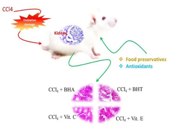
iv. Histological evaluations:
The histological changes observed were ranged from none (control group) to severe (CCl4 treated group). Administration of food preservatives and vitamins to food formulations helped to ameliorate such severe damage (Fig.4; Table.1). The assessment of the structure of kidney sections of control rats showed normal architectural
integrity. Administration of CCl4 caused significant
morphological damage to the renal cortex, affected glomeruli were observed with dilatation of Bowman’s space and renal tubules. There was an evident infiltration of lipid molecules into the cell as a key indicator of nephro- fibrosis. Supplementation of selected antioxidants leads to no or less degenerative changes in renal glomeruli as well as less sign of accumulation of fat droplets, cellular necrosis and
nephrotic vein disruption, compared to CCl4 treated group.
D
E
F
Fig.4: histological structure of kidney tissue Section A- Group I: Control, Section B- Group II: CCl4 treated, Section C-
Group III: CCl4 + BHA, Section D- Group IV: CCl4 + BHT, Section E- Group V: CCl4 + Vit. C, Section F-
Group VI: CCl4 + Vit. E . (Duration- 28 Days)
A
C
Table.1. Histological changes in different experimental groups (The Ishak system (a six-point scale) was applied for scoring nephrotoxicity. Group I:Control, Group II: CCl4 treated, Group III: CCl4 + BHA, Group IV: CCl4 + BHT, Group V: CCl4+Vit C, Group
VI: CCl4+Vit E. (Duration- 28 Days).
Parameters Group I Group II Group III Group IV Group V Group VI
Lipid Accumulation
0 6 2 2 2 2
lipid infiltration 0 6 2 2 2 2
Degeneration of glomeruli
0 6 1 1 1 1
Renal cell degeneration
0 6 1 1 1 1
Cellular necrosis 0 6 1 1 1 1
4.
DISCUSSION
CCl4 is a well known environmental pollutant specifically
hepatotoxin possesses the ability to induce nephrotoxicity through the generation of huge amount of reactive oxygen species. The generation of reactive oxygen species is the prime mechanism by to induce nephrotoxicity which can be contributed by any xenobiotic component from nature. In this study, we hypothesized that proposed food preservatives would effectively protect kidneys by its
antioxidant properties against CCl4 -induced injury. To the
best of our knowledge, this is the first study to evaluate the comparative antioxidative potential of food preservative
against CCl4 induced nephrotoxicity. CCl4 lowers intracellular
partial pressure of oxygen which favors reductive dehalogination of the said and produces trichloromethyl
radicals (•CCl3) with the help of nicotinamide adenine
dinucleotide phosphate (NADPH) dependent CYP450 enzyme. trichloromethyl radical in presence of oxygen converts into more toxic compound i.e. trichloro methyl
peroxyl radical (Cl3COO-) in mesangial cells of kidney29. Such
alteration in intracellular redox status leads to development of renal ascites with increased abdominal size together with congestion, edema, hypertension and acidosis, and eventually reaches to a state of total sodium and water
excess30. These types of toxic effects may be prevented by
supplementation of antioxidants. The efficacy of these food preservatives as antioxidant was determined by its degree of amelioration against altered intracellular redox status. The serum levels of urea, creatinine, blood urea nitrogen are often considered as reliable indices for the purpose of
measurement of in vivo renal impairment. In this study, it
was observed that the experimental group treated with CCl4
showed a significant increase in serum levels of urea and creatinine following exposure to CCl4. A significant reduction
in the plasma levels of urea is due to the decreased levels of protein in rats treated with carbon tetrachloride. Creatinine is a product of protein metabolism which is excreted in the urethra by glomerular filtration in higher concentration in comparison with its level in the blood. Therefore it can be
Fig. 5. Amelioration through food preservatives and vitamins in CCl4 induced
ISSN: 2250-1177 [418] CODEN (USA): JDDTAO
considered as a sign of kidney dysfunction31, 32. This kind of
altered renal function was re-established toward normal after pretreatment with BHA, BHT, Vit C and Vit E with food formulations.
Alterations in the concentration of electrolytes are important factors in disturbances in kidney function. An abrupt increase in the concentration of sodium ion (Na+) and a
decrease in potassium ion (K+) concentration in CCl4 treated
group successfully established failure in the renal system in experimental rats33. Alteration in the concentration of Na+
and K+ may alter the trans-epithelial transport of solutes and
water in the kidney. Although the treatment with antioxidants effectively reduced such alterations within a significant level.
Our earlier works had established strong free radical
scavenging activity against the same dose of CCl4 induced
toxicity in hepatic system by inhibition of lipid peroxidation by pre-treatment with synthetic antioxidants like butylated
hydroxyanisole and butylated hydroxytoluene18. CCl4
induces membrane-bound lipid peroxidation (specifically
PUFA) thus produces trichloromethyl radical (CCl3˙). Due to
alteration in intracellular partial pressure of oxygen later converts to a more toxic radical trichloromethyl peroxyl
radical (CCl3O2˙) which leads to detrimental organ damage
liver, Kidney, brain33, 34, 35. Production of increased
intracellular malondialdehyde (MDA) actually evidenced
CCl4 induced hepatonephro cellular damage due to abrupt
level of intracellular lipid peroxidation (LPO) which was
later replenished by administration of food
preservatives36,9,17,18. This free radical-induced intracellular
damage may be replenished by compounds with anti-oxidative properties. Superoxide dismutase (SOD) and catalase (CAT) are important detoxifying enzymes, which
protect against the free radical-mediated injury36. CCl4
induced oxidative stress (OS) was responsible for the decreased concentration of these above said intracellular antioxidant enzymes renal cells due to high scavenging activity37, 38. These free radical driven changes were recovered and intracellular SOD, as well as CAT, was restored by supplementation of BHA, BHT, Vit C, Vit E9,18. Subcutaneous administration of CCl4 was also responsible for the altered concentration of intracellular thiol (-SH) defense machinery. Increased LPO drove OS lead to a decreased level of reduced glutathione (GSH) as well as glutathione peroxidase (GPx) into the cell17 and there was the successful restoration of these above said oxidative markers by supplementation of food preservative antioxidants9,18. Thus, it may conclude that supplementation of synthetic as well as vitamin food preservative antioxidants mixed with food formulations able to provide protection against
nephrotoxicity induced by CCl4.
Conflict of interest statement
The authors declare that there are no conflicts of interest.
Acknowledgement
The authors thank the Department of Science and Technology (DST), Govt. of India, Raja N L Khan Women’s College, Midnapore, University College of Science and Technology, University of Calcutta, Midnapore College, West Bengal, India for providing the facilities to execute these studies
REFERENCES
(1) Schetz M, Dasta J, Goldstein S, Golper T. Druginduced acute kidney injury.Curr Opin Crit Care.2005;11:555-65.
(2) Rossberg M, Gerhartz W, Yamamoto YS, Campbell FT. 5th edition. Chlorinated hydrocarbons, Ullmann’s encyclopedia of industrial chemistry, VCH Publishers, New York. 2002;370 (3) Dassarma B, Samanta S, Nandi DK. Toxicokinetics of
carbontetrachloride (CCl4) exposure on hepatorenal system. Euro. J. Biomed. Pharmace. Sci. 2017a; 4(1):520-5.
(4) Bahcecioglu, I.H., Ustundag, B., Ozercan, I., Ercel, E., Baydas, G.,Akdere, T., Demir, A., Protective effect of Ginkgo biloba extracton CCI4-induced liver damage. Hepatology Research 1999; 15:215–224.
(5) Fadhel, Z.A., Amran, S., Effects of black tea extract on carbon tetrachloride-induced lipid peroxidation in liver, kidneys, and testes of rats. Phytotherapy Research 2002; 16:28–32. (6) Kahl R, Kappus H. Toxicology of the synthetic antioxidants
BHA and BHT in comparison with the natural antioxidant vitamin E. Zeitschrift fur Lebensmittel-untersuchung und-forschung. 1993; 196(4):329-38.
(7) Yehye WA, Rahman NA, Ariffin A, Hamid SB, Alhadi AA, Kadir FA, Yaeghoobi M. Understanding the chemistry behind the antioxidant activities of butylated hydroxytoluene (BHT): A review. European journal of medicinal chemistry. 2015; 101:295-312.
(8) Burton G, Ingold KU. Autoxidation of biological molecules. 1. Antioxidant activity of vitamin E and related chain-breaking phenolic antioxidants in vitro. Journal of the American Chemical Society. 1981; 103(21):6472-7.
(9) Elgazar AF. Effects of butylated hydroxytoluene and butylated hydroxyanisole against hepatotoxicity induced by carbon tetrachloride in rats. World Applied Sci J. 2013; 22(1):63-9. (10) Landi M, Degl'Innocenti E, Guglielminetti L, Guidi L. Role of
ascorbic acid in the inhibition of polyphenol oxidase and the prevention of browning in different browning‐sensitive Lactuca sativa var. capitata (L.) and Eruca sativa (Mill.) stored as fresh‐cut produce. Journal of the Science of Food and Agriculture. 2013; 93(8):1814-9.
(11) Institute of Medicine (2000). "Vitamin E". Dietary Reference Intakes for Vitamin C, Vitamin E, Selenium, and Carotenoids. Washington, DC: The National Academies Press. pp. 186– 283. (12) Galli F, Azzi A, Birringer M, Cook-Mills JM, Eggersdorfer M,
Frank J, Cruciani G, Lorkowski S, Özer NK. Vitamin E: Emerging aspects and new directions. Free Radical Biology and Medicine. 2017; 102:16-36.
(13) Dalton L. Food Preservatives. Chemical and Engineering News. 2002; 80(45):40.
(14) USDA Food Composition Databases.United States Department of
Agriculture, Agricultural Research Service. Release 28. 2015.
Retrieved 18 August 2018.
(15) https://www.accessdata.fda.gov/scripts/fdcc/?set=SCOGS (16) Olfert ED, Cross BM, McWilliam AA, editors. Guide to the care
and use of experimental animals. Ottawa: Canadian Council on Animal Care; 1993.
(17) Dassarma B, Samanta S, Nandi D K, Gangopadhyay S. Carbon tetrachloride induced hepatorenal failure in male albino rats: A dose response study. International Journal of Recent Scientific Research. 2017b; 8(3): 15885-15892.
(18) Dassarma B, Nandi DK, Gangopadhyay S, Samanta S. Hepatoprotective effect of food preservatives (butylated hydroxyanisole, butylated hydroxytoluene) on carbon tetrachloride-induced hepatotoxicity in rat. Toxicology reports. 2018a; 5:31-7.
(19) Al-Attar AM. Vitamin E attenuates liver injury induced by exposure to lead, mercury, cadmium and copper in albino mice. Saudi journal of biological sciences. 2011 Oct 1;18(4):395-401.
(20) Asuku O, Atawodi SE, Onyike E. Antioxidant, hepatoprotective, and ameliorative effects of methanolic extract of leaves of Grewia mollis Juss. on carbon tetrachloride–treated albino rats. Journal of medicinal food. 2012; 15(1):83-8.
(21) Coulambe CG, Favrean LA. Quaternary ammonium compound toxicity in chicken. Clinical Chemistry. 1965; 11(17):624. (22) Bosnes RW, Taussky HH. On the colorimetric determination of
creatinine by the Jaffe reaction. J Biol Chem 1945; 158: 581. (23) Ohkawa H, Ohishi N, Yagi K. Assay for lipid peroxides in animal
(24) Maestro RF, McDonald W. Oxidative enzymes in tissue homogenates. Handbook of methods for oxygen radical research. 1985:291-6.
(25) Luck HA. Spectrophotometric method for the estimation of catalase. Methods of enzymatic analysis. 1963.
(26) Moron MS, Depierre JW, Mannervik B. Levels of glutathione, glutathione reductase and glutathione S-transferase activities in rat lung and liver. Biochimica et Biophysica Acta (BBA)-General Subjects. 1979; 582(1):67-78.
(27) Paglia DE, Valentine WN. Studies on the quantitative and qualitative characterization of erythrocyte glutathione peroxidase. The Journal of laboratory and clinical medicine. 1967; 70(1):158-69.
(28) Iranloye BO, Bolarinwa AF. Effect of nicotine administration on weight and histology of some vital visceral organs in female albino rats. Nigerian Journal of Physiological Sciences. 2009; 24(1).
(29) Dassarma B, Jana S, Rakshit S, Nandi D K, Gangopadhyay S, Samanta S. An overview of systemic effects of carbon tetrachloride (CCl4) exposure. Indian Science Cruiser. 2018b; 32(3): 36-40.
(30) Adewole S, Salako A, Doherty O, Naicker T. Effect of melatonin on carbon tetrachloride-induced kidney injury in Wistar rats. African Journal of Biomedical Research. 2007; 10(2).
(31) Rafieian-Kopaei M, Baradaran A. Teucrium polium and kidney. Journal of renal injury prevention. 2013; 2(1):3-4.
(32) Nasri H. On the occasion of the world diabetes day 2013; diabetes education and prevention; a nephrology point of view. Journal of renal injury prevention. 2013;2(2):31. (33) Pradhan S, Mandal S, Roy S, Mandal A, Das K, Nandi DK.
Attenuation of uremia by orally feeding alpha-lipoic acid on acetaminophen induced uremic rats. Saudi Pharmaceutical Journal. 2013; 21(2):187-92.
(34) Ritesh KR, Suganya A, Dileepkumar HV, Rajashekar Y, Shivanandappa T. A single acute hepatotoxic dose of CCl4 causes oxidative stress in the rat brain. Toxicology reports. 2015; 2:891-5.
(35) Tirkey N, Pilkhwal S, Kuhad A, Chopra K. Hesperidin, a citrus bioflavonoid, decreases the oxidative stress produced by carbon tetrachloride in rat liver and kidney. BMC pharmacology. 2005; 5(1):2.
(36) Oyinbo CA, Dare WN, Okogun GR, Anyanwu LC, Ibeabuchi NM, Noronha CC, Okanlawon OA. The hepatoprotective effect of vitamin C and E on hepatotoxicity induced by ethanol in Sprague Dawley rats. Pak J Nutr. 2006; 5(6):507-11.
(37) Sreelatha S, Padma PR. Antioxidant activity and total phenolic content of Moringa oleifera leaves in two stages of maturity. Plant foods for human nutrition. 2009; 64(4):303.