Original Research Article
Clinical, laboratory and radiological profile of dengue among pediatric
patients admitted in tertiary care hospital
Sangeetha Jairaj
1, Sridhar D.
2,
Mettu Pradeep Reddy
3*
INTRODUCTION
Dengue fever (DF) is the commonest of the arboviral infections in humans.1 Dengue is found in tropical and subtropical regions around the world, predominantly in urban and semi-urban areas. Dengue is a mosquito-borne viral infection causing a severe flu-like illness and, sometimes causing a potentially lethal complication called severe dengue. Over the past three decades, there
has been a dramatic global increase in the frequency of dengue fever (DF). Up to 50-100 million infections are now estimated to occur annually in over 100 endemic countries, putting almost half of the world’s population at risk.2
The viruses that cause dengue fever are Flaviviruses. There are four closely related but antigenically distinct virus serotypes: DEN-1, DEN- 2, DEN-3, and DEN-4.3 Dengue viruses cause symptomatic infections or
ABSTRACT
Background: Dengue is found in tropical and subtropical regions around the world, predominantly in urban and semi-urban areas. Dengue mortality can be reduced by implementing early case detection and referral systems for patients; managing severe cases with appropriate treatment; reorienting health services to cope with dengue outbreaks; and training health personnel at all levels of the health system. Many studies that focus on the difference between the frequency of clinical findings in DHF and dengue shock syndrome (DSS) with respect to classical DF has been published. The objectives of the study were to assess signs and symptoms of dengue among patients with severe dengue and patients with dengue fever and to evaluate laboratory and radiological profile among dengue patients.
Methods: This Hospital based cross sectional study was conducted on 70 seropositive cases, admitted in the Paediatrics Department of Gandhi hospital during the period of July to December 2017 for 6months. Data was analyzed by using SPPS Version 17 and student t test was used for inferential statistics.
Results: Out of 70 cases 38 were presented with severe dengue and 32 presented with dengue fever. Average age of presentation is 7.24 years among severe dengue group, 5.52 years among dengue fever group. In group of severe dengue, 60.5% of the cases were female, which was insignificantly more as compared to 40.6% of the cases from group of dengue fever.
Conclusions: All levels of health personnel must be aware of clinical signs and symptoms of all dengue types. Careful monitoring of unusual presentations early recognition severe manifestation and timely intervention can reduce disease specific mortality rate.
Keywords: Dengue fever, Severe dengue, Clinical laboratory and profiles
Department of Community Medicine, 1Gandhi Medical College, Secunderabad, 2ESIC Medical College, Hyderabad, Telangana, India
3
Department ofPediatrics, ESIC Medical College, Hyderabad, Telangana, India
Received: 05 April 2018
Revised: 23 April 2018
Accepted: 24 April 2018
*Correspondence:
Dr. Mettu Pradeep Reddy,
E-mail: [email protected]
Copyright: © the author(s), publisher and licensee Medip Academy. This is an open-access article distributed under the terms of the Creative Commons Attribution Non-Commercial License, which permits unrestricted non-commercial use, distribution, and reproduction in any medium, provided the original work is properly cited.
asymptomatic seroconversion. Symptomatic dengue infection is a systemic and dynamic disease. It has a wide clinical spectrum that includes severe and non-severe clinical manifestations. After the incubation period, the illness begins abruptly and, in patients with moderate to severe disease, is followed by three phases − febrile, critical and recovery. Due to its dynamic nature, the severity of the disease will usually only be apparent around defervescence i.e. during the transition of the febrile to the afebrile phase, which often coincides with the onset of the critical phase.4 Many DF cases are self-limiting but its complications like haemorrhage and shock can be life-threatening. If untreated, mortality from the complications of DF is as high as 20%, whereas if recognized early and managed properly, mortality is less than 1%. Hence, it will be useful if certain symptoms, signs and laboratory parameters associated with the development of complications are identified so that such cases would receive more attention.1 For a disease that is complex in its manifestations, management is relatively simple, inexpensive and very effective in saving lives, so long as correct and timely interventions are instituted. The key to a good clinical outcome is understanding and being alert to the clinical problems that arise during the different phases of the disease, leading to a rational approach in case management.4
Dengue mortality can be reduced by implementing early case detection and referral systems for patients; managing severe cases with appropriate treatment; reorienting health services to cope with dengue outbreaks; and training health personnel at all levels of the health system.2 Many studies that focus on the difference between the frequency of clinical findings in DHF and dengue shock syndrome (DSS) with respect to classical DF has been published.
Objectives
To assess signs and symptoms of dengue among patients with severe dengue and patients with dengue fever.
To evaluate laboratory and radiological profile among dengue patients.
METHODS
This hospital based cross sectional study was conducted on seropositive cases of dengue in 70 patients admitted in the Paediatrics Department of Gandhi hospital during the period of July to December 2017 for 6months.A protocol was drafted according to the specifications of the Ethics Committee of the hospital. Informed written consent of the parents was obtained before starting the study. 1to14years age group was included with clinical suspicion of dengue fever i.e. acute febrile illness with any of the associated symptoms like: myalgia, headache, retro-orbital pain, bleeding manifestations, abdominal pain, drowsiness, low platelets count, evidence of shock and dengue sero-positive. Dengue sero-negative and all
children with identified bacterial or other focus of infection were excluded. Data was analyzed by using SPPS version 17 and student t test was used for inferential statistics.
RESULTS
Out of 70 cases 38 were presented with severe dengue and 32 presented with dengue fever. Average age of presentation is 7.24 years among severe dengue group, 5.52 years among dengue fever group the difference was not significant. In group of severe dengue, 60.5% of the cases were female, which was insignificantly more as compared to 40.6% of the cases from group of dengue fever.
Clinical profile
single patient with dengue fever had prolonged capillary refill time which was significantly less as compared to 34.2% of cases with severe dengue. This study states that, 65.8% of cases of severe dengue had warm pink extremities which were significantly less as compared to
100.0% of cases with dengue fever. This data indicates that, 42.1% of cases with severe dengue showed tourniquet test positive which was comparable with 40.6% of cases with dengue fever and the difference was not significant (Table 1).
Table 1: Clinical profile of study subjects.
Parameters Severe dengue (n=38)
(%)
Dengue fever (n=32)
(%) P value
Headache Absent 19 (50) 17 (53.1) >0.05
Present 19 (50) 15 (46.9)
Mayalgia Absent 14 (36.8) 11 (34.4) >0.05
Present 24 (63.2) 21 (65.6)
Retro orbital pain Absent 15 (39.5) 21 (65.6) <0.05
Present 23 (60.5) 11 (34.4)
Vomiting Absent 11 (28.9) 16 (50) >0.05
Present 22 (57.9) 16 (50)
Abdominal pain Absent 3 (8.1) 26 (81.2) <0.05
Present 30 (81.1) 6 (18.8)
Consciousness
Clear and lucid 21 (55.2) 27 (84.4)
<0.05
Irritable 5 (13.2) 4 (12.5)
Lethargy 7 (18.4) 1 (3.1)
Restless 5 (13.2) 0 (0)
Rash/petechie Absent 20 (52.6) 19 (59.4) >0.05
Present 18 (47.4) 13 (40.6)
Blood pressure Normal 35 (92.1) 32 (100) >0.05
Hypotension 3 (7.9) 0 (0)
Tachy cardia Good volume 25 (65.8) 32 (100) <0.05
Feeble and weak thready 13 (34.2) 0 (0)
Capillary refill time Brisk 25 (65.8) 32 (100) <0.05
Prolonged 13 (34.2 0 (0)
Extremities between Cold, clammy 13 (34.2) 0 (0) <0.05
Warm Pink 25 (65.8) 32 (100)
Tourniquet test Absent 22 (57.9) 19 (59.4) >0.05
Present 16 (42.1) 13 (40.6)
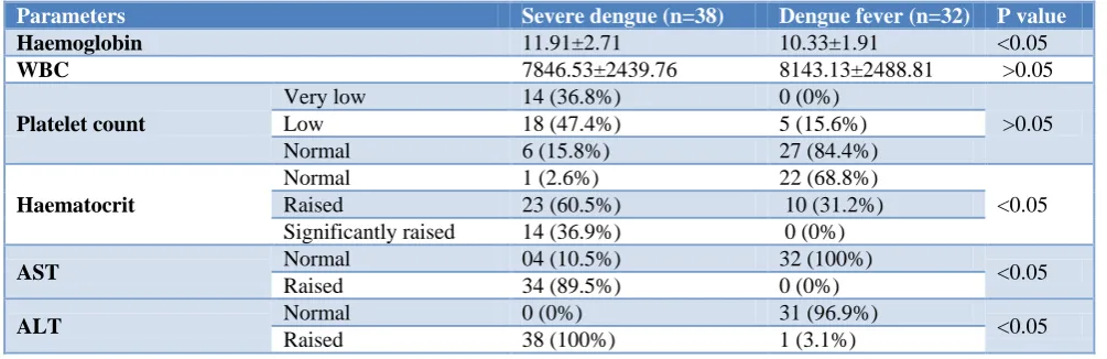
Table 2: Laboratory profiles of study subjects.
Parameters Severe dengue (n=38) Dengue fever (n=32) P value
Haemoglobin 11.91±2.71 10.33±1.91 <0.05
WBC 7846.53±2439.76 8143.13±2488.81 >0.05
Platelet count
Very low 14 (36.8%) 0 (0%)
>0.05
Low 18 (47.4%) 5 (15.6%)
Normal 6 (15.8%) 27 (84.4%)
Haematocrit
Normal 1 (2.6%) 22 (68.8%)
<0.05
Raised 23 (60.5%) 10 (31.2%)
Significantly raised 14 (36.9%) 0 (0%)
AST Normal 04 (10.5%) 32 (100%) <0.05
Raised 34 (89.5%) 0 (0%)
ALT Normal 0 (0%) 31 (96.9%) <0.05
Raised 38 (100%) 1 (3.1%)
Laboratory profile
Mean HB was 11.91 in group of severe dengue, which was more as compared to 10.33 of dengue fever group and difference was statistically significant. According to
significantly more as compared to 15.6% of patients with dengue fever. HMCRT was raised to significantly raised in 97.4% of patients having severe dengue, which was significantly more as compared to 31.2% of patients with dengue fever. This study profile states that, not a single patient with dengue fever had AST rose, which was less as compared to 89.5% of patients with severe dengue who had raised AST value and the difference was statistically significant. ALT value was observed raised in 100.0% of patients having severe dengue, which was significantly more as compared to 3.1% of patients with dengue fever (Table 2).
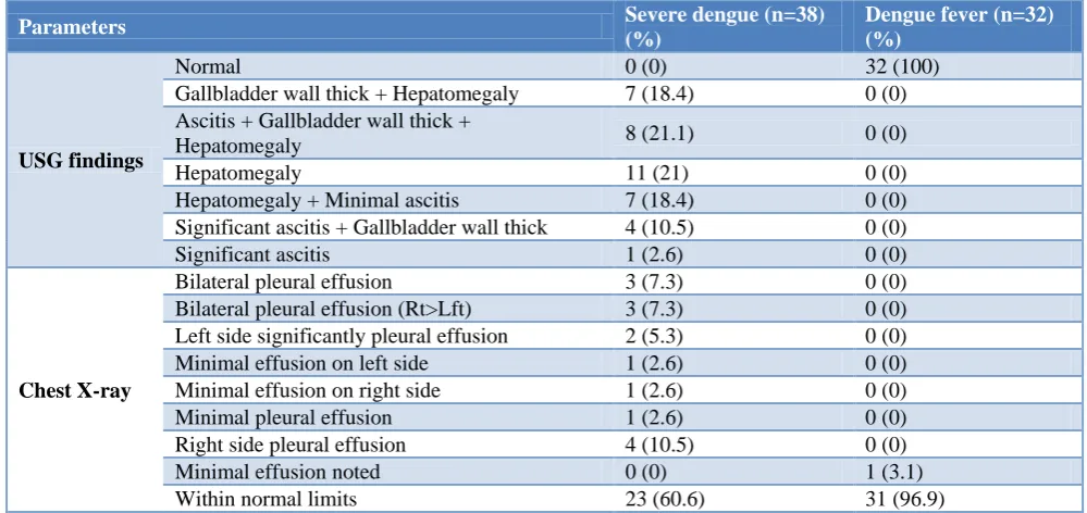
Radiological profile
Present study states that, CRX was not within normal limit in 39.4% of cases with severe dengue which was more as compared to 3.1% of cases with dengue fever. This study profile states that, not a single patient with dengue fever had ultrasonography abdomen abnormal, which was less as compared to 100.0% of patients with severe dengue who had ultrasonography abdomen abnormal (Table 3).
Table 3: Radiological findings among study subjects.
Parameters Severe dengue (n=38)
(%)
Dengue fever (n=32) (%)
USG findings
Normal 0 (0) 32 (100)
Gallbladder wall thick + Hepatomegaly 7 (18.4) 0 (0)
Ascitis + Gallbladder wall thick +
Hepatomegaly 8 (21.1) 0 (0)
Hepatomegaly 11 (21) 0 (0)
Hepatomegaly + Minimal ascitis 7 (18.4) 0 (0)
Significant ascitis + Gallbladder wall thick 4 (10.5) 0 (0)
Significant ascitis 1 (2.6) 0 (0)
Chest X-ray
Bilateral pleural effusion 3 (7.3) 0 (0)
Bilateral pleural effusion (Rt>Lft) 3 (7.3) 0 (0)
Left side significantly pleural effusion 2 (5.3) 0 (0)
Minimal effusion on left side 1 (2.6) 0 (0)
Minimal effusion on right side 1 (2.6) 0 (0)
Minimal pleural effusion 1 (2.6) 0 (0)
Right side pleural effusion 4 (10.5) 0 (0)
Minimal effusion noted 0 (0) 1 (3.1)
Within normal limits 23 (60.6) 31 (96.9)
DISCUSSION
Dengue is most epidemiologically important mosquito born disease in tropical countries like India. It is causing major outbreaks leading to mortality and morbidity in the continent
70 cases were included in the study. Majority males infected with dengue compared to females. This is similar with many studies, simple attribution to this preponderance is, females covered with clothes in Indian
scenario and male children given importance
sociologically to bring hospital.5 The most common age group affected was 8-11 years indicating the school activity of these group and exposure to breeding places of mosquitoes during playing. Severe dengue presented with 7 -8 yrs age group which is similar to Ahmed et al.6
Results were analysed and presented according to WHO TDR guidelines 2009. All cases were categorised into severe dengue and dengue fever. In our study 45.71% were non severe and 54.28% as severe dengue cases.
Symptoms like headache and myalgia were almost equal in both groups marginally high percentage of above symptoms has seen in severe dengue group. Retro orbital pain was seen with higher percentage among severe dengue patients. Mishra et al presented similar results.7
However headache was found with higher prevalence among non-severe dengue patients in Rehman et al.8 This variation is because of subjective feeling of patients.
Vomiting, abdominal pain symptoms were marginally
high among severe dengue patients. Dengue
haemorrhagic symptoms like consciousness, hypotension, feeble pulse, cold calmy extremities were seen among severe dengue group. Similar findings had been observed by Ahmed et al.5
non-severe dengue cases. According to this observation probably there is no demarcation between severe and non-severe; health personal must aware with all signs and symptoms and able to tackle all situations.
Majority of severe dengue patients were presented with thrombocytopenia compared to non-severe dengue patients. Studies by Harris et al, Murge et al reported no significant correlation between platelet count and bleeding manifestations.11,12 In contrast Mittal et al found significant co relation.13 Raised haematocrit and decreased white blood cell count was observed significantly among severe dengue cases. Agarwal et al reported contrast results.14 Type of study, subjects involved in the study and drawing of blood sample plays pivotal role for contrast results.
ALT and AST levels were significantly increased among severe dengue cases compared to non-severe cases, similar results were observed by Kalyanarooj et al mayosites involvements leads increase of AST and ALT.15
Pleural effusion was noted in all cases of severe dengue cases. Either it may bilateral or unilateral. Majority of non-severe dengue cases didn’t show any plural effusion. These findings are similar with studies of Pushpa et al and Malavige et al.16,17 Hepatomegaly was found in severe dengue cases and ascetic fluid was also seen among severe dengue cases these findings are similar with findings of Joshi et al and Srivastava et al.18,19
CONCLUSION
All levels of health personnel must be aware of clinical signs and symptoms of all dengue types. Careful monitoring of unusual presentations early recognition severe manifestation and timely intervention can reduce disease specific mortality rate.
Funding: No funding sources Conflict of interest: None declared
Ethical approval: The study was approved by the Institutional Ethics Committee
REFERENCES
1. Narayanan M, Aravind MA, Thilothammmal N,
Prema R, Sargunam CSR, Ramamurty N. Dengue fever epidemic in Chennai – A study of clinical profile and outcome. Indian Pediatr. 2002;39:1027-33.
2. Special programme for research, training in tropical diseases, and World Health Organization, dengue: guidelines for diagnosis, treatment, prevention and control, World Health Organization, Geneva, Switzerland; 2009.
3. Alberta Health Public Health Notifiable Disease Management Guidelines. 2012.
4. World Health Organization, Regional Office for South-East Asia. Comprehensive Guidelines for Prevention and Control of Dengue and Dengue Hemorrhagic Fever, Revised and Expanded Edition.
WHO-SEARO 2011. (SEARO Technical
Publication Series No. 60); 2011.
5. Ahmed FU, Mahmood BC, Sharma JD, Hoque SM,
Zaman R, Hasan MS. Dengue and dengue haemorrhagic fever in children during the 2000 outbreak in Chittagong, Bangladesh. Dengue Bulletin. 2001;25:33-9.
6. Ahmed S, Arif F, Yahya Y, Rehman A, Abbas K, Ashraf S, et al. Dengue fever outbreak in Karachi 2006: a study of profile and outcome of children under 15 years of age. J Pakistan Med Assoc. 2008;58(1):4-8.
7. Mishra S, Ramanathan R, Agarwalla SK. Clinical profile of dengue fever in children: a study from
Southern Odisha, India. Scientifica.
2016;6391594:6.
8. Rahman M, Rahman K, Siddque AK, Shoma S,
Kamal AH, Ali KS, et al. First outbreak of dengue hemorrhagic fever, Bangladesh. Emerg Infect Dis. 2002;8:738-40.
9. Kobilan L, Balasubramanian S, Keshava SM.
Dengue disease spectrum among infants in the 2001 dengue epidemic in Chennai, Tamilnadu, India. J Clin Microbiol. 2003;41:3919-21.
10. Mishra B, Itsstho RK. Virological interpretations of dengue disease spectrum in infants in Chennai, Tamil Nadu, India, need re-evaluation. J Clin Microbiol. 2004;42:23-7.
11. Harris E, Videa E, Perez L, Sandoval E, Tellez Y, Perez ML, et al. Clinical, epidemiologic, and virologic features of dengue in the 1998 epidemic in Nicaragua. Am J Trop Med Hyg. 2000;63:5-11. 12. Murgue B, Deparis X, Chungue E, Cassar O, Roche
C. Dengue: an evaluation of dengue severity in French Polynesia based on an analysis of 403 laboratory- confirmed cases. Trop Med Int Health. 1999;4:765-73.
13. Mittal HM, Faridi MA, Arora SK, Patil R, Clinicohematological profile and platelet trends in children with dengue during 2010 epidemic in North India. Indian J Pediatr. 2012;79(4):467-71.
14. Aggarwal A, Chandra J, Aneja S, Patwari AK, Dutta AK. An epidemic of dengue hemorrhagic fever and dengue shock syndrome in children in Delhi. Indian Pediatr. 1998;35:727-32.
15. Kalayanarooj S, Vaughn DW, Nimmannitya S.
Early clinical and laboratory indicators of acute dengue illness. J Infect Dis. 1997;176(2):313-21. 16. Pushpa V, Venkatadesikalu M, Mohan S, Cherian T,
John TJ, Ponnuraj EM. An epidemic of dengue haemorrhagic fever/dengue shock syndrome in tropical India. Ann Trop Pediatr. 1998;18:289-93. 17. Malavige GN, Ranatunga PK, Velathanthiri VGNS,
18. Joshi R, Baid V. Profile of dengue patients admitted to a tertiary care hospital in Mumbai. Turkish J Pediatr. 2011;53(6):626-31.
19. Srivastava VK, Suri S, Bhasin A, Srivastava L, Bharadwaj M. An epidemic of dengue hemorrhagic fever and dengue shock syndrome in Delhi: a clinical study. Ann Trop Pediatr. 1990;10:329-34.