Prateek Chaudhury et al JMSCR Volume 06 Issue 09 September 2018 Page 715
A Prospective Study to Evaluate Cardiac Functional Status in Permanent
Pacemaker Implanted Patients at a Tertiary Care Centre
Authors
Prateek Chaudhury
1, Lipika Adhikari
2*, Biswajit Majumdar
3, Abhay Chaturbedi
41,4
DM Senior Resident, Cardiology Department, RG Kar Medical College, Kolkata
2
Assistant Professor, Cardiology Department, RG Kar Medical College, Kolkata
3
Associate Professor, Cardiology Department, RG Kar Medical College, Kolkata *Corresponding Author
Lipika Adhikari
Assistant Professor, Cardiology Department, RG Kar Medical College, Kolkata
Address: C/O Manoj Adhikari, School Danga, post & district Bankura, West Bengal, PIN 722101, India
Abstract
Our study aimed at finding the incidence of overt heart failure or asymptomatic left ventricular systolic/diastolic dysfunction and cardiac dyssynchrony in patients undergoing single or dual chamber cardiac pacing and its impact on exercise capacity. 80 patients undergoing permanent pacemaker implantation (single or double chamber) were examined for various predetermined echocardiographic parameters of left ventricular systolic function, diastolic function and dyssynchrony before and one year after implantation. Also the impact of pacing on the exercise capacity is assessed by six minute walk test. The various echocardiographic parameters of left ventricular systolic function, diastolic function and dyssynchrony namely, Ejection Fraction (EF), E´ velocity, Interventricular Mechanical Delay (IVMD), Septum to Posterior Wall Motion Delay (SPWMD), Time to peak Systolic velocity between the septal and lateral wall (Ts-SL), showed no significant variation between before and after pacemaker implantation values. In patients with normal ejection fraction and with no structural heart disease there was no significant difference in the systolic, diastolic function and various dyssynchrony indices after one year of right ventricular pacing.Also right ventricular pacing did not affect the functional capacity of the patient as assessed by six minute walk test.
Keywords: Cardiac pacing, Echodoppler parameters, Left ventricular systolic function, Left ventricular diastolic function, Six minute walk test.
Introduction
Cardiac pacing at any point of the ventricle alters the natural heart activation and contraction pattern, as the stimulus conduction velocity is slower across the ventricular myocardium when compared with that resulting from the specialized His-Purkinje system[1,2]. RV apical pacing can
induce both interventricular dyssynchrony [between the RV and the LV] and intraventricular dyssynchrony (within the LV)[3].
It has been demonstrated that the presence of ventricular dyssynchrony is associated with an increased risk of cardiac morbidity[4] and mortality[5] in heart failure patients. In addition, it
www.jmscr.igmpublication.org Impact Factor (SJIF): 6.379
Prateek Chaudhury et al JMSCR Volume 06 Issue 09 September 2018 Page 716 has been suggested that the presence of
mechanical dyssynchrony after long-term RV apical pacing is associated with reduced LV systolic function and deterioration in the functional capacity[3].
Left ventricular diastolic function is an equally important component of the cardiac cycle, which is closely coupled with systolic function. In particular, early diastolic function is an energy-dependent process consuming ATP, and yet is also dependent on the mechanical properties of the ventricle such as chamber pressure, annulus recoil, and stiffness[6]. However, there is little information on the effects of RVA pacing on diastolic function. Early studies were unable to draw a clear conclusion on the effect of RVA pacing on LV diastolic function, and its relationship with changes in systolic function[7-10]. In the background of this knowledge we seek to find the prevalence of new onset LV systolic dysfunction and cardiac dyssynchrony and its impact on the functional status in patients undergoing single or dual chamber PPM implantation
Materials and Methods
A total of 80 patients undergoing single/dual pacemaker implantation at R. G. Kar Medical College over a span of 1 year from JAN 2016 to JAN 2017 formed the study population.
Inclusion Criteria
1. All patients undergoing PPI for Atrioventricular node or Sinus node dysfunction.
2. No structural heart disease at the time of implantation.
3. Ability to provide written consent.
Exclusion Criteria
1. Comorbidities that themselves might cause LV remodelling
a. History of chemotherapy with cardiotoxic agents,
b. Hypertensive heart disease, c. Chronic alcohol abuse, and d. Valvular heart disease
2. Chronic disesases like CKD, CLD, malignancies etc.
3. H/o ACS in past or during study period
Study Protocol
All patients underwent echocardiography, ECG and detailed clinical examination before and after 1 year of endovenous single/dual chamber pacing. 6 minute walk test (6MWT) was also performed before or within 1 month and after 1 year of endovenous single/dual chamber pacing (if there is no physical disability or other contraindications to 6MWT).
Before Pacemaker Implantation Echocardiography
Transthoracic echocardiography examination was done using Philips EPIQ7C machine to define cardiac anatomy, ventricular function, and valvular competence. LV posterior and septal wall thickness and LV end-diastolic and end-systolic diameters were obtained by averaging the measurements over 3 consecutive cardiac cycles with M-mode echocardiography.
Prateek Chaudhury et al JMSCR Volume 06 Issue 09 September 2018 Page 717 dyssynchrony in patients with non-ischemic
cardiomyopathy. Time-to-peak velocity between the septal and lateral wall was measured using the 4-chamber color TDI images from the beginning of the QRS complex, and the maximal time-to-peak difference between the basal segments of the septal and the lateral walls was calculated (Ts-SL). A predefined cut off ≥65 was considered dyssynchrony.
Electrocardiography
Baseline ECG was done during index admission before and after PPI and then at 2 months, 6 months and 1 year after implantation to detect new, delayed and late onset rhythm abnormalities. 6 Minute Walk Test:
6 minute walk test was done before or within 1 month of PPI.
After Pacemaker Implantation
ECHO, ECG and 6 min walk test will be repeated after 1 year ofPPI for reassessment of the above mentioned parameters.
Statistical Analysis
The collected data were tabulated and statistically analyzed using the SPSS version 20.0 for Windows (SPSS Inc., Chicago, Illinois, USA). Comparisons between the baseline and at one year parameters were performed using the Wilcoxon
Signed Ranks Test. A probability value of 0.05 was considered to be statistically significant
Results and Observations
In the study population, 52(65%) were male and 28(35%) were female subjects. Mean age of the study population was 66.57 ± 7.031 years. Out of the total 80 patients undergoing permanent pacemaker implantation, 48(60%) were hyperten-sive and 21(26.2%) patients were diabetic.
In the patients undergoing permanent pacemaker implantation 49(61.3%) had complete heart block, 21(26.3%) had sick sinus syndrome, 8(10%) had trifasicular block and 2(2.5%) had bifasicular block, with all patients having class 1 indication for permanent pacing.
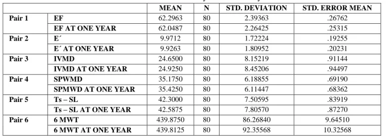
Out of the total 80 patients, 61 patients (76.3%) underwent single chamber permanent pacemaker implantation and the rest 19(23.8%) underwent dual chamber permanent pacemaker implantation. There were various variables, namely Ejection Fraction (EF), E´ velocity, Interventricular Mechanical Delay (IVMD), Septum to Posterior Wall Motion Delay (SPWMD), Time to peak Systolic velocity between the septal and lateral wall(Ts-SL) and 6 Minute Walk Test, which were examined at baseline and at one year to assess the impact of right ventricular pacing(Table 1).
Table 1: Various variables studied at baseline and at one year in the study
MEAN N STD. DEVIATION STD. ERROR MEAN
Pair 1 EF 62.2963 80 2.39363 .26762
EF AT ONE YEAR 62.0487 80 2.26425 .25315
Pair 2 E´ 9.9712 80 1.72224 .19255
E´ AT ONE YEAR 9.9263 80 1.80952 .20231
Pair 3 IVMD 24.6500 80 8.15219 .91144
IVMD AT ONE YEAR 24.9250 80 8.45206 .94497
Pair 4 SPWMD 35.1750 80 6.18855 .69190
SPMWD AT ONE YEAR 35.4250 80 6.11447 .68362
Pair 5 Ts – SL 42.3000 80 7.50595 .83919
Ts – SL AT ONE YEAR 42.5875 80 7.80570 .87270
Pair 6 6 MWT 439.8750 80 86.26840 9.64510
6 MWT AT ONE YEAR 439.8125 80 92.35568 10.32568
Prateek Chaudhury et al JMSCR Volume 06 Issue 09 September 2018 Page 718
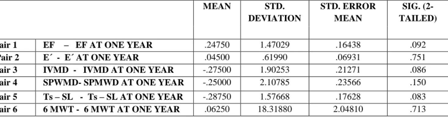
Table 2 Showing variation of different parameters studied in pacemaker implanted patients at baseline and at one year (Wilcoxon Signed Ranks Test)
MEAN STD.
DEVIATION
STD. ERROR MEAN
SIG. (2-TAILED)
Pair 1 EF – EF AT ONE YEAR .24750 1.47029 .16438 .092
Pair 2 E´ - E´ AT ONE YEAR .04500 .61990 .06931 .751
Pair 3 IVMD - IVMD AT ONE YEAR -.27500 1.90253 .21271 .086
Pair 4 SPWMD- SPMWD AT ONE YEAR -.25000 2.10785 .23566 .150
Pair 5 Ts – SL - Ts – SL AT ONE YEAR -.28750 1.57668 .17628 .083
Pair 6 6 MWT - 6 MWT AT ONE YEAR .06250 18.31880 2.04810 .713
Discussion
Long-term effects of RV apical pacing have been studied, and not much information is available on the acute and early effects of RV pacing on LV function and LV dyssynchrony[11]. Our study aimed to demonstrate the early impact of RV apical pacing induced by single chamber and dual-chamber pacemakers on LV systolic and diastolic functions in patients with preserved EF
Effect on left ventricular systolic functions
In our study, there was no statistically significant difference within repeated measurements of the LV EF over time (p =0.136). Mohan et al.[12] in their study found no significant difference in the resting LV EF in the long-term follow-up of AAI-paced and VVI-AAI-paced patients of sinus node dysfunction without structural heart disease. Similarly, Anderson et al.[13] in their study of sick sinus syndrome patients, found no difference in the incidence of clinical congestive heart failure or LV dimensions between AAI and VVI pacing modes on long-term follow-up. In contrast, Pehrsson et al.[14] found an increase in the end-diastolic volume in VVI-paced patients after 3 months. Likewise, Faerestrand et al.[15] found greater resting LV volumes and lesser EF in patients paced on VVI rather than with the physiological pacing mode. Dwivedi et al.[16] studied 48 patient with VVI pacing and found that the LV EF decreased progressively from baseline (61.82 ± 10.36%) and was statistically significant at 6 months (52.52 ± 12.11%, P < 0.05). The cardiac dimensions, the LV end diastolic dimension and the LV end-systolic dimension
increased significantly over their corresponding baseline values by 6 months (P < 0.05). However, the aforementioned study used a higher percentage of pacing (>90%) in all patients, and this may be the cause of early effects on the LV dimension and systolic functions.
The effect on left ventricular diastolic functions
We did not find any statistically significant difference between both groups regarding septal and lateral E′ waves by pulsed-tissue Doppler. This was the same as in previous studies; they also found no significant changes in the diastolic function. Brandon K. Fornwalt[17] et al. acute RVA pacing does not affect diastolic synchrony (Diastolic dyssynchrony was 11 ± 13 ms, 15 ± 18 ms and 15 ± 27 ms for sinus rhythm, atrial pacing and right ventricular pacing respectively “p” = 0.375). Naegeli et al.[18] conducted a single-blind, randomized crossover study evaluating the impact of the DDD(R) mode as against the VVI(R) mode on objective and functional parameters. They found no significant changes in the transmitral flow propagation rate and the E/E´ ratio. Kojuri et al.[19] also found no significant changes in E and A′by pulsed-tissue Doppler between DDD and VVI.
Prateek Chaudhury et al JMSCR Volume 06 Issue 09 September 2018 Page 719 hypoxia suppresses the load dependence of
relaxation[22] suggesting that patients with coronary artery disease may have a different response to acute pacing relative to the healthy patients in our study.
Interventricular mechanical dyssynchrony
In our study there was no significant difference within the repeated measurement of IVMD at baseline (24.650 ± 8.152 ms) and at one year (24.925 ± 8.452), supported by a “p” value of .086. Similarly Tops et al.[3] showed that even after long-term RV pacing, mean interventricular delay did not increase significantly in the patients without LV dyssynchrony (24 ± 14 ms vs. 35 ± 25 ms, “p” > .05). In contrast, in the same study interventricular delay revealed a significant increase in the patients with LV dyssynchrony after long-term RV pacing (25 ± 12 ms vs. 49 ± 19 ms, “p” = 0.05).
Intraventricular dyssynchrony
Both M-mode using SPWMD and TDI using the septal-to-lateral delay have proven to be effective in the detection of LV dyssynchrony[23,24]. In our study, change in SPWMD and Ts-SL from baseline to one year was not significant (“p” = .150 and .083 repectively).
Previous studies have shown that ventricular dyssynchrony may be present in up to 50% of the patients after long-term RV apical pacing[25-27], which is in contrast to our findings. This can be partially explained by the shorter follow up period in our study (1 year) which was more (3 to 5 years) in the studies where ventricular dyssynchrony was seen associated with right ventricular pacing. In the study by Schmidt M et al.[25] where significant LV dyssynchrony developed after right ventricular pacing, had patients with severely depressed left ventricular ejection fraction, whereas in our study no patient had any structural heart disease. This can again explain the discrepancy of results between the two studies. .
It remains, however, undetermined why some patients develop LV dyssynchrony during RV pacing while other patients do not.
Functional assessment
In accordance with the other parameters of systolic and diastolic function, the functional capacity of the subjects as assessed by six minute walk test also did not show any statistically significant (“p” = .713) difference between baseline (439.875 ±86.268m) and at one year (439.812 ± 92.356m) measurements.
Limitations
The present analysis focuses on the short-term impact of RV apical pacing on LV functions and LV dyssynchrony and does not provide information about the long-term consequences. Also the number of patients was relatively small in this study. There was no control population to assess normal variation of various parameters with age. Electrocardiographic parameters like QRS duration and percent of paced beats was not determined and the site of RV pacing was not taken into account. Comparative analysis between patients undergoing single and double chamber pacemaker cannot be done because of difference in the baseline characters of the two groups. Finally better techniques, like 3D speckle tracking may have assessed intraventricular dyssynchrony better than the techniques used in the current study.
Conclusion
Right ventricular apical pacing is an integral part of the treatment of brady-arrhythmias for the majority of patients receiving pacemakers. Right ventricular apical pacing is, however, an often pathologic substitute for intrinsic ventricular activation over the His-Purkinje system. Several reports indicate that this form of pacing is detrimental potentially increasing the risk of heart failure episodes and death, particularly in patients with abnormal LV function.
Prateek Chaudhury et al JMSCR Volume 06 Issue 09 September 2018 Page 720 Various parameters of inter and intra ventricular
dyssynchrony also showed non-significant variation between baseline.
Also right ventricular pacing did not affect the functional capacity of the patient as assessed by six minute walk test in our study.
Although much has been said regarding the detrimental effects of right ventricular pacing, in patients with good left ventricular function it is still a good and cost effective treatment for bradyarrythmias.
References
1. Prinzen FW, Augustijn CH, Arts T, Allessie MA, Reneman RS. Redistribution of myocardial fiber strain and blood flow by asynchronous activation. Am J Physiol 1990; 259:H300–H308.
2. Manolis AS. The deleterious consequences of right ventricular apical pacing: time to seek alternate site pacing. Pacing ClinElectrophysiol 2006;29:298–315. 3. Tops LF, Schalij MJ, Holman ER, van
Erven L, van der Wall EE, Bax JJ. Right ventricular pacing can induce ventricular dyssynchrony in patients with atrial fibrillation after atrioventricular node ablation. J Am CollCardiol 2006;48:1642– 8.
4. Bader H, Garrigue S, Lafitte S, Reuter S, Reuter S, Jaïs P, et al. Intra-left ventricular electromechanical asynchrony. A new independent predictor of severe cardiac events in heart failure patients. J Am Coll Cardiol 2004;43:248–56.
5. Cho GY, Song JK, Park WJ, Han SW, Choi SH, Doo YC, et al. Mechanical dyssynchrony assessed by tissue Doppler imaging is a powerful predictor of mortality in congestive heart failure with normal QRS duration. J Am Coll Cardiol 2005; 46:2237–2243.
6. Gibson DG, Francis DP. Clinical assessment of left ventricular diastolic function. Heart 2003;89:231–8.
7. Litwin SE, Gorman G, Huang SK. Effects of different pacing modes on left ventricular relaxation in closed-chest dogs. Pacing ClinElectrophysiol 1989;12: 1070 –6.
8. Zile MR, Blaustein AS, Shimizu G, Gaasch WH. Right ventricular pacing reduces the rate of left ventricular relaxation and filling. J Am CollCardiol 1987;10: 702 –9.
9. Bedotto JB, Grayburn PA, Black WH, Raya TE, McBride W, Hsia HH, Eichhorn EJ. Alterations in left ventricular relaxation during atrioventricular pacing in humans. J Am CollCardiol 1990;15:658 – 64.
10.Betocchi S, Piscione F, Villari B, Pace L, Ciarmiello A, Perrone-Filardi P, Salvatore C, Salvatore M, Chiariello M. Effects of induced asynchrony on left ventricular diastolic function in patients with coronary artery disease. J Am CollCardiol 1993;21:1124 –31.
11.Fornwalt BK, Cummings RM, Arita T, Delfino JG, Fyfe DA, Campbell RM, et al. Acute pacing-induced dyssynchronous activation of the left ventricle creates systolic dyssynchrony with preserved diastolic synchrony. J Cardiovasc Electrophysiol 2008; 19:483– 8
12.Mohan JC, Sethi KK, Arora R, Khalilullah M. Comparative evaluation of left ventricular function in sick sinus syndrome on different long-term pacing modes. Indian Heart J 1994; 46:303–6.
13.Andersen HR, Thuesen L, Bagger JP, Vesterlund T, Thomsen PE. Prospective randomised trial of atrial versus ventricular pacing in sicksinus syndrome. Lancet 1994; 344:1523–8.
Prateek Chaudhury et al JMSCR Volume 06 Issue 09 September 2018 Page 721 15.Faerestrand S, Ohm OJ. A time related
study by Doppler and M mode echocardiography of haemodynamics, heart size and AV valvular functions during activity sensing rate responsive ventricular pacing. Pacing Clin Electrophysiol 1987; 10:507–18.
16.Dwivedi SK, Bansal S, Puri A, Makharia MK, Narain VS, Saran RK, et al. Diastolic and systolic right ventricular dysfunction precedes left ventricular dysfunction in patients
17.Brandon K. Fornwalt, B.S., Rose M. Cummings et al. Acute Pacing-Induced Dyssynchronous Activation of the Left Ventricle Creates Systolic Dyssynchrony with Preserved Diastolic Synchrony. J CardiovascElectrophysiol. 2008 May ; 19(5): 483–488.
18.Naegeli B, Kurz DJ, Koller D, Straumann E, Furrer M, Maurer D, et al. Single chamber ventricular pacing increases markers of left ventricular dysfunction compared with dual-chamber pacing. Europace 2007; 9:194–9.
19.J Kojuri, E Atabati, S Moslemi. Assessment of BNP level in patients with single chamber and dual chamber pacemakers. IntCardiovasc Res J 2010; 4:118–22.
20.Theroux P, Ross J Jr, Franklin D, Kemper WS, Sasyama S. Regional myocardial function in the conscious dog during acute coronary occlusion and responses to morphine, propranolol, nitroglycerin, and lidocaine. Circulation. 1976; 53:302–14. 21.Wiegner AW, Allen GJ, Bing OH. Weak
and strong myocardium in series: Implications for segmental dysfunction. Am J Physiol. 1978; 235:H776–H783. 22.Brutsaert DL, Housmans PR, Goethals
MA. Dual control of relaxation. Its role in the ventricular function in the mammalian heart. Circ Res. 1980; 47:637–52.
23.Pitzalis MV, Iacoviello M, Romito R, et al. Cardiac resynchronization therapy tailored by echocardiographic evaluation of ventricular asynchrony. J Am CollCardiol 2002;40:1615–22.
24.Bax JJ, Bleeker GB, Marwick TH, et al. Left ventricular dyssynchrony predicts response and prognosis after cardiac resynchronization therapy. J Am CollCardiol 2004;44:1834 – 40.
25.Schmidt M, Bromsen J, Herholz C, et al. Evidence of left ventricular dyssynchrony resulting from right ventricular pacing in patients with severely depressed left ventricular ejection fraction. Europace 2007; 9: 34–40.
26.Thambo JB, Bordachar P, Garrigue S, et al. Detrimental ventricular remodeling in patients with congenital complete heart block and chronic right ventricular apical pacing. Circulation 2004; 110: 3766 –72. 27.Tops LF, Suffoletto MS, Bleeker GB, et al.