A perpetual switching system in pulmonary capillaries
Wiltz W. Wagner, Jr.,1,2,4Eric M. Jaryszak,2Amanda J. Peterson,1Claire M. Doerschuk,3 H. Glenn Bohlen,2Judy A. C. King,4Judith A. Tanner,2Edward S. Crockett,4Robb W. Glenny,5 and Robert G. Presson, Jr.1
1Department of Anesthesiology, Indiana University School of Medicine, Indianapolis, Indiana;2Department of Cellular and
Integrative Physiology, Indiana University School of Medicine, Indianapolis, Indiana;3Center for Airways Disease,
Department of Medicine, University of North Carolina, Chapel Hill, North Carolina;4Department of Molecular and Cellular Pharmacology, Department of Physiology and Cell Biology, Center for Lung Biology, University of South Alabama, Mobile, Alabama; and5Departments of Medicine and of Physiology and Biophysics, University of Washington, Seattle, Washington
Submitted 7 June 2018; accepted in final form 17 December 2018
Wagner WW Jr, Jaryszak EM, Peterson AJ, Doerschuk CM, Bohlen HG, King JA, Tanner JA, Crockett ES, Glenny RW, Presson RG Jr.A perpetual switching system in pulmonary capil-laries.J Appl Physiol126: 494 –501, 2019. First published December 20, 2018; doi:10.1152/japplphysiol.00507.2018.—Of the 300 billion capillaries in the human lung, a small fraction meet normal oxygen requirements at rest, with the remainder forming a large reserve. The maximum oxygen demands of the acute stress response require that the reserve capillaries are rapidly recruited. To remain primed for emergencies, the normal cardiac output must be parceled throughout the capillary bed to maintain low opening pressures. The flow-distributing system requires complex switching. Because the pulmo-nary microcirculation contains contractile machinery, one hypothesis posits an active switching system. The opposing hypothesis is based on passive switching that requires no regulation. Both hypotheses were tested ex vivo in canine lung lobes. The lobes were perfused first with autologous blood, and capillary switching patterns were recorded by videomicroscopy. Next, the vasculature of the lobes was saline flushed, fixed by glutaraldehyde perfusion, flushed again, and then reperfused with the original, unfixed blood. Flow patterns through the same capillaries were recorded again. The 16-min-long videos were divided into 4-s increments. Each capillary segment was recorded as being perfused if at least one red blood cell crossed the entire segment. Otherwise it was recorded as unperfused. These binary measurements were made manually for each segment during every 4 s throughout the 16-min recordings of the fresh and fixed capillaries (⬎60,000 mea-surements). Unexpectedly, the switching patterns did not change after fixation. We conclude that the pulmonary capillaries can remain primed for emergencies without requiring regulation: no detectors, no feedback loops, and no effectors—a rare system in biology.
NEW & NOTEWORTHYThe fluctuating flow patterns of red blood cells within the pulmonary capillary networks have been assumed to be actively controlled within the pulmonary microcirculation. Here we show that the capillary flow switching patterns in the same network are the same whether the lungs are fresh or fixed. This unexpected observation can be successfully explained by a new model of pulmo-nary capillary flow based on chaos theory and fractal mathematics.
capillaries; chaos theory; fractals; lung; red blood cells
INTRODUCTION
Red blood cells, the transporters of oxygen, pass endlessly through an extensive network of capillaries in the lung (47, 48). The blood cells often switch between capillary branches as they reach junctions within the network (17, 43). The perfusion of a single alveolar network can fluctuate from partial recruit-ment to no flow and then to full recruitrecruit-ment in a matter of seconds. This causes the flow pattern variations to appear so complex that the mechanisms of switching and its biological importance have eluded explanation since first observed more than three centuries ago (12, 19, 45, 47). For practical purposes the chaotic pulmonary capillary flow patterns have been sim-plified to a one-capillary, one-alveolus, one-airway model. That concept is so readily taught that it has become the literal interpretation of the pulmonary gas exchange unit. The one-capillary model, however, has no reserve capillaries to recruit, which makes it impossible to meet the increased demand for oxygen during fight or flight. To remain constantly at the ready, all reserve capillaries must have a low opening pressure that will immediately permit the passage of red blood cells. Be-cause the pulmonary microcirculation contains the machinery for contraction (15), it is plausible that the highly complex switching system is an active one. The opposing hypothesis is that the switching operates successfully without requiring any form of regulation. This study aims at testing the active versus passive switching hypotheses.
METHODS
Animal preparation.The Animal Care Committee at Indiana Uni-versity School of Medicine approved these animal studies. All animals were handled according to guidelines established by the National Institutes of Health and Institute of Laboratory Animal Resources. The protocols were based on techniques previously developed in our laboratory (3, 4, 6, 36, 37, 38, 39, 40, 41, 42, 43) and extended by others who studied the pulmonary microcirculation in vivo (2, 5, 20, 21, 22, 29, 30, 33, 34).
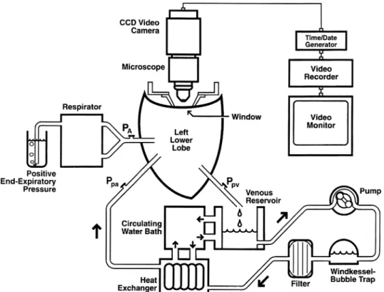
Purpose-bred healthy adult male dogs (18 –27 kg, n ⫽ 5) were deeply anesthetized with pentobarbital sodium (30 – 40 mg/kg iv), intubated, and mechanically ventilated (Harvard 607D animal venti-lator; Harvard Apparatus, Holliston, MA). After intravenous heparin-ization (1,000 U/kg), the animals were rapidly exsanguinated via the left common carotid artery, and the blood was stored. Following a left thoracotomy in the fifth intercostal space, with the lungs inflated to a constant pressure of 5 mmHg, the left upper lobe was excised to
provide access to the left lower lobe. The left lower lobe pulmonary artery was cannulated with a 6-mm-inner diameter Teflon cannula (Chemours, Wilmington, DE), and the left lower lobe bronchus was cannulated to maintain inflation. The lobe was excised along with a cuff of the left atrium and placed on a microscope stand. The left atrial cuff was secured around another Teflon cannula (9-mm inner diam-eter). The lobe was perfused with the heparinized autologous whole blood (hematocrit, 32– 43%). The time interval from thoracotomy to reperfusion was⬍30 min. Care was taken to exclude all air bubbles from the circuit before perfusion was initiated. Blood was pumped by a Masterflex 7522-10 pump drive 7-24-20 pump head (Cole-Parmer, Vernon Hills, IL) that was controlled by a feedback system that kept flow constant (⬍1% variation). After leaving the pump, the blood flowed through a windkessel to dampen high-frequency oscillations from the pump as well as to trap bubbles, then through a filter (model 4C7700, 20-m pore size; Baxter Fenwal, Deerfield, IL) to remove microaggregates, and finally through a heat exchanger (model HE-30; Baxter Bentley, Deerfield, IL) to warm the blood to 38°C before entering the lobe (Fig. 1). Venous blood drained passively from the lobe into a reservoir. The lobe was ventilated (model 607D; Harvard Apparatus) with 6% CO2-17% O2-77% N2at a tidal volume of 100
ml, 2 breaths/min, with an inspiratory time⬍1 s.
Blood gases were sampled from the pulmonary venous catheter and measured with a blood analysis system (Diametrics IRMA SL series; Diametrics, Roseville, MN). The blood gas tensions were kept in the normal range for arterial blood by periodically adding sodium bicar-bonate (1 meq/ml) to the venous reservoir to neutralize metabolic acid produced by the lung tissue. End-expiratory pressure was set at 5 mmHg by a water column on the expiratory outlet of the ventilator. Pulmonary arterial and venous pressures and airway pressures were monitored with pressure transducers (Statham P23 XL; Gould Elec-tronics, Bilthoven, The Netherlands) that were zeroed at the site at which microcirculatory observations were to be made. These pres-sures were monitored continuously by use of a personal computer and monitoring software. Pulmonary venous pressure was set at ~1 mmHg by adjusting the height of the reservoir. Pump flow rate was set at ~300 ml/min, which resulted in a pulmonary arterial pressure of
10 –15 mmHg. These zone 2 settings perfused approximately half of the capillaries thereby providing an opportunity to study blood flow switching among capillary segments.
The lobe was suspended by two small spring-backed paper clips attached to opposite edges of the lobe. The lobe was raised until the uppermost pleural surface (in this case the diaphragmatic surface) came into contact with a transparent window (Fig. 1). Suspending the lobe against the window in this manner prevented compression of the subpleural alveoli by the window and allowed free downward expan-sion of the lobe during ventilation. The remainder of the lobar surface was covered with a plastic sheet to prevent drying and to slow transpleural diffusion of gases. A 1.3-cm2area on the surface of the
lobe was observed through the window, which was surrounded by a vacuum ring to arrest all lateral tissue movement (35, 37). This arrangement permitted the same alveolar capillary network to be recorded throughout the study.
Data acquisition.The capillary circulation in a single subpleural alveolar wall under the window was illuminated by a 200-W mercury arc lamp, heavily filtered to prevent tissue damage with a combination of dichroic infrared reflecting filters, broad band-pass ultraviolet absorbing filters, and a narrow band-pass interference filter to illumi-nate the field using the mercury green line (546 nm). That light is partially absorbed by hemoglobin, thereby increasing the contrast between the red blood cells and the surrounding tissue (35, 36, 37, 40). Video recordings of the capillary flow patterns within a single subpleural wall were made with a video recorder (SVO-5800 SVHS; Sony, Tokyo, Japan) and an intensified charge-coupled device (CCD camera, model 5500; CohuHD, San Diego, CA) attached to a surface-illuminating microscope (⫻11 objective, Leitz Ultropak; Leitz, Wet-zlar, Germany). The magnification on the microscope screen was ⫻560. Because the lung was held in exactly the same place through-out the experiment, the same capillary network was always video recorded. The alveolus was selected so that it lay between a single arteriole and a single venule, which served as the only input and single output for the capillary network. Every capillary segment in the alveolar wall was identified by briefly raising capillary pressure to 30 mmHg, 5 times the normal pressure, and video was recorded. This
pressure exceeded the opening pressure of all of the segments. During recording of this state of total capillary recruitment, every segment was traced and numbered onto a sheet of clear acetate placed over the video monitor (Fig. 2).
Experimental design.These experiments were designed to test two hypotheses. First, to test the active hypothesis, in which capillary switching is caused by active processes, fresh autologous, heparinized blood was pumped through the lung lobe when fresh, and a video recording was made of capillary flow. Second, testing of the passive hypothesis required blocking all known and unknown mechanisms that could biologically alter capillary diameters. The only feasible test was to render all pulmonary vessels inert by glutaraldehyde fixation and then record the capillary flow patterns in the same network.
The experimental design consisted of sequentially perfusing the lobe with fluids contained in one of three reservoirs (Fig. 3) and following a protocol consisting of six steps.1) Autologous, heparin-ized whole blood was perfused through the fresh lobe. Capillary flow patterns were video recorded for 16 min.2) Perfusion was changed to saline (Dulbecco’s phosphate-buffered saline) to flush the lobe free of blood. The blood was stored for later use. (In some cases, there was a time delay of up to an hour betweensteps 2and3; these delays acted as time controls and showed no change in the capillary flow in either the flow level or patterns.)3) Perfusion was changed to 1% buffered glutaraldehyde for 20 min to fix the lung. 4) Perfusion with fresh saline was resumed for another 20 min to thoroughly flush the fixative from the lung to avoid fixation of the red blood cells.5) Perfusion was changed to the original autologous unfixed blood with the same hematocrit. The same capillary network flow patterns were video recorded for 16 min. 6) Tissue was taken directly from the site of observation and studied by electron microscopy to determination the state of fixation.
Data analysis. Measuring switching rate required recording whether red blood cells switched directions as they exited each segment. That measurement required determining the transit time of red blood cells through a single capillary segment. If the estimate was too short, then there would not be sufficient time for a red blood cell to complete its transit and switch or not switch; if too long, many switches would not be counted. On the basis of the pump rate and microhematocrit in the present study, experienced investigators were eventually able to agree that a 4-s observation interval provided an acceptable estimate of switching rate. The video recordings were replayed until each segment in the recording was observed during all consecutive 4-s periods (0 – 4 s, 4 – 8 s, 8 –12 s, ...). The computer had a 4-s timer that prompted the observer to record whether the segment was not perfused (score, 0) or perfused (score, 1).
To test the active versus passive hypotheses, the following com-parisons were made on the basis of pilot data showing that red blood cells flowed through all fresh and fixed pulmonary capillaries. The first comparison was to measure the total number of switches for each condition, to determine whether the switching rates were different. In
the second comparison, the total numbers of perfused segments were tabulated to determine whether the gas exchange surface areas were different. These analyses provided numerical comparisons between the conditions.
The third test provided an analysis of the switching patterns over the entire 16-min recording to compare self-similarity. The flow patterns in the 16-min video recordings produced data streams of sufficient length to permit pattern analysis by computing the fractal dimensions for the fresh and the fixed lungs. The fractal dimensions were measured in the following way (10, 11, 43). The relative dispersion (standard deviation divided by the arithmetic mean) of the number of perfused segments was repeated for progressively longer frames by combining multiples of the 4-s periods using all integer multiples that when divided into 240, produced a whole number (i.e., 2, 3, 4, ..., 80, 120). At each step, the relative dispersions were plotted against the length of the time frames. The slope of the relationship was determined using a weighted least squares fit (KaleidaGraph; Synergy Software, Reading, PA). When plotted on a log-log scale, the fractal dimension was computed as Dt⫽1⫺slope. This analysis provided
pattern comparisons between fresh and fixed conditions.
To test for the stability of the preparation, we used paired, two-tailed t-tests to compare blood gas and pressure measurements that were made at the beginning of the study with those made at the end of the study. Pressure measurements were made every 5 min. Changes over time were analyzed with a two-way analysis of variance. Statis-tical significance was defined by P ⱕ 0.05. Tissue samples were removed and stored for transmission electron microscopy.
RESULTS
Tracking of capillary switching patterns was feasible be-cause our 16-min-long recording period provided enough time for every segment to receive red blood cell flow. A total of 240 observations were made for each segment (16 min⫻60 s⫼4 s ⫽ 240 observations). Recordings during the fresh (active) state produced ~27,000 observations (the total number of all observed segments⫻5 animals⫻240 intervals). Recordings during the fixed (passive) state permitted an analysis of the same microscopic field in all 5 animals and produced ~32,000 observations (~27,000 ⫹ ~32,000 ⬇ 60,000 total measure-ments). The higher number during the fixed state was caused
Fig. 2. A typical subpleural alveolar capillary network from this study. These capillaries accurately reflect the perfusion characteristics of interior capillaries (27). Numbers representcapillary segments 1–20.
Fig. 3. Simplified schema of perfusion system. The pump was switched between three reservoirs for perfusion with fluids in the following order:1) fresh heparinized autologous blood,2) buffered saline to wash out the blood,
by the pump not always being set to produce the same level of capillary perfusion in every animal; nevertheless, the differ-ence of 18% did not have an effect on the comparisons of switching analysis.
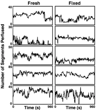
During the 16-min recording, red blood cell flow within the capillary networks constantly varied. Rapid switching between segments produced complex flow patterns whether the tissue was fresh (Fig. 4,left) or fixed (Fig. 4,right). Each data point represents the total number of perfused segments during each 4-s interval.
Here we show that flow patterns continued unabated follow-ing fixation by comparfollow-ing the perfusion patterns in three ways. First, the total number of perfused segments in the fresh versus fixed states was not statistically different. Second, the total number of switches between perfused segments in the fresh versus fixed states was not statistically different. Third, this test provided an analysis of temporal switching patterns. The pat-terns had fractal dimensions of 1.09 (fresh) and 1.11 (fixed). Because both fractal dimensions were close to 1.0 (perfect self-similarity), both fresh and fixed lungs were following similar repeating patterns and were not statistically different (2-tailedt-test, Table 1).
To test whether the preparations were adequately fixed, we used pharmacological challenges with potent vasoconstrictors (100 mM boluses each of KCl and CaCl2) injected into the
pulmonary artery following fixation. Both failed to elicit a pressure rise. Transmission electron micrographs of tissue samples from the observed fields provided evidence of ade-quate fixation as shown by the following criteria: normal cell shapes and sizes, intact organelles, uniform membrane struc-tures, no blebbing of cellular membranes, no tissue separation from basement membranes, no large vacuoles in the endothe-lial cytoplasm, and (Fig. 5,inset) intact double membranes of the mitochondria and the well-defined cristae (Fig. 5). In sum, these independent techniques support the conclusion that fix-ation was complete and thereby blocked active alterfix-ations in capillary cell size and shape.
DISCUSSION
These experiments were designed to determine whether red blood cell switching between pulmonary capillaries is an active or a passive process. These data support the hypothesis that capillary switching patterns can be explained by a passive system, because flow switching continued unchanged after lung fixation. The number of perfused segments, the total number of switches, and the fractal dimensions were not
Fig. 4. Relation between total number of capillary segments perfused during each 4-s period vs. time. Duration of each recording, 16 min (960 s). Each row shows data from 1 alveolus in each of the 5 dogs. Each data point equals the total number of perfused segments during each 4-s interval.Left: data from networks in fresh alveolar walls.Right: same alveolar networks after fixation.
Table 1. Switching of pulmonary capillary flow in the fresh and fixed lung
State of Left Lower Lobe
Fresh Fixed P
No. of perfused segments 22⫾6 27⫾8 NS
No. of switches 935⫾140 1,250⫾282 NS
Fractal dimension 1.09⫾0.06 1.11⫾0.06 NS
Values are means⫾SE;n⫽5 specimens. NS, not significant (P⬎0.05).
Fig. 5. Electron micrograph of pulmonary lobe tissue directly at the site of observation. Complete fixation is confirmed by intact organelles, uniform membrane structure, absence of tissue separation from basement membrane, intact double membranes of mitochondria (inset), and well-defined mitochon-drial cristae (inset). An occasional red blood cell observed in the specimens was further evidence of fixation.
Table 2. Physiological variables at the beginning and end of the study
Variable Before After P
PaO
2, Torr 118⫾7 115⫾6 NS
PaCO
2, Torr 36⫾2 36⫾2 NS
pHa 7.37⫾0.01 7.38⫾0.01 NS
different before and after fixation, evidence that fresh and fixed lungs had similar, repeating patterns.
Several issues need to be considered in interpreting these data. A critical issue is to determine whether the protocol altered the preparation. This issue was tested in the following ways. Perivascular cuffing and alveolar flooding were absent throughout the videotaped periods, evidence that microvascu-lar membranes were intact. Lobar blood flows, venous and arterial pressures, and arterial blood gases (beginning vs. end) were not statistically different and were in the normal physio-logical range, showing that the preparation was stable (Table 2). The fixed lobe unexpectedly exchanged gases as well after fixa-tion as it did when it was fresh.
After fixation, we used vasoconstrictor challenges injected into the main pulmonary artery (100 mM boluses each of KCl and CaCl2) to determine whether the pulmonary vessels were
capable of constricting. There were no pressure changes fol-lowing the injections. Transmission electron microscopy sam-ples were taken from the regions of the lobes that had been observed. All were well fixed (Fig. 5). The stored red blood cells during reperfusion remained fresh throughout the exper-iments, because they were flexible, did not occlude the capil-laries, and did not cause a rise in pulmonary arterial pressure. These data support the conclusion that the lobe was well fixed and incapable of vasomotion and that the red blood cells remained fresh and flexible.
To investigate the unexpected result that fixed rigid cap-illaries could produce the same kind of switching patterns as
fresh capillaries, we modeled passive switching using sim-ple elements. First, consider a theoretical, frictionless metal ball rolling down an inclined plane until it collides with a metal post (as in the pachinko game). Using the identical release point, the same ball always bounces off the post in the same way. Following that collision, the ball then rolls through a maze of posts, bouncing off of post after post until it rolls out of the maze. During repeated trials with the same entry point, the ball will always follow the same path through the maze and exit at the same point. This model is a deterministic system that has a predictable outcome re-gardless of the number of trials.
In the next experiment, the release point is altered randomly between trials but is constrained so that the ball always strikes the first post. After each release, the ball will strike the first post randomly and therefore will bounce in a random direction compared with previous trials. As the ball rolls through the maze, the collisions with each new post become increasingly altered from previous trials. The multiplier effect of the in-creasing numbers of collisions will cause the ball to switch through different pathways anywhere in the maze. During repeated trials, the route of the ball becomes virtually impos-sible to predict because of the unfathomable number of vari-ables and is classified as a nondeterministic system that has unpredictable outcomes. This model, being highly sensitive to initial conditions, fits the chaos theory of chaotic, complex systems (8, 25, 26, 27, 44).
In a more realistic physiological model, consider that a thin flexible disk (a red blood cell) is substituted for the rigid ball. The inclined plane is replaced by a network of hollow capillary tubes. The red blood cell is propelled by the heart. The posts are replaced by the apexes of capillary junctions. As the red blood cell enters the capillary maze, ahead lies a network of
⬎100 tubes (7, 31, 34, 46, 47) with differing diameters, branching angles, and opening pressures. As the red blood cell moves through the capillary network, the collisions at each branch point can be altered by the roll, pitch, and yaw of the disk-shaped cell, as well as by its impact point. Any of these random flow factors affecting the angles of incidence can lead to a switch.
There are additional variables that increase the probability of switching. As red blood cells flow through the capillary net-work, they frequently form trains of cells of varying lengths that are followed by gaps filled with plasma. The lead cell in a train that is trailing a plasma gap is not influenced by the closely spaced cells in the train ahead. That permits the
Fig. 6. Red blood cell flow through alveolar capillaries. Yellow indicates a switching of flow to a different capillary branch. See Supplemental Video S1 available online at https://zenodo.org/record/2255411/files/RBC_Capillary_ Switching_Animation.mp4, doi:10.5281/zenodo.2255411.
following train to switch directions anywhere in the network depending on the characteristics of a particular lead cell-junction collision (4; Fig. 6). As these direction changes occur and the driving pressure within segments alters, multiple flu-idic switching systems can become engaged that cause contin-uously altering switching patterns (1).
Another confounding variable is that the red blood cells enter the network not from a single point as in the ball-and-inclined plane models, but from numerous locations along the feeding arteriole, which acts as a manifold (32). Multiple entry points can readily scramble established flow patterns. White blood cells, which are less deformable than red blood cells, can temporarily plug a segment and force a switch (16, 23, 25). The relatively small number of circulating white blood cells, how-ever, and the large number of capillary segments, make this kind of switching-by-plugging a relatively minor factor (14, 18). With each new variable, the probability of switching increases to the point where it is virtually impossible that blood cells could avoid switching (Fig. 7), even in a passive network that travels across several alveolar walls before finally draining into a venule (31, 47).
These stochastic flow patterns (sequences of random vari-ables) will occur endlessly as red blood cells flow across capillary networks. The networks have finite albeit statistical boundaries because red blood cells become less and less likely to stray far from their arteriolar starting point and, in an absurd example, reverse direction and swim upstream to a distant part of the lobe fed by a different arteriole. These statistical bound-aries will lead to the emergence of repeating flow patterns. In this study, the average fractal dimension of 1.1 in either case provides strong evidence for self-similar patterns. The video recordings of individually numbered segments showed that every segment received flow multiple times during the 16-min-long measurement under these zone 2 conditions (pulmonary arterial pressure ⬎ alveolar pressure ⬎ pulmonary venous pressure). The result is that the underlying patterns are self-similar and fall into the particularly interesting class of patterns that are fractal in nature (25, 26, 27, 28). The perfusion patterns result in more unexpected results when comparing perfusion patterns of adjoining alveolar walls, each of which has its own fractal dimension (43).
These repeating switching patterns can be explained as a consequence of the physics of an emergent system of particu-late fluid flowing in a complex, passive network. With physics determining the outcome, there might be no biologic advan-tage; it would simply be a result of the innate, natural outcome of an emergent process (9). To explore whether these perfusion patterns can lead to complete perfusion of a capillary bed, we developed computer simulations of flow through networks modeled after pulmonary capillary geometry (13). The flow choices at every junction in the program were random as long as they did not flow backward. These simulations produced wide distributions, but in all models, every segment was perfused.
On the basis of these observations, we hypothesized that rapidly repeating fractal flow patterns will maintain opening pressures so low that they approach zero in all capillaries, the result of the constantly repeating perfusions. We have tested this low-opening pressure hypothesis by suddenly doubling flow to the lobe (17). In those preparations, the lobe was perfused the same system as shown in Fig. 1, with
the addition of a second parallel pump. In the control, the lobe was perfused by one pump. Turning on the second pump caused a sudden increase of flow. The majority of these peripheral capillaries were recruited in under 1 s, and all capillaries were perfused in⬍4 s (Fig. 8). We think that this experiment demonstrates that the massive reserve of capillaries is constantly at the ready for immediate recruit-ment. The rapid recruitment response in these controlled experimental conditions does not mimic exercise. Heavy work elicits many responses throughout the body, which would have confounded the simple question that we were asking. Nevertheless, the rapid recruitment response is cer-tain to be part of the acute stress response.
Here we have shown that switching of pulmonary capil-lary blood flow patterns is unchanged following fixation of the lung. This demonstrates that flow switching patterns can be explained entirely by passive processes, a surprising finding that ran contrary to our initial predictions. The switching can be explained as the result of stochastic pro-cesses, yet offers the important physiologic advantage of maintaining all capillaries continuously ready for emer-gency recruitment. We posit that such a system can operate without a feedback loop and provides a new model of pulmonary capillary blood flow. The many redundant, au-tonomous, and independent passive components result in a switching system of simple and robust design that can function reliably for prolonged periods requiring no regula-tion—an elegant engineering system in biology.
ACKNOWLEDGMENTS
We thank B. B. Mandelbrot for encouragement and helpful critique. G. Schmidt and C. Brown at Indiana University made significant contributions to the figures and animations. We also thank G. Tanner, M. Petrun, J. Kuck, S. Gairhe, J. McClendon, J. Downey, B. Wagner, and E. Trepman for providing helpful critiques and editing.
GRANTS
This work was supported by NIH Grant RO1-HL-36033-25 and NASA Graduate Student Research Program Grant 01-GSRP-054.
DISCLOSURES
No conflicts of interest, financial or otherwise, are declared by the authors.
AUTHOR CONTRIBUTIONS
W.W.W., E.M.J., A.J.P., C.M.D., and R.W.G. conceived and designed research; W.W.W., E.M.J., and A.J.P. performed experiments; W.W.W., E.M.J., A.J.P., C.M.D., H.G.B., J.A.C.K., J.A.T., E.S.C., R.W.G., and R.G.P. analyzed data; W.W.W., E.M.J., A.J.P., C.M.D., H.G.B., J.A.C.K., J.A.T., R.W.G., and R.G.P. interpreted results of experiments; W.W.W., E.M.J., C.M.D., J.A.C.K., and E.S.C. prepared figures; W.W.W., E.M.J., A.J.P., C.M.D., H.G.B., J.A.C.K., J.A.T., R.W.G., and R.G.P. drafted manuscript; W.W.W., E.M.J., A.J.P., C.M.D., H.G.B., J.A.C.K., J.A.T., E.S.C., R.W.G., and R.G.P. edited and revised manuscript; W.W.W., E.M.J., A.J.P., C.M.D., H.G.B., J.A.C.K., J.A.T., E.S.C., R.W.G., and R.G.P. approved final version of manuscript.
REFERENCES
1. Angrist SW.Fluid control devices.Sci Am211: 81–88, 1964. doi:10. 1038/scientificamerican1264-80.
2. Ayappa I, Brown LV, Wang PM, Lai-Fook SJ.Arterial, capillary, and venous transit times and dispersion measured in isolated rabbit lungs.J Appl Physiol (1985)79: 261–269, 1995. doi:10.1152/jappl.1995.79.1.261. 3. Baumgartner WA Jr, Jaryszak EM, Peterson AJ, Presson RG Jr, Wag-ner WW Jr.Heterogeneous capillary recruitment among adjoining alveoli.J Appl Physiol (1985)95: 469–476, 2003. doi:10.1152/japplphysiol.01115.2002. 4. Baumgartner WA Jr, Peterson AJ, Presson RG Jr, Tanabe N,
Jaryszak EM, Wagner WW Jr.Blood flow switching among pulmonary capillaries is decreased during high hematocrit.J Appl Physiol (1985)97: 522–526, 2004. doi:10.1152/japplphysiol.00068.2003.
5. Bhattacharya J, Staub NC.Direct measurement of microvascular pres-sures in the isolated perfused dog lung. Science210: 327–328, 1980. doi:10.1126/science.7423192.
6. Capen RL, Wagner WW Jr.Intrapulmonary blood flow redistribution during hypoxia increases gas exchange surface area.J Appl Physiol52: 1575–1580, 1982. doi:10.1152/jappl.1982.52.6.1575.
7. Fung YC.Blood flow in the lung. In:Biodynamics: Circulation. New York: Springer, 1984, p. 290 –369.
8. Gleick J.Chaos: Making a New Science. New York: Penguin, 1987. 9. Glenny RW. Emergence of matched airway and vascular trees from
fractal rules.J Appl Physiol (1985)110: 1119 –1129, 2011. doi:10.1152/ japplphysiol.01293.2010.
10. Glenny RW, Robertson HT.Fractal properties of pulmonary blood flow: characterization of spatial heterogeneity.J Appl Physiol (1985)69: 532– 545, 1990. doi:10.1152/jappl.1990.69.2.532.
11. Glenny RW, Robertson HT, Yamashiro S, Bassingthwaighte JB.
Applications of fractal analysis to physiology.J Appl Physiol (1985)70: 2351–2367, 1991. doi:10.1152/jappl.1991.70.6.2351.
12. Hales S.Statical Essays: Containing Haemastaticks. London: Innys and Manby, 1733. [Reprinted. New York: Hafner, 1964.]
13. Hanger CC, Presson RG Jr, Okada O, Janke SJ, Watkins JJ, Wagner WW Jr, Capen RL. Computer determination of perfusion patterns in pulmonary capillary networks. J Appl Physiol (1985) 82: 1283–1289, 1997. doi:10.1152/jappl.1997.82.4.1283.
14. Hanger CC, Wagner WW Jr, Janke SJ, Lloyd TC Jr, Capen RL.
Computer simulation of neutrophil transit through the pulmonary capillary bed.J Appl Physiol (1985)74: 1647–1652, 1993. doi:10.1152/jappl.1993. 74.4.1647.
15. Hillier SC, Graham JA, Hanger CC, Godbey PS, Glenny RW, Wag-ner WW Jr. Hypoxic vasoconstriction in pulmonary arterioles and venules.J Appl Physiol (1985)82: 1084 –1090, 1997. doi:10.1152/jappl. 1997.82.4.1084.
16. Hogg JC. Neutrophil kinetics and lung injury.Physiol Rev67: 1249 – 1295, 1987. doi:10.1152/physrev.1987.67.4.1249.
17. Jaryszak EM, Baumgartner WA Jr, Peterson AJ, Presson RG Jr, Glenny RW, Wagner WW Jr. Selected contribution: measuring the response time of pulmonary capillary recruitment to sudden flow changes.
J Appl Physiol (1985)89: 1233–1238, 2000. doi:10.1152/jappl.2000.89. 3.1233.
18. Kiani MF, Pries AR, Hsu LL, Sarelius IH, Cokelet GR.Fluctuations in microvascular blood flow parameters caused by hemodynamic mecha-nisms. Am J Physiol Heart Circ Physiol 266: H1822–H1828, 1994. doi:10.1152/ajpheart.1994.266.5.H1822.
19. Krogh A.The Anatomy and Physiology of Capillaries. New Haven, CT: Yale University Press, 1929.
20. Kuebler WM, Kuhnle GE, Groh J, Goetz AE.Leukocyte kinetics in pulmonary microcirculation: intravital fluorescence microscopic study.J Appl Physiol (1985)76: 65–71, 1994. doi:10.1152/jappl.1994.76.1.65. 21. Kuebler WM, Parthasarathi K, Lindert J, Bhattacharya J.Real-time
lung microscopy.J Appl Physiol (1985)102: 1255–1264, 2007. doi:10. 1152/japplphysiol.00786.2006.
22. Kuebler WM, Parthasarathi K, Wang PM, Bhattacharya J.A novel signaling mechanism between gas and blood compartments of the lung.J Clin Invest105: 905–913, 2000. doi:10.1172/JCI8604.
23. Lien DC, Henson PM, Capen RL, Henson JE, Hanson WL, Wagner WW Jr, Worthen GS.Neutrophil kinetics in the pulmonary microcircu-lation during acute inflammation.Lab Invest65: 145–159, 1991. 24. Lien DC, Wagner WW Jr, Capen RL, Haslett C, Hanson WL,
Hofmeis-ter SE, Henson PM, Worthen GS.Physiological neutrophil sequestration in the lung: visual evidence for localization in capillaries.J Appl Physiol (1985)
62: 1236 –1243, 1987. doi:10.1152/jappl.1987.62.3.1236.
25. Lorenz EN.Deterministic nonperiodic flow.J Atmos Sci20: 130 –141, 1963. doi:10.1175/1520-0469(1963)020⬍0130:DNF⬎2.0.CO;2.
26. Lorenz EN.Predictability: does the flap of a butterfly’s wings in Brazil set off a tornado in Texas? American Association for the Advancement of Science, 139th Meeting. Washington, DC, Dec. 29 1972. [Available at http://eaps4.mit.edu/research/Lorenz/Butterfly_1972.pdf].
27. Lorenz EN.The Essence of Chaos. Seattle, WA: University of Washing-ton Press, 1995.
28. Mandelbrot BB.The Fractal Geometry of Nature. New York: Freeman, 1977.
29. Shepard JM, Gropper MA, Nicolaysen G, Staub NC, Bhattacharya J.
Lung microvascular pressure profile measured by micropuncture in anes-thetized dogs.J Appl Physiol (1985) 64: 874 –879, 1988. doi:10.1152/ jappl.1988.64.2.874.
30. Short AC, Montoya ML, Gebb SA, Presson RG Jr, Wagner WW Jr, Capen RL.Pulmonary capillary diameters and recruitment characteristics in subpleural and interior networks.J Appl Physiol (1985)80: 1568 –1573, 1996. doi:10.1152/jappl.1996.80.5.1568.
31. Staub NC, Schultz EL.Pulmonary capillary length in dogs, cat and rabbit.
Respir Physiol5: 371–378, 1968. doi:10.1016/0034-5687(68)90028-5. 32. Tabuchi A, Kuppe H, Pries AR, Kuebler WM.Intravital microscopy of
the murine pulmonary microcirculation (Abstract).J Vasc Res43,Suppl1: 7, 2006. doi:10.1159/000094937.
33. Tabuchi A, Styp-Rekowska B, Slutsky AS, Wagner PD, Pries AR, Kuebler WM. Precapillary oxygenation contributes relevantly to gas exchange in the intact lung.Am J Respir Crit Care Med188: 474 –481, 2013. doi:10.1164/rccm.201212-2177OC.
34. Townsley MI.Structure and composition of pulmonary arteries, capillaries, and veins.Compr Physiol2: 675–709, 2012. doi:10.1002/cphy.c100081. 35. Wagner WW Jr.Pulmonary microcirculatory observations in vivo under
physiological conditions.J Appl Physiol26: 375–377, 1969. doi:10.1152/ jappl.1969.26.3.375.
36. Wagner WW Jr, Barker DB, Filley GF.A photographic method for quantitating blood flow in the pulmonary microcirculation.J Biol Photogr Assoc35: 95–108, 1967.
37. Wagner WW Jr, Brinkman PD, Baker DB, Filley GF. Erythrocyte photomicrography: contrast control by monochromatic transillumination.
J Biol Photogr Assoc37: 156 –162, 1969.
38. Wagner WW Jr, Filley GF.Microscopic observation of the lung in vivo.
Vasc Dis2: 229 –241, 1965.
39. Wagner WW Jr, Latham LP.Pulmonary capillary recruitment during airway hypoxia in the dog.J Appl Physiol39: 900 –905, 1975. doi:10. 1152/jappl.1975.39.6.900.
40. Wagner WW Jr, Latham LP, Brinkman PD, Filley GF.Pulmonary gas transport time: larynx to alveolus.Science163: 1210 –1211, 1969. doi:10. 1126/science.163.3872.1210.
41. Wagner WW Jr, Latham LP, Capen RL.Capillary recruitment during airway hypoxia: role of pulmonary artery pressure. J Appl Physiol47: 383–387, 1979. doi:10.1152/jappl.1979.47.2.383.
42. Wagner WW Jr, Latham LP, Gillespie MN, Guenther JP, Capen RL.
Direct measurement of pulmonary capillary transit times.Science218: 379 –381, 1982. doi:10.1126/science.7123237.
44. Waldrop MM.Complexity: The Emerging Science at the Edge of Order and Chaos. New York: Simon & Schuster, 1992.
45. Wearn JT, Ernstene AC, Bromer AW, Barr JS, German WJ, Zschi-esche LJ. The normal behavior of the pulmonary blood vessels with observations on the intermittence of the flow of blood in the arterioles and capillaries. Am J Physiol 109: 236 –256, 1934. doi:10.1152/ajplegacy. 1934.109.2.236.
46. Weibel ER.Morphometry of the Human Lung. New York: Academic, 1963.
47. Weibel ER. The Pathway for Oxygen: Structure and Function in the Mammalian Respiratory System. Cambridge: Harvard University Press, 1984, p. 291.