Original Research Article
Cross sectional study evaluating the correlation of thyroid dysfunction
with severity of disease in rheumatoid arthritis
Vinoth Kumar D., Aruna R.*
INTRODUCTION
Rheumatoid arthritis (RA) is a chronic inflammatory arthritis involving 0.5 to 1% of the general population globally and the Indian prevalence ranges from 0.2-1%.1,2
Though RA is considered primarily a disease of the joints, it involves abnormal immune responses and involves multiple organs. RA is highly prevalent among those aged 25-55 years. The classical symptoms include ‘morning stiffness’, constitutional symptoms such as fatigue and malaise with extra articular manifestations
such as subcutaneous nodules, neurological and hematological abnormalities, pulmonary manifestations, peripheral neuropathy and vasculitis. Autoimmune response, both systemic and articular are the key factors in the destructive progression of the disease. The presence of autoantibodies, such as rheumatoid factor (RF) and anticitrullinated protein antibody (anti-CCP) are considered diagnostic but anti-CCP has comparatively more specificity. Autoimmune response with high levels of proinflammatory cytokines such as tumor necrosis factor α (TNF-α) is seen in RA and these cytokines has Department of Medicine, Government Medical College, Thiruvananthapuram, Kerala, India
Received: 14 May 2020
Accepted: 19 May 2020
*Correspondence:
Dr. Aruna R.,
E-mail: [email protected]
Copyright: © the author(s), publisher and licensee Medip Academy. This is an open-access article distributed under the terms of the Creative Commons Attribution Non-Commercial License, which permits unrestricted non-commercial use, distribution, and reproduction in any medium, provided the original work is properly cited.
ABSTRACT
Background: The present study was conducted to evaluate the correlation of disease severity in RA and thyroid dysfunction.
Methods: The present cross-sectional descriptive study enrolled 164 participants aged 12 years and above diagnosed as having RA. Use of drugs causing thyroid dysfunction, malignancy, diabetes mellitus, systemic hypertension, pregnancy and prior thyroidectomy were the criteria for exclusion. Data was analyzed using R and tests of significance were Chi square test and independent sample t-test and Pearson correlation. Institutional ethics committee approved the study and written informed consent was obtained from all study participants.
Results: Serum TSH positively correlated with DAS 28 (r=0.2, p=0.005), ESR (r=0.2, p=0.03), CRP (r=0.2, p=0.006), RA factor (r=0.2, p=0.003), subjective assessment (r=0.3, p= 0.001) and anti TPO antibodies (r=0.7, p=0.001). Free T4 negatively correlated with DAS28 (r=-0.2, p=0.006), ESR (r=-0.2, p=0.02), CRP (r=-0.2, p=0.01). RA factor (r=-0.2, p=0.01), subjective assessment (r=-0.2, p= 0.01), anti TPO (r=-0.6, p=0.001) and Free T3 negatively correlated with DAS28 score (r=-0.2, p=0.02) , ESR (r=-0.2, p=0.03), RA factor (r=-0.3, p=0.001) and anti TPO antibodies (r=- 0.3, p=0.001).
Conclusions: Hypothyroidism was significantly associated with disease severity of RA with linear positive correlation of TSH with DAS28 score, ESR, CRP, RA factor, subjective assessment and anti TPO antibodies, linear negative correlation of serum free T4 with DAS 28 score, ESR, CRP, RA factor, subjective assessment and anti TPO antibody and linear negative correlation of free T3 with DAS28 score, ESR, RA factor and anti TPO antibody was observed.
Keywords: DAS-28 score, Disease activity, Hypothyroidism, Rheumatoid arthritis, Thyroid dysfunction
also been implicated in the pathogenesis of thyroid dysfunction. Due to overlapping symptomatology, thyroid dysfunction may be masked in patients with RA. The prevalence of thyroid dysfunction is 1-2% and is 10 times more common in females.3
The autoimmune response mediated reduction in thyroid function occurs gradually and, in the time, compensatory feedback process activates resulting in normal serum levels of thyroid hormones (T3 and T4) due to elevation in plasma TSH. In this stage, patients will develop mild symptoms (subclinical hypothyroidism), later developing into clinical hypothyroidism with significant elevations in TSH (usually >10 IU/L) with clinical manifestations. Thyroid dysfunction can also manifest as hyperthyroidism (subclinical or overt) in patients with RA. The present study was conducted to evaluate the correlation between thyroid hormones and disease severity in participants with RA.
METHODS
The present cross-sectional descriptive study enrolled 164 participants aged 12 years and above diagnosed as having RA using ACR/EULAR criteria in a period of 1 year attending rheumatology outpatient and those admitted in internal medicine department of Government Medical College, Thiruvananthapuram. Participants with history of use of drugs known to cause thyroid dysfunction (e.g. lithium), with evidence of malignancy, other connective tissue disorders, chronic liver or renal disorders, diabetes mellitus, systemic hypertension, pregnancy and those who had history of thyroidectomy were excluded.
Sample size was calculated based on the prevalence from previous published literature using formula for calculating sample size for qualitative data. Collected details included history (duration, morning stiffness, chest symptoms, list of painful joints, other systemic diseases, extra articular manifestations and thyroid swelling), sociodemographic parameters (age, gender), examination findings (tenderness, swelling, deformity and range of movements, examination for thyroid swelling, cardiovascular, gastrointestinal, nervous and respiratory system and extraarticular manifestations) and investigations (sonography for confirming thyroid swelling, erythrocyte sedimentation rate (ESR), rheumatoid factor IgG, C-reactive protein (CRP), thyroid function test, hemoglobin, total and differential leukocyte count, blood urea, serum creatinine and plasma glucose). Disease activity score (DAS-28) was used to assess the severity of disease.4
Statistical analysis
Data was analyzed using R and tests of significance were Chi square test, Fischer exact, independent sample and paired t-test. Correlation between parameters were estimated using Pearson correlation. Institutional ethics committee approved the study protocol and written
informed consent was obtained from all study participants. Values are expressed as mean [standard deviation (SD)] or as proportions after rounding off to single decimal and a p<0.05 is considered statistically significant.
RESULTS
Mean age of the study participants was 43.3 (11.7) years. 90.2% (n=148) were females. Morning stiffness lasting for more than 1 hour in a day was present in 90.9% (n=149) participants. 93.9% (n=154) had constitutional symptoms (fever, anorexia, tiredness) associated with RA. Among study participants, 62.2% (n=102) had involvement of all the limbs, 33.5% (n=55) had involvement of both upper limbs and 4.3% (n=7) had involvement of both lower limbs. The mean number of tender joints among study participants was 11.3 (6.5%) and swollen joints was 12.4 (6.3%). Subcutaneous nodules were seen in 12.2% (n=20) and purpura was seen in 1.8% (n=3) participants. 11% (n=18) had joint deformity and 5.5% (n=9) had restriction of movements. Disease severity among study participants is demonstrated in Table 1. 42.1% (n=69) were on treatment for RA
Table 1: Proportion of participants with various disease severities.
Disease severity according to DAS
28 scoring n (%)
Mild (≥2.6 and ≤3.2) 0
Moderate (>3.2 and ≤5.1) 57 (34.8%) High (>5.1) 107 (65.2%)
Among participants receiving treatment, 49.3% (n=34) were receiving non-steroidal anti-inflammatory drugs (NSAIDs) alone, 15.9% (n=11) were on a combination of NSAIDs and hydroxychloroquine, 8.7% (n=6) were on combination of NSAIDs, hydroxychloroquine and sulfasalazine, and 2.9% (n=2) participants were receiving a combination of methotrexate, NSAIDs, hydroxychloroquine and systemic corticosteroids. The mean serum thyroid stimulating hormone (TSH) was 4.1 (3.1), free T4 was 0.99 (0.5) and free T3 was 2.3 (0.8). No difference in thyroid function was observed between age groups [TSH (p=0.8), free T4 (p=1) and free T3 (p=0.3)].
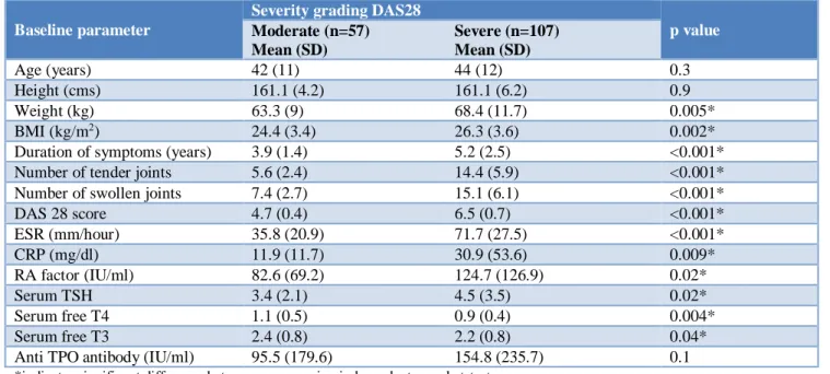
Comparison of baseline parameters between moderate and severe disease activity is demonstrated in Table 2,
and comparison of disease parameters between moderate and severe disease activity in Table 3.
Table 2: Comparison of baseline parameters between participants with moderate and severe disease activity.
Baseline parameter
Severity grading DAS28
p value
Moderate (n=57) Mean (SD)
Severe (n=107) Mean (SD)
Age (years) 42 (11) 44 (12) 0.3
Height (cms) 161.1 (4.2) 161.1 (6.2) 0.9 Weight (kg) 63.3 (9) 68.4 (11.7) 0.005* BMI (kg/m2) 24.4 (3.4) 26.3 (3.6) 0.002*
Duration of symptoms (years) 3.9 (1.4) 5.2 (2.5) <0.001* Number of tender joints 5.6 (2.4) 14.4 (5.9) <0.001* Number of swollen joints 7.4 (2.7) 15.1 (6.1) <0.001* DAS 28 score 4.7 (0.4) 6.5 (0.7) <0.001* ESR (mm/hour) 35.8 (20.9) 71.7 (27.5) <0.001* CRP (mg/dl) 11.9 (11.7) 30.9 (53.6) 0.009* RA factor (IU/ml) 82.6 (69.2) 124.7 (126.9) 0.02*
Serum TSH 3.4 (2.1) 4.5 (3.5) 0.02*
Serum free T4 1.1 (0.5) 0.9 (0.4) 0.004* Serum free T3 2.4 (0.8) 2.2 (0.8) 0.04* Anti TPO antibody (IU/ml) 95.5 (179.6) 154.8 (235.7) 0.1
*indicates significant difference between groups using independent sample t-test.
Table 3: Comparison of disease parameters between moderate and severe disease.
No association of type of thyroid dysfunction with gender (p=0.7), age (p=0.4) and morning stiffness (0.05) was observed. Significant association was observed between duration of symptoms and thyroid dysfunction (p=0.049) (Table 4).
A significant positive correlation of DAS28 score was observed with serum TSH (r=0.2, p=0.005) (Figure 1),
ESR (r=0.6, p=0.001), CRP (r=0.5, p= 0.001), RA factor (r=0.3, p=0.001), subjective assessment (r=0.6, p=0.001), tender joints (r=0.8, p=0.001), swollen joints (r=0.8, p=0.001), and anti TPO antibodies (r=0.2, p=0.002) (Figure 2).
Significant negative correlation of DAS 28 score was observed with serum free T4 (r=-0.2, p=0.006) (Figure 3) and serum free T3 (r=-0.2, p= 0.02) (Figure 4).
Parameter Severity grading DAS28 p value
Moderate (n=57) Severe (n=107)
Gender; female: male 50:7 98:9 0.4
Joints involved; QL: BUL: BLL 11:41:5 91:14:2 0.001* Morning stiffness >60 mins, n (%) 53 (35.6%) 96 (64.4%) 0.5 Constitutional symptoms, n (%) 49 (31.8%) 105 (68.2%) 0.002* Deformed joints, n (%) 3 (16.7%) 15 (83.3%) 0.09 Joint restriction of movements, n (%) 4 (44.4%) 5 (55.6%) 0.04 Rheumatoid nodules, n (%) 5 (25%) 15 (75%) 0.3 Purpura, n (%) 2 (66.7%) 1 (33.3%) 0.6 Symptoms of hypothyroidism, n (%) 3 (11.5%) 23 (88.5%) 0.007* No thyroid swelling, n (%) 49 (35%) 91 (65%) 0.001* Multinodular goiter, n (%) 3 (30%) 7 (70%) 0.04* Diffuse enlargement, n (%) 5 (35.7%) 9 (64.3%) 0.3 Hypothyroid, n (%) 11 (27.5%) 29 (72.5%) 0.2 Subclinical hypothyroid, n (%) 2 (33.3%) 4 (66.7%) 0.4 Subclinical hyperthyroid, n (%) 0 (00.0%) 2 (100%) 0.2
Table 4: Duration of symptoms and thyroid dysfunction.
Duration of symptoms (years)
Thyroid dysfunction p value
Absent Present
≤3 34
(64.2%) 19 (35.8%)
0.049 >3 to ≤6 65
(79.3%) 17 (20.7%) >6 17
(58.6%) 12 (41.4%)
Figure 1: Correlation of DAS28 score with serum TSH.
Figure 2: Correlation of DAS28 score with anti TPO antibody titre.
Figure 3: Correlation of DAS28 score with free T4.
Serum TSH positively correlated with DAS 28 score (r=0.2, p=0.005) (Figure 5),ESR (r=0.2, p=0.03) (Figure 6), CRP (r=0.2, p=0.006) (Figure 7), RA factor (r=0.2, p=0.003) (Figure 8), subjective assessment (r=0.3, p=0.001) (Figure 9) and anti TPO antibodies (r=0.7, p=0.001) (Figure 10). The correlation of serum TSH with tender and swollen joints is demonstrated in Figure 11 and 12.
Figure 4: Correlation of DAS28 score with free T3.
Figure 5: Correlation between serum TSH with DAS28 score.
Figure 6: Correlation between serum TSH with ESR.
assessment 0.2, p= 0.01) and anti TPO antibodies (r=-0.6, p=0.001). Negative correlation of FT3 with DAS28 score (r=-0.2, p=0.02) (Figure 14), ESR (r=-0.2, p=0.03), RA factor(r=-0.3, p=0.001) and anti TPO antibody (r=- 0.3, p=0.001) was observed.
Figure 7: Correlation of serum TSH with CRP.
Figure 8: Correlation of serum TSH with RA factor.
Figure 9: Correlation between serum TSH and subjective assessment.
Figure 10: Correlation of serum TSH with anti TPO.
Figure 11: Correlation between serum TSH and tender joints.
Figure 12: Correlation between TSH and swollen joints.
Figure 13: Correlation of free T4 with DAS 28 score.
DISCUSSION
RA has been described as a disease affecting women in the 40’s and 50’s, a similar observation was made in the present study.5 The present study reports a female: male
ratio of 9:1 which is higher than previous reports which has reported a maximum proportion of 8:1. The duration of symptoms was ~5 years which is considerably high considering the educational status of the state. 91% participants had morning stiffness which is extremely common in RA. 94% participants had constitutional symptoms which has been described as occasional in RA.6 This discrepancy could be due to the higher
prevalence of thyroid dysfunction among Keralites, or could be due to the noncompliance in medications which would unmask the constitutional symptoms. 62% participants had involvement of all the limbs which is probably one of the typical presentations of RA, ‘palindromic rheumatism’ in which multiple joints may be involved in episodic events lasting from hours to days.6 The mean number of tender and swollen joints
were 11 and 12 respectively which could indicate the above mentioned presentation or could indicate the higher severity of the disease among our study participants or the noncompliance to treatment. The proportion of participants with subcutaneous nodules were higher than previous reports and those with purpura was lower compared to previous reports which could be due to the enrollment of higher number of participants with dermatological manifestations in the previous report.7 65% participants had severe disease and 35%
participants had disease of moderate severity which could be due to high number of severe and referred cases attending the tertiary care institution. The drugs used by the study participants were NSAIDs and DMARDs which are the standard of care in RA. As the severity of the disease increases the TSH values increase (p=0.02) and free T3 (0.04) and T4 (p=0.004) values reduce which is similar to previous studies which could indicate that thyroid hormone defects especially findings similar to hypothyroidism are associated with the increasing severity of disease.8 Thyroid dysfunction correlated
significantly with ESR (p=0.004) which is similar to reports from previous study.9 Significantly higher CRP
was observed in participants with thyroid dysfunction (p=0.02) which is in contrast to previous reports.9 Since
the thyroid dysfunction and RA are both of inflammatory pathology and the thyroid dysfunction being associated with the severity of the disease, elevations in inflammatory markers such as ESR and CRP could be justified.10 Similar to previous reports, the serum RA
factor was significantly higher in participants with thyroid dysfunction.9 Thyroid dysfunction is higher in
more severe RA, which could translate to the laboratory finding of higher elevations of RA factor. This could also explain the elevated levels of anti TPO antibodies in participants with thyroid dysfunction. Observed between participants with and without thyroid dysfunction which could be an area of focus for future research.8
Significantly higher DAS 28 score was observed among
participants with thyroid dysfunction (p=0.03) similar to previous studies.11 The presence of thyroid dysfunction
indicates more severe inflammatory process and hence more severe RA which could manifest as symptoms and could increase the DAS 28 score. Significantly higher weight and BMI was observed in participants with severe disease indicating more severe hypothyroidism among these patients which could produce weight gain and increase in BMI. Significantly higher number of swollen and tender joints were observed in participants with severe disease which is an expected result in severe disease. The duration of symptoms was longer in participants with severe disease, as the duration of disease increases, so does the progression. There were higher levels of inflammatory markers such as ESR, CRP and RA factor among participants with severe disease which is also an expected finding in participants with severe disease. Significantly higher proportion of participants with quadratus lumborum involvement was seen in severe RA which requires further evaluation. Significantly higher proportion of participants with severe RA had constitutional symptoms. Constitutional symptoms such as aching muscles, tiredness, generalized weakness, low mood, fever, weight loss and loss of appetite has been linked to subclinical arterial inflammation and is believed to predominate early stages of RA. These constitutional symptoms are more commonly seen in elderly RA patients, the evidence on the correlation with severity of RA is limited. Significantly higher proportion of participants with severe RA had symptoms of hypothyroidism with higher proportion of participants without thyroid swelling. Significantly higher proportion of participants who had the duration of disease less than 6 years did not have evidence of hypothyroidism.
There was significant positive correlation of serum TSH with DAS28, ESR, CRP; RA factor, subjective assessment and anti TPO antibodies indicating linear elevations in these parameters with elevations in TSH. Positive correlation of these parameters except RA factor in thyroid dysfunction associated with RA has been published.12,13 There was significant negative correlation
between free T4 with DAS28 score, ESR, CRP, RA factor, subjective assessment, anti TPO antibody. These results are similar to previous reports indicating that lower levels of free T4 is associated with higher grades of RA disease activity.8,12 There was significant negative
correlation between free T3 with DAS28 score, ESR, RA factor and anti TPO antibodies. These results are also similar to previous reports indicating that lower levels of free T3 is associated with higher grades of RA disease activity.12 However, this association has not been
observed in other studies which could be due to the short duration of disease among those patients.14,15 Also,
published literature concludes that thyroid hormone defects to be related to the duration of disease and not disease activity.8 Anti TPO antibody had a significant
free T3 similar to previous reports of association with DAS 28 score.16 This could be due to the high rates of
drug default (especially anti-inflammatory and immunosuppressants) among the study participants which lead to increase in anti TPO antibodies in these patients. Published literature could not conclusively establish the relationship probably due to the ethnic and geographical differences in the study population and a wide range of anti TPO antibodies from 0.5 to 27% has been reported.17
This should be explored further since elevated anti TPO antibody is a risk for developing diabetes mellitus and cardiovascular disease. The prevalence of silent autoimmune thyroid disorders is controversial since a wide range has been described.18,19 Reversible subclinical
thyroid disorders may remain unnoticed in RA and the thyroid disorders are thought to be due to one of the autoantibodies in RA.18 The involvement of HLA DR in
patients with RA would explain the involvement of thyroid gland as well.20
Significant negative correlation of serum free T4 with DAS 28 score, ESR, CRP, RA factor, subjective assessment and anti TPO antibody was observed indicating reduction in free T4 levels would produce a linear increase in these parameters. Free T3 was also negatively correlated with DAS28 score, ESR, RA factor and anti TPO antibody which indicates that reduction in free T3 levels would produce linear elevations of these parameters.
CONCLUSION
Hypothyroidism was significantly associated with disease severity of RA. Elevations in TSH was associated with linear elevations of DAS28 score, ESR, CRP; RA factor, subjective assessment and anti TPO antibodies. Elevations in serum free T4 was associated with linear reductions of DAS 28 score, ESR, CRP, RA factor, subjective assessment and anti TPO antibody. Elevations in serum free T3 was associated with linear reductions of DAS28 score, ESR, RA factor and anti TPO antibody.
Funding: No funding sources Conflict of interest: None declared
Ethical approval: The study was approved by the Institutional Ethics Committee
REFERENCES
1. Alivernini S, Tolusso B, Petricca L, Ferraccioli G, Gremese E. Chapter 46 - Rheumatoid Arthritis. In: Perricone C, Shoenfeld Y, editors. Mosaic of Autoimmunity. Academic Press; 2019:501-526. 2. Akhter E, Bilal S, Kiani A, Haque U. Prevalence of
arthritis in India and Pakistan: a review. Rheumatol Int. 2011;31(7):849-55.
3. Tunbridge WM, Evered DC, Hall R, Appleton D, Brewis M, Clark F, et al. The spectrum of thyroid disease in a community: the Whickham survey. Clin Endocrinol (Oxf). 1977;7(6):481-93.
4. Jensen Hansen IM, Asmussen Andreasen R, van Bui Hansen MN, Emamifar A. The reliability of disease activity score in 28 joints-c-reactive protein might be overestimated in a subgroup of rheumatoid arthritis patients, when the score is solely based on subjective parameters. J Clin Rheumatol. 2017;23(2):102-6.
5. Vollenhoven RF van. Sex differences in rheumatoid arthritis: more than meets the eye. BMC Med. 2009;7:12.
6. Ngian GS. Rheumatoid arthritis. Aust Fam Physician. 2010;39(9):626-8.
7. Ghosh SK, Bandyopadhyay D, Biswas SK, Darung I. Mucocutaneous manifestations in patients with rheumatoid arthritis: a cross-sectional study from eastern India. Indian J Dermatol. 2017;62(4):411-7. 8. Singh B, Mittal B, Bhattacharya A, Deodhar S. A
cross sectional evaluation of circulating thyroid hormonal profile in patients with rheumatoid arthritis. Indian J Nucl Med. 2002;17:68-72. 9. Ghitany MK, Soliman EA, Bondok ME,
Elmaadawy SA. Autoimmune thyroid disorders in seropositive versus seronegative rheumatoid arthritis. Egypt J Obes Diabet Endocrinol. 2015;1(1):53.
10. Gupta G, Sharma P, Kumar P, Itagappa M. Study on subclinical hypothyroidism and its association with various inflammatory markers. J Clin Diagn Res. 2015;9(11):BC04-6.
11. Joshi P, Agarwal A, Vyas S, Kumar R. Prevalence of hypothyroidism in rheumatoid arthritis and its correlation with disease activity. Trop Doct. 2017;47(1):6-10.
12. Elattar EA, Younes TB, Mobasher SA. Hypothyroidism in patients with rheumatoid arthritis and its relation to disease activity. Egypt Rheumatol Rehabil. 2014;41(2):58.
13. Mousa AA, Ghonem M, Hegazy A, El-Baiomy AA, El-Diasty A. Thyroid function and auto-antibodies in Egyptian patients with systemic lupus erythematosus and rheumatoid arthritis. Trends Med Res. 2012;7(1):25-33.
14. Punzi L, Betterle C. Chronic autoimmune thyroiditis and rheumatic manifestations. Joint Bone Spine. 2004;71(4):275-83.
15. El-Sherif WT, El Gendi SS, Ashmawy MM, Ahmed HM, Salama MM. Thyroid disorders and autoantibodies in systemic lupus erythematosus and rheumatoid arthritis patients. Egypt J Immunol. 2004;11(2):81-90.
16. Chen YL, Lin JZ, Mo YQ, Liang JJ, Li QH, Zhou CJ, et al. Joint damage is amplified in rheumatoid arthritis patients with positive thyroid autoantibodies. Peer J. 2018;6:e4216.
18. Ilias I, Mastorakos G, Mavrikakis M, Papazoglou S, Karamitsos D, Ntantis P, et al. Thyroid disease associated with rheumatoid arthritis is not adequately screened with a sensitive chemiluminescence thyrotrophin assay. Acta Med Austriaca. 1999;26(1):26-8.
19. Innocencio RM, Romaldini JH, Ward LS. Thyroid autoantibodies in autoimmune diseases. Medicina. 2004;64(3):227-30.
20. Raterman HG, Jamnitski A, Lems WF, Voskuyl AE, Dijkmans BAC, Bos WH, et al. Improvement of thyroid function in hypothyroid patients with
rheumatoid arthritis after 6 months of adalimumab treatment: a pilot study. J Rheumatol. 2011;38(2):247-51.