Dr Meenakshi Gundewar el al JMSCR Volume 07 Issue 09 September 2019 Page 418
Comparative Cytological Techniques in the Diagnosis of Non Neoplastic and
Neoplastic Bronchopulmonary Lesions
Authors
Dr Meenakshi Gundewar
1, Dr Pragati Karmarkar
2*, Dr Sadhana Mahore
31
Consultant pathologist, 2Associate Professor, 3Professor
Department of Pathology, NKP Salve Institute of Medical Science and Research Centre, Nagpur, India *Corresponding Author
Dr Pragati Karmarkar
Department of Pathology, NKP Salve Institute of Medical Science and Research Centre, Nagpur, India
Abstract
Objective: Diagnostic cytology is one of the suitable modality for both non-neoplastic and neoplastic conditions of the lung. The aim is to use different cytological methods in the diagnosis of lesions of the respiratory tract. It has been generally acclaimed as one of the most successful application in the evaluation of patient with suspected lung malignancy.
Materials and Method: It was a prospective study of 127 cases. The present study was conducted in the department of pathology at a tertiary care hospital. It is a cross- sectional study, carried out on all patients who were admitted with lung diseases during a period of two years . Both male and female patients of all age group with the specified respiratory tract complaints like cough, breathlessness, hemoptysis, fever and chest pain were subjected to bronchoscopic examination.
Results: There were 81 males and 46 females. Male : female ratio was 1.76 : 1. The mean age was 50±16.14 years. The lowest age was 14 years and the oldest age was 85 years. It was included in both the non- neoplastic and neoplastic study groups. Sensitivity of BB was 91.8%; while that of BAL was only 59.2%. specificity of BB was 87.5% and that of BAL was 75%. Accuracy of BB was 90.7% while that of BAL was 63.1%.
Conclusions: Bronchial brushing is a much superior technique in the diagnosis of bronchopulmonary lesions. It demonstrates far better sensitivity, specificity and accuracy. Combination of various cytohistological techniques complements each other and enhances the diagnostic efficacy of various non- neoplastic and neoplastic lung diseases.
Keywords: cytology, lung, techniques.
Introduction
Cytology of the pulmonary lesions provides valuable diagnostic information. These cytological procedures constitute the most useful and least expensive investigative tools available for the detection of pulmonary diseases. Respiratory tract cytology is well established throughout the world as a vital diagnostic procedure in the evaluation of
any patient with suspected pulmonary lesions especially lung cancer. The aim of this study is to assess the efficacy and diagnostic importance of BAL/ bronchial washings, brushings when compared to biopsy in the investigations of patients who are suspected of having any bronchopulmonary lesion both clinically and radiologically.
http://jmscr.igmpublication.org/home/ ISSN (e)-2347-176x ISSN (p) 2455-0450
Dr Meenakshi Gundewar el al JMSCR Volume 07 Issue 09 September 2019 Page 419
Materials and Method
This study was carried out prospectively in the department of Pathology at a tertiary care hospital. It was a hospital based cross-sectional study. The patients had one or more of the following features; growing peripheral lesion on chest X- ray, positive sputum cytology, and clinical symptom refractory to medication or visible endobronchial mass. Detailed clinical history, physical examination, hemogram, chest X-ray and bronchoscopy was performed on all the cases. All the subjects were informed about the study and a written consent was obtained from all.
Paediatric patients, patients with bleeding disorders or recent myocardial infarction were excluded from the study.
Bronchoscopy was performed through the transnasal approach, using an Olympus BF- 2TR fibreoptic bronchoscope.
BAL was obtained by introducing a bronchoscope in the lower respiratory tract and specimens obtained by means suction apparatus after infusing 60ml or more saline. A minimal volume of 5 ml of a pooled BAL sample was needed for BAL cellular analysis.
Bronchial washings was obtained by repetitive instillation of 3-5 ml of a sterile balanced salt solution through the bronchoscope and
reaspiration of fluid. Brushings (BB) are obtained by the use of a small circular stiff-bristle brush. Materials obtained by broncho-alveolar lavage and brushing were fixed by various fixatives The slides were stained with May-Grünwald-Giemsa (MGG), Papanicolaou (Pap), and hematoxylin and eosin (H and E) stains. The overall cytodiagnostic efficacy as well as that of various cytological samples were analyzed. Endobronchial biopsy was performed using a flexible long biopsy forceps and tissue bits were fixed in 10% formalinand processed for histopathological examination.
Results
The present study group comprised of 127 patients. The age of patients ranged from 14 yrs to 85 yrs. There were 81 males and 46 females. Male: female ratio was 1.76:1. There were 73 smokers and 08 non- smokers.
The most common presenting complaint was cough followed by breathlessness. The laterality of lung lesion, most commonly was in right lung and right upper lobe was most commonly involved.
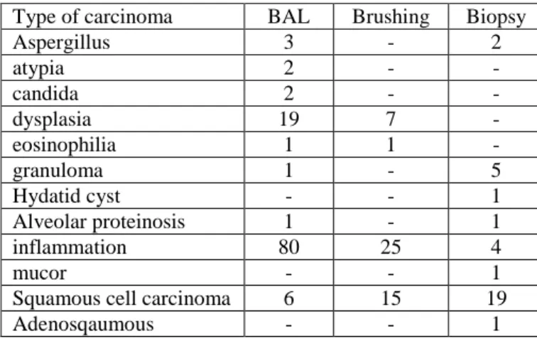
Neoplastic lesions was diagnosed in 49 patients and non-neoplastic lesions were seen in 78 patients. Biopsy was done in 65 cases (TABLE I).
Table no. I Diagnostic accuracy of different modalities in different lung lesions
Type of carcinoma BAL Brushing Biopsy
Aspergillus 3 - 2
atypia 2 - -
candida 2 - -
dysplasia 19 7 -
eosinophilia 1 1 -
granuloma 1 - 5
Hydatid cyst - - 1
Alveolar proteinosis 1 - 1
inflammation 80 25 4
mucor - - 1
Squamous cell carcinoma 6 15 19
Adenosqaumous - - 1
Bronchial washing showed 29 true positive (TP), 12 true negative (TN), 4 false positive (FP) and 20 false negative (FN) cases whereas bronchial
Dr Meenakshi Gundewar el al JMSCR Volume 07 Issue 09 September 2019 Page 420
Table No. II Diagnostic evaluation of various lesion confirmed on Biopsy Bronchial washing and bronchial brushing
Sample Test result
TP TN FP FN Total
Bronchial washing 29 12 4 20 65
Bronchial Brushing 45 14 2 4 65
Bronchial brushing showed good sensitivity (91.8%) and specificity (87.5%) compared to bronchial washing which had sensitivity of 59.2% and specificity of 75%. Similarly, the positive predictive value (PPV), negative predictive value (NPV), false negative index (FNI) and false
positive index (FPI) of BB were better in brush samples than washings. The accuracy of BB was 90.7 while that of washing was 63.1. morphologic preservation was better in brushing specimens compared to washings.(TABLE III)
Table no. III Comparison of Indices of Bronchial washing and brush cytology.
Indices Washing Brushing
Sensitivity 59.2 (0.45 – 0.70) 91.8 (0.83 – 0.97)
Specificity 75 (0.63 – 0.85) 87.5 (0.77 – 0.94)
PPV 87.8 (0.77 – 0.94) 95.7 (0.87 – 0.99)
NPV 37.5 (0.25 – 0.50) 77.8 (0.66 – 0.87)
Accuracy 63.1 (0.50 – 0.74) 90.7 (0.81 – 0.96)
Bracketed figure indicate 95% confidence interval.
The sample size was taken as 65 as only 65 patients underwent all the three interventions BAL/BW, BB AND biopsy. Hence, Mcnemars chi-square sample test was applied.
The cancer detection by BAL/BW gave mcnemars chi-square = 11.56 and p-value =0.0007. (Table IV)
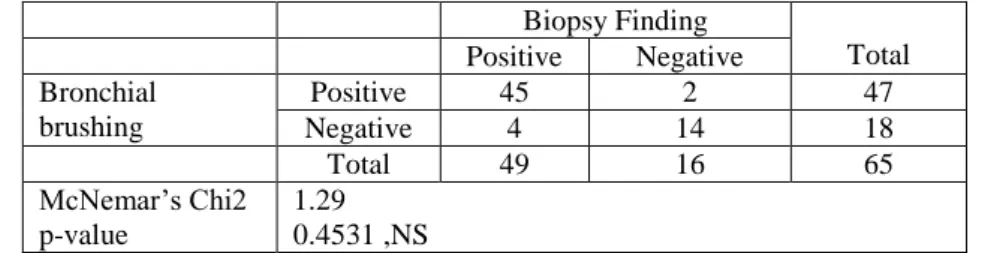
The cancer detection by BB gave mcnemars chi-square=1.29 and p-value = 0.4531. (TABLE V)
Table No. IV Comparison of diagnostic accuracy of Bronchial washing and Biopsy finding.
Biopsy Finding Total
Positive Negative Bronchial
washing
Positive 29 4 33
Negative 20 12 32
Total 49 16 65
McNemar’s Chi2 p-value
11.56 0.0007, HS
Table V Comparison of Diagnostic Accuracy of Bronchial Brushing and Biopsy Findings
Biopsy Finding
Total Positive Negative
Bronchial brushing
Positive 45 2 47
Negative 4 14 18
Total 49 16 65
McNemar’s Chi2 p-value
Dr Meenakshi Gundewar el al JMSCR Volume 07 Issue 09 September 2019 Page 421
Fig I photomicrograph showing eosinophils on brush (H&E, 40X)
Fig II photomicrograph showing eosinophilic granular material in lavage (H&E, 40X)
FIG III Photomicrograph showing carcinoid tumour on bb(PAP, 40X)
Fig IV Photomicrograph showing adenocarcinoma with glandular acini on bb (H&E, 10X)
Fig V photomicrograph showing tumour cells with large nuclei and macronucleoli on bb (H&E, 40X)
Discussion
Due to new therapeutic options in thoracic oncology, pathological diagnosis of different bronchopulmonary lesions has become more challenging.
With the advent of flexible fiber-optic bronchoscope, respiratory cytology has taken a new turn as samples like bronchoalveolar lavage, bronchial washings and bronchial brushings could be collected from the respiratory tract which yield significant amount of cytological material.
Dr Meenakshi Gundewar el al JMSCR Volume 07 Issue 09 September 2019 Page 422 The present study was comprised of 127 patients.
The age of the patients ranged from 14-85 yrs. Most of the patients were in the age group of 61-70 yrs (22.83%) with mean age of 50.15 ±16.14. There were 81 males and 46 females and the M:F ratio was 1.76:1. In a similar study by Choudhary m et al(1) the age ranged from 18 – 88 yrs and M : F ratio was 3.3:1.
Non- neoplastic lesions
In this study there were a total of 78 non-neoplastic lesions in which there are 45 males and 33 females and the M : F is 1.36:1. Most of the patients were in the age group of 41-50 yrs with mean age of 45.30±16.54 yrs. In a similar study by Tuladhar et al patients ranged in the age group from 19 to 74 yrs with a mean of 50.80±7.7 yrs and M : F ratio was 3.7:1. Similarly, Shiner et al found the male : female ratio to be 1:1 in non-neoplastic patients in their study.(2,3)
In our study we came across three cases of aspergillosis. Out of three there were two cases diagnosed on bal/wash specimens in which septate hyphae with acute angles were seen and they were later confirmed on biopsy. There was one case detected with squamous cell carcinoma on biopsy. The bb specimens only showed inflammation. There were two cases of candida diagnosed on bal/bw and bb only showed inflammation. There was a case of mucor which was diagnosed on biopsy consisting of non-septate, obtuse angle hyphae. The bal/bw and bb showed only inflammatory infiltrate.
In our study, 30 cases (38.46%) were of acid-fast bacillus positive tuberculosis. There were no case identified by bronchial brushings and all showed inflammatory exudates. However, 2 out of 30 cases of tuberculosis were identified by washing and rest only showed inflammation. In a study by Wallace et al on proven cases of tuberculosis he found bronchoscopic specimens mostly have a non- specific chronic inflammatory reaction(4,1). In Altaf bach et al.(5,1) study bronchial wash smear was positive for acid fast bacilli in 35% of cases. Similarly, Purohit et al.(6,1) and Kulpati et al.(7, 1)
demonstrated acid fast bacilli in 42% and 40% resply(1).
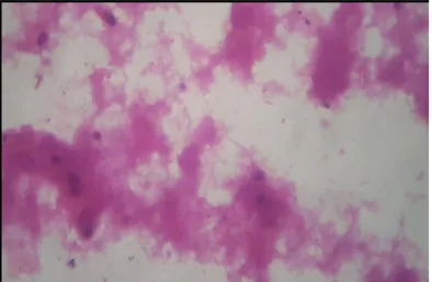
There were one each case of hydatid cyst, Loefflers syndrome (FIG I) and Alveolar proteinosis (FIG II). Both bronchial washings and brushings was normal but biopsy showed hydatid cyst with laminated membrane and hooklets. A patient, 22 yrs old came with the history of cough and cold since five days which was serous initially but later turned out to be mucoid. Her bal/bw specimen consist of pink, eosinophilic, granular material where as brush only showed inflammation with bronchial epithelial cells. Later biopsy was done which confirmed alveolar proteinosis which consists of PAS positive pink lipoproteinaceous, granular and eosinophilic material.
Eosinophilia was seen both in wash and brush which was diagnosed as Loefflers syndrome.
NEOPLASTIC lesions
There were 49 malignant neoplastic lesions. The diagnosis of the patients were confirmed by cytological and histological examinations. Male patients accounted for 73.46% and female patients for 26.53% with male:female ratio was 2.8:1. There was a male preponderance in the present study.
In similar studies done by Andrews et al(8), M:F ratio was 6.8:1 where as Mc Duffiie et al(9) reported M:F ratio of 3.7:1.
The age of the clinically confirmed lung cancer patients ranged from 29 to 85 years, with mean age of 58.48±11.21. The youngest patient was a male aged 29 years and the oldest was also male aged 85 years.
Dr Meenakshi Gundewar el al JMSCR Volume 07 Issue 09 September 2019 Page 423 (1986) reported that majority of the lung tumours
occurred in fifth, sixth and seventh decades with the peak incidence in sixth decade(11, 10). Similarly in the recent studies done by Al Rawi f. a. z. (2004)(12) and Gaur ds et al (2007)(13), majority of the cases were in the age group of 61-70 yrs. In a study by Vivekanand et al (2014), the majority of the cases were in fifth and sixth decade and the peak incidence was seen in sixth and seventh decade(10).
In the present study there were total of 73 smokers all were male patients with 27 male patients having smoking history of >40 pack years with p-value of 0.0001. In SPORE (specialized program of research excellence) study with a high risk cohort of patients with chronic obstructive pulmonary disease and >40 pack year smoking history, dysplasia was present in 25% of cases and subsequent follow up study showed malignancy in 36% of cases. So smoking history of >40 pack years is very often associated with risk of carcinoma. There were 8 male patients and all the female patients were non- smokers. A. Vigg and associates reported male to female ratio of about 6:1 and 62% ex-smokers, 10% current smokers and 28% non-smokers amongst males[10]. This high incidence in males could be due to the higher prevalence of smoking in males.
The most common clinical manifestation of lung cancer in present study is cough followed by breathlessness. In a similar study by Arora et al[14] (1990), the common clinical manifestation reported was cough (92%), hemoptysis (29%), chest pain (52%), breathlessness (40%) . Similarly in a study by Dr. Srividhya et al(15), the common clinical manifestation was cough (100%) followed by fever (75%). Vivekananda et al(1) presented with common symptoms of cough of long duration and chest pain, and very few cases presented with hemoptysis as seen in our study where hemoptysis was presented by only 7 cases of lung cancer.[16, 10]
In the current study, right lung is more affected than left lung in which right upper lobe was commonly affected. In a study by Vivekananda et
al[10], lung cancer mostly occurred in the right lung (56.6%) as compared to the left lung (43.30%) with a right lung, left lung ratio of 1.3:1. Again in a study by Dr. Srividya et al[15], right lung was more affected than left. In the right lung it was the right main bronchus that was largely affected.
In the diagnosis of 49 malignant lesions, bronchial washings yielded 17 positive cases and the remaining 32 cases showed dysplasia(11 cases), atypia (2 cases) and inflammatory smear(19cases) where as bb gave positive results in 42 cases and dysplasia was seen in 4 cases, inflammation was seen in (4 cases). There was a case in which squamous cell carcinoma was diagnosed by both bal/wash and bb but only necrotic tissue was seen. One case of squamous cell carcinoma also had aspergillus. It was included in both the neoplastic and non- neoplastic study group. In a case of adenosquamous carcinoma which was confirmed by biopsy both bal/wash and bb specimens showed adenocarcinoma.
In the present study the diagnostic accuracy of bronchial washings is low when compared to brushings. Which coincides with the study of Miep A, Vander Drift et al. in 2005[17]. The low diagnostic results in washing is due to the following reasons. 1.) Tumors just confined to bronchial wall not infiltrating into lumen. 2) Tumor necrosis 3) Lack of exfoliation due to poor communication of tumor with bronchus secondary to bronchial stenosis. . 4) Exclusion of atypical or highly suspicious cell from positive criteria. 5) Inadequate collection of material.
Dr Meenakshi Gundewar el al JMSCR Volume 07 Issue 09 September 2019 Page 424 by biopsy. This is closely compared with a study
by Vivekananda et al[10], where adenocarcinoma was the most commonly diagnosed tumor at 34.82% followed by SCC 31.02%. But in many other studies like in Anupam Sharma et al(18), Choudhary m et al(1), dr. Srividhya et al(15) and many more, SCC was the most common carcinoma followed by adenocarcinoma.
Tamboli P and Ro. J.Y (Lung cancers, M.D. Anderson Cancer care series, 2003) stated squamous cell carcinoma used to be the most common type of lung cancer accounting for 25-45% of all lung tumors but the incidence of adeno-carcinoma has significantly has increased in the last two decades; 25-40% of lung carcinoma are now classified as adeno-carcinoma and this tumor is now the most common form of lung cancer in women and in many studies, in men as well(19).
In a study by Tuladhar et al(2), the most common carcinoma was SCC (51%), followed by small cell carcinoma (19%) and then adenocarcinoma (11%).
In our study, the Sensitivity, Specificity and Accuracy of BAL/bw samples were 59.2%, 75% and 63.1% respectively. The sensitivity of bronchial aspirates in diagnosing lung cancers has been 75 to 88.1 % at various centres.[20,21,22].Truong et al[23,13] reported Sensitivity of 66.0%; while Ng. & Horak(24) reported a Sensitivity as high as 74.0% for BAL. Studies have shown that increasing the number of attempts obtaining BAL/bw sampling can improve its Sensitivity, Specificity and Accuracy.[23,20,25). The sensitivity, specificity and accuracy of bb were reported as 91.8%, 87.5% and 90.7%. Studies from various authors show the range of sensitivity for bb from 67% to 97.3%.[26,27,28] Similarly various other workers in the past, like Chopra et al[36] (86.3%); Zavala et al[29] (88.5%); and Solomon et al[30] (89.1%), Shroff et al[28] reported the Sensitivity of BB to be as high as 97.3% in their study. Hence, they reported the Sensitivity of BB to be much higher (98%) which was comparable to our study.
The specificity of bal/bw in other studies like Choudhary m et al[1] is 47.6%, Gaur ds et al[13] is 89.6% and accuracy is 57.1% and 71.4% resply comparable to our study. Similarly in bb the specificity and accuracy in the study of Choudhary m et al is 85.7% and 82.8% resply. The two studies of Gaur ds et al have reported similar results in which the first study gave specificity and accuracy of 77.8% and 80.6% and the second study gave the specificity and accuracy as 97.6% and 93.9% resply as seen in our study. In another study by Anupam sarma et al[18] the specificity and accuracy of bal/bw was 96.6% and 87.3% resply.
With a good Sensitivity (87.3 %), Specificity (97.6 %) and Accuracy (93.9 %), bronchial brushing promises to be a very convenient cytological technique that can be confidently utilized for screening of doubtful cases and early diagnosis of lung cancer, as it saves the time needed for the processing of biopsy specimens.
Conclusion
The advent of fiberoptic bronchoscopy has greatly facilitated the detection and diagnosis of bronchopulmonary lesions. Although it has been shown that the combined use of cytology and biopsy renders the highest probability of detecting malignancy, the merit of routine brush cytology has been questioned since it appears to duplicate biopsy.
With increased experience and adherence to strict cytological criteria for malignancy, it can be effectively detected in early stages and possibly cured.
Bibliography
1. Choudhury M., Singh S., Agarwal S. Efficacy of Bronchial Brush Cytology and Bronchial Washings in Diagnosis of Non Neoplastic and Neoplastic Bronchopulmonary Lesions. Turk Patoloji Derg 2012, 28:142-146.
Dr Meenakshi Gundewar el al JMSCR Volume 07 Issue 09 September 2019 Page 425 techniques in diagnoses of lung lesions.
Journal of Pathology of Nepal 2011, 1:126-130.
3. Shiner RJ, Rosenman J, Katz I, Reichert N, Hershko E, Yellin A. Bronchoscopic evaluation of peripheral lung tumors. Thorax 1988;43:887-9.
4. Wallace JM, Deutsch AL, Harrell JH, Moser KM: Bronchoscopy and transbronchial biopsy in evaluation of patients with suspected active tuberculosis. Am J Med 1981, 70:1189–1194
5. Altaf Bachh A, Gupta R, Haq I, Varudkar HG:Diagnosing sputum /smear-negative pulmonary tuberculosis: Does fibreoptic bronchoscopy play a significant role? Lung India 2010, 27:58–62
6. Purohit SD, Sisodia RS, Gupta PR, Sarkar SK, Sharma TN: Fibreoptic bronchoscopy in the diagnosis of smear negative pulmonary tuberculosis. Lung India 1983, 1:143–146
7. Kulpati DD, Heera HS: Diagnosis of smear negative pulmonary tuberculosis by flexible fibreoptic bronchoscopy. Indian J Tuberc 1986, 33:179–182
8. Andrews1985Andrews, L. Joseph Jr, Sarahbloom, Karohy Balogh (1985): Lung cancer in women, Lahey Clinic Experience, 1957-1980: Cancer 55 : 2894-2898.
9. Mc. Duffie H. et al 1987Mc. Duffie H. Helen David J. Klaasen & James A. Dosman (1987): Female Male differences in patients with primary lung cancer 59 : 1825 - 1830.
10.Reddy A.S., Vivekanand N., Durga K. Efficacy of bronchial wash and brush cytology in the diagnosis of lung cancers. Sch. J. App. Med. Sci.,, 2014; 2(2D):816-820.
11.Malhotra V, Malik. R. Beohar P.C, Gondal R (1986): Tumors of the lung – A histomorphological study, Indian Journal
of Chest Diseases and Allied Sciences 28: No. 1, 28 - 40.3.
12.Faiza a. z. al-rawi msc. Ficpath. Bronchial wash in the diagnosis of lung lesions. Iraqi j med sci, 2004; vol. 3(2): 125-127
13.Gaur DS, Thapiyal NC, Kishore S, Pathak VP: Efficacy of bronchial alveolar lavage and bronchial brush cytology in diagnosing lung cancers. J Cytol 2007, 24:73-77. (17)
14.Arora VK, Seetharaman ML, Ramkumar S, Mamatha TV, Subbarao KSVK, Banerjee A et al.; Bronchogenic carcinoma, Clinicopathological pattern in South Indian Population. Lung India, 1990; 8(3): 133-136.
15.Dr. Srividya. V.V.L, Dr.Sailendra.Vaddadi, Dr.Dost Mohmad khan. Correlative study of lung tumors by bronchial washings, brush biopsy & foreceps biopsy. Int J Biol Med Res. 2014; 5(1): 3817-3826.(56)
16.Hensing T. A. Clinical evaluation and staging of patients who have Lung cancer. Hematol Oncol Clin N Am. 2005; 19:219-23.
17.van der Drift MA, van der Wilt GJ, Thunnissen FB, Janssen JP. A prospective study of the timing and cost-effectiveness of bronchial washing during bronchoscopy for pulmonary malignant tumors. Chest 2005; 128 (1): 394- 400.
18.Sarma A, Sharma JD, Bhuyan C, Kataki AC, Sangma RA; A study of cytological evaluation of bronchial washing and brushing in Bronchogenic Carcinoma. International Journal of Scientific and Research Publications, 2013; 3(8): 1-7. 19.Tamboli P & Ro.J.Y: Pathologic
evaluation of lung cancer. Lung Cancer. M.D. Anderson cancer cares series; M.D. Anderson cancer center, Texas, 2003. 20.Ahmad M, Afzal S, Saeed W, Mubarik A,
Dr Meenakshi Gundewar el al JMSCR Volume 07 Issue 09 September 2019 Page 426 with biopsy in lung tumours. J Pak Med
Assoc. 2004;54 : 13–6.
21.Di Bonito L, Colautti I, Patriarca S, et al. Cytological typing of primary lung cancer: study of 100 cases with autopsy confirmation. Diagn Cytopathol 1991;7:7-10. (28)
22.Piaton E, Djelid D, Duvert B, Perrichon M, Saugier B. Sequential use of bronchial aspirates, biopsies and washings in the preoperative management of lung cancers. CytoJournal,2007 jun 4;4:11
23.Truong LD, Underwood RD, Greenberg SD, McLarty JW. Diagnosis and typing of lung carcinomas by cytopathologic methods. A review of 108 cases. Acta Cytol 1985; 29:379-84.
24.Ng ABP, Horak GC. Factors significant in the diagnostic accuracy of lung cytology in bronchial washing and sputum samples: II. Sputum samples. Acta Cytol 1983; 27 : 397-402.(14)
25.Naryshkin S, Daniels J, Young NA. Diagnostic correlation of fiberoptic bronchoscopic biopsy and bronchoscopic cytology performed simultaneously. Diagn Cytopathol 1992; 8:119-23.
26.Chopra SK, Genvesi MG, Simmons DH, Gothe B. Fibreoptic bronchoscopy in the lung cancer. Comparison of pre and post bronchoscopic sputa, washings, brushings and biopsies. Acta Cytol 1977; 21(4) : pp 524 -527 (36)
27.Pilotti, Rilke F, Gribaudi G, Ravasi GL. Sputum cytology for the diagnosis of carcinoma of the lung. Acta Cytol 1982; 26: 649-54. (122)
28.Shroff CP. Abrasive bronchial brushing cytology. A preliminary study of 200 specimens for the diagnosis of neoplastic and non-neoplastic broncho- pulmonary lesions. Acta Cytol 1985; 29 : 101-7. (123)
29.zavala DC. Diagnostic fiberoptic bronchoscopy: techniques and results of biopsy in 600 patients.Chest. 1975;68:12– 19. (38)
30.Solomon DA, Solliday NH, Gracey DR. Cytology in fiberoptic bronchoscopy: comparison of bronchial brushing, washing and post-bronchoscopy sputum.