© 2019 by the Serbian Biological Society 475
Growth performance and biochemical profile of
Azolla pinnata
and
Azolla caroliniana
grown under greenhouse conditions
Taylan Kösesakal1,* and Mustafa Yıldız2
1Department of Botany, Faculty of Science, Istanbul University, Istanbul, Turkey
2Department of Aquaculture, Faculty of Aquatic Sciences, Istanbul University, Istanbul, Turkey
*Corresponding author: [email protected]
Received: January 31, 2019; Revised: March 22, 2019; Accepted: April 25, 2019; Published online: May 10, 2019
Abstract: This study aimed to evaluate the growth performance, pigment content changes, essential amino acids (EAAs),
fatty acids (FAs), and proximate composition of Azolla pinnata and Azolla caroliniana grown in a greenhouse. Plants were grown in nitrogen-free Hoagland’s solution at 28±2°C/21±2°C, day/night temperature and 60-70% humidity and examined on the 3rd, 5th, 10th and 15th days. The mean percentage of plant growth and relative growth rate for A. pinnata were 119%
and 0.148 gg-1day-1, respectively, while for A. caroliniana these values were 94% and 0.120 gg-1day-1, respectively. Compared
to day 3, the amount of total chlorophyll obtained on day 15 decreased significantly (p<0.05) for A. pinnata while the total phenolic and flavonoid contents increased significantly (p<0.05) from the 3rd to the 15th day. However, the total phenolic
and flavonoid contents did not differ (p>0.0.5) in A. caroliniana. The crude protein, lipid, cellulose, ash values and the amounts of EAAs were higher in A. pinnata than A. caroliniana. Palmitic acid, oleic acid, and lignoceric acid were found to be predominant in A. pinnata and A. caroliniana. From the plant growth and pigment contents, we concluded that A. pinnata grew faster than A. caroliniana and its photosynthetic efficiency was more effective.
Keywords:Azolla; chlorophyll; fatty acids; phenolics; essential amino acids
How to cite this article: Kösesakal T, Yıldız M. Growth performance and biochemical profile of Azolla pinnata and Azolla caroliniana grown under greenhouse conditions. Arch Biol Sci. 2019;71(3):475-82.
INTRODUCTION
Azolla is a floating fern that can grow in the absence of nitrogen in freshwater because of the symbiotic relationship between the heterocyst-forming, filamen-tous, nitrogen-fixing cyanobacteria Anabaena azollae, which lives in the dorsal lobe cavity of the leaves [1]. This symbiotic association has recently gained con-siderable importance due to its potential for use as an alternative to nitrogenous chemical fertilizers and animal feeding [2-4]. Azolla is very important for the agricultural activities of developed and developing countries [5]. Azolla has great potential for biological N fixation (30-100 kg N ha−1) and thus Azolla species can be used effectively as a biofertilizer for paddy fields [6]. Furthermore, the use of Azolla species as a biofertilizer in rice fields improves soil fertility by increasing the organic matter in the soil, thus improv-ing soil structure and environmental safety [7,8]. In addition, Azolla species are rich in proteins, essential amino acids, minerals, vitamins, carotenoids and
growth promoter intermediaries. Therefore, with these nutritional values, Azolla species are a good source of feed for livestock [9].
In addition, Azolla species have been proposed as good candidates for phytoremediation of polluted freshwater areas [14-17]. Because of the multifaceted uses of Azolla species, especially in food, feed, biofuel production, agriculture and phytoremediation, it would be an ideal and environmentally-friendly factor in sustainable agriculture [2].
The main purpose of this study was to elucidate growth performance, pigment content changes, proxi-mate composition, fatty acids and essential amino acids of Azolla pinnata and Azolla caroliniana grown under greenhouse conditions. Also, the potential use of A. pinnata and A. caroliniana for further experimental studies has also been evaluated.
MATERIALS AND METHODS
Azolla pinnata and Azolla caroliniana plants were used, which were grown in modified Hoagland’s nutrient solution containing (in mg L-1): KCl, 74.55; KH
2PO4, 136.08; CaCl2•2H2O, 147.02; MgSO4•7H2O, 246.08; ZnSO4•7H2O, 0.22; H3BO3, 2.86; Na2MoO4•2H2O, 0.09; CuSO4•5H2O, 0.09; MnCl2•4H2O, 1.82; FeCl3•6H2O, 4.84 and Na2EDTA, 15. The pH value of the nutrient solution was adjusted to 6.0. Plants were grown at 28±2°C/21±2°C, day/night temperature and 60-70% humidity under greenhouse conditions. To ensure the transfer of only healthy and young plantlets, the procedure described in [18] was applied. The percent-age of plant growth, relative growth rate (RGR), total chlorophyll, carotenoid, total phenolic and flavonoid content changes were examined on the 3rd, 5th, 10th and 15th days.
Plant growth
Prior to application, the plants were weighed. The growth rate was measured by comparing the weight of the plants before and after the experimental times. Alsorelative growth rate (RGR) (g g−1 d−1) of the plants was calculated using the formula:
RGR = (ln W2−ln W1)/t,
where W1 and W2 are the initial and final fresh weights, respectively, and t is the experimental time [19].
Determination of chlorophyll and carotenoid contents
To determine the chlorophyll and carotenoid contents, 200 mg of leaves were extracted in 80% acetone (Merck) and the samples were centrifuged (Heraeus Labofuge 400 R) at 3000 x g (4°C) for 15 min. The pigment contents (chlorophyll a and b, total chlorophyll, and carotenoid) were measured using a Shimadzu 1601 UV-Visible Spectrophotometer) and expressed in µg/g fresh weight [20].
Analysis of total phenolics and flavonoids
Total phenolic and flavonoid contents of Azolla were measured as described [21]. The leaves were extracted with 1% HCl-methanol (5 mL), the extract was filtered and the filtrate was diluted with 1% HCl-methanol to 10 mL. Absorbance of the solution was measured at 280 nm for total phenolic and at 325 nm for the flavonoid contents. The total phenolic content was calculated from a standard curve made with gallic acid as a standard and expressed in GAeq. The flavonoid content was expressed as the absorbance at 325 nm/g fresh weight of Azolla.
Crude protein, lipid and cellulose analysis
The crude protein, lipid, cellulose and ash contents were determined according to the standard methodology [22]. Crude protein was determined as total nitrogen (N) using a semi-automatic Kjeldahl (Gerhardt VA-PODEST, 45s) technique (N×6.25). The lipid content was determined by ether extraction using a Soxtherm Multistat/SX PC (Gerhardt, Germany). The ash content was obtained from the weight loss after incineration of dried samples in a muffle furnace. Cellulose was determined using sulfuric acid, then sodium hydroxide (12.5%, w/w), and the final residue was washed with 5% HCl and water, then filtered, dried, and weighed. All samples were analyzed in triplicate.
Analysis of amino acids
The samples were then filtered and the excess acid from the hydrolysate was removed by flash evapora-tion under reduced pressure and resuspended in 0.02 N HCl. Amino acid analysis was performed using HPLC (Agilent 1100) [24].
Fatty acid (FA) analysis
Fatty acid methyl esters were transmethylated with 2 M potassium hydroxide (KOH) (Merck, Germany) in methanol and n-hexane (Sigma-Aldrich, Germany) [25], with minor modification. Ten mg of extracted oil were dissolved in 2 mL of hexane followed by the addition of 4 mL of 2 M methanolic KOH. By vor-texing the tube for 2 min at room temperature and a centrifugation at 4000 x g for 10 min, the resulting hexane layer was taken for GC analyses. By means of a gas chromatograph (Auto System XL Perkin Elmer, FID detector), using a 30 m x 0.25 mm x 0.25 µm capillary column (CP-2380 Supelco, USA), the FA composition was analyzed. The conditions of the method were as follows: carrier gas, helium; flame ionization detection temperature, 260°C; split rate was 1/0, oven tempera-ture programmed to rise from 120°C/2 min to 220°C/ 15 min at a rate of 5°C min−1; injector temperature was 240°C. Fatty acid methyl esters were identified by comparison to external standards (Sigma, USA). All FA analyses were performed on triplicate samples.
Statistical analysis
All the experimental data were obtained in 3 repli-cates. The experimental results are expressed as the mean±standard deviation. Statistical analysis was performed using GraphPad Prism version 5.2 for windows (GraphPad Software, San Diego, CA, USA). Statistically significant differences between the means were determined by post-hoc Tukey’s multiple com-parison test.
RESULTS Plant Growth
Fig. 1. shows plant growth percentages and RGR’s of A. pinnata and A. caroliniana respectively. Plant growth percentages of A. pinnata on the 3rd, 5th, 10th and 15th
days were 44%, 107%, 129% and 194%, respectively. The RGR’s of the A. pinnata plants at the experimental times were determined as 0.137, 0.180, 0.139 and 0.135 gg-1day-1, respectively. The average percentage growth and RGR for A. pinnata were 119% and 0.148 gg-1day-1, respectively. According to the plant growth percentage and RGR values, the biomass-doubling time of A. pin-nata is 5.6 days (Fig. 1A). The percentage growth of A. caroliniana on days 3, 5, 10, and 15 was 36%, 82%, 104% and 154%, respectively, whereas the RGRs were 0.111, 0.146, 0.113 and 0.109 gg-1day-1 (Fig. 1B). The mean percentage growth and RGR for A. caroliniana were 94% and 0.120 gg-1day-1, respectively. The biomass-doubling time of A. caroliniana was 6.8 days.
Photosynthetic pigment contents
Table 1 shows the changes in the photosynthetic pig-ment contents of Azolla pinnata and Azolla caroliniana on days 3, 5, 10, and 15. For A. pinnata, the amount of chlorophyll a decreased significantly (p<0.05) from day 3 to day 15. The chlorophyll a/b ratio did not differ (p>0.05), even though the chlorophyll/carotenoid ratio decreased (Table 1). In addition, the total chlorophyll and carotenoid amounts on the 15th day decreased significantly (p<0.05) compared to the 3rd day (Table 1, Fig. 2A). The pigment contents of Azolla caroliniana on days 3, 5, 10 and 15 are given in Table 1. The pho-tosynthetic pigment contents for A. caroliniana were slightly different from day 3 to day 15 (Table 1, Fig. 2B). Fig. 1. Plant growth percentages and RGRs of A. pinnata (A) and
Total phenolic and flavonoid contents
The amounts of total phenolics and flavonoids of A. pinnata on days 3, 5, 10 and 15 are given in Fig. 3A. The total phenolic content increased significantly (p<0.05) from day 3 to 15 (Fig. 3A). The mean total phenolic amount was 2.24 μg/g FW on the 15th day. The amount of total flavonoid content increased sig-nificantly from day 3 to 15 (Fig. 3A). However, the values obtained between day 5 and day 10 did not exhibit any significant difference (p>0.05). The mean total flavonoid content was 2.59 μg/g FW (Fig. 3A). The total phenolic content of A. caroliniana increased with time; however, this increase was not significant (p>0.05). The mean total phenolic content was 1.74 μg/g FW, while the total flavonoid content was 1.96 μg/g FW for the 15-day experimental period (Fig. 3B).
The proximate composition, essential amino acids and FA
The amounts of crude protein, lipid, ash and cellulose of A. pinnata and A. caroliniana plants are given in Table 2 and the amounts of EAAs are given in Table 3. Crude protein, lipid, cellulose and ash amounts were higher in A. pinnata than in A. caroliniana (Table 2). Analysis of the amino acid concentrations of A. pinnata showed higher amounts of EAAs than A. caroliniana (Table 3). On the other hand, arginine and leucine concentrations in A. pinnata and A. caroliniana were higher whereas histidine and methionine concentrations were lower at the end of the 15-day growth period (Table 3).
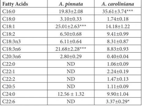
The main FA composition shows quantitative vari-ations in Azolla species (Table 4). Oleic acid (C18:1), Table 1. Photosynthetic pigment contents of A. pinnata and A. caroliniana on days 3, 5, 10 and 15.
Days 3 5 10 15
A.
p
in
na
ta Chlorophyll a (µg/g FW) 325.8±6.7 317.0±1.4 318.3±5.0 307.2±10.8
Chlorophyll b(µg/g FW) 74.4±6.6 67.8±4.9 67.0 ±3.7 71.5±11.8
Chlorophyll a/b 4.40±0.45 4.69±0.32 4.76±0.30 4.38±0.77
Total Chlorophyll/Carotenoid 1.77±0.03 1.69±0.06 1.75±0.04 1.82±0.04
A.
ca
ro
lini
an
a Chlorophyll a (µg/g FW) 289.7±5.8 292.5±15.6 292.5±11.2 266.4±9.3
Chlorophyll b (µg/g FW) 62.2±1.2 61.8±0.5 62.2±3.0 58.0±3.3
Chlorophyll a/b 4.66±0.16 4.73±0.27 4.71±0.13 4.60±0.10
Total Chlorophyll/Carotenoid 1.90±0.03 1.84±0.03 1.90±0.13 1.96±0.14
Fig. 2. Total chlorophyll and carotenoid contents of A. pinnata
(A) and A. caroliniana (B) on days 3, 5, 10 and 15. Bars represent the standard deviation. Significant differences determined by the Tukey’s multiple comparison test (p<0.05) are indicated by different letters.
gamma-linolenic acid (C18:3n6, GLA), docosahex-aenoic acid (C22:6, DHA), lignoceric acid (C24:0), and palmitic acid (C16:0), linoleic acid (C18:2, LA), alpha-linolenic acid (C18:3n3, ALA) were dominant FAs in A. pinnata and A. caroliniana, respectively. Oleic acid and gamma-linolenic acid amounts were higher in A. pinnata than in A. caroliniana. In addi-tion, palmitic acid amounts were significantly higher in A. caroliniana.
DISCUSSION
Azolla is one of the fastest growing plants capable of doubling its biomass in 5-6 days [11]. In the present study, A. pinnata showed higher growth performance than A. caroliniana considering the average plant growth percentage and RGR values and the biomass-doubling time. In [25] it was reported that the RGR in control Azolla filiculoides plants was 0.148 gg-1day-1. In [26] it was stated that the RGR for A. microphylla and A. caroliniana was 0.085 and 0.087 gg-1day-1, respectively, whereas biomass-doubling time for the same plants were 8.29 and 7.98 days, respectively. In [27] it was demonstrated that in control Azolla microphylla plants, the RGR was 0.133 gg-1day-1 and the biomass-doubling time was 8.6 days. Also, it was reported that the RGR values obtained on day 14 for A. filiculoides, A. micro-phylla, A. pinnata, A. rubra, A. mexicana and A. caro-liniana grown under greenhouse conditions were 0.11, 0.13, 0.06, 0.11, 0.10, and 0.11 gg-1day-1, respectively [28]. In the same study, the biomass-doubling times of A. filiculoides, A. microphylla, A. pinnata, A. rubra, A. mexicana and A. caroliniana plants were 6.3, 5.4, 11.1, 6.1, 6.6, and 6.1 days, respectively. The RGR is one of the important components of plant health and theoretically, plant RGR is closely related to biomass [29]. RGR values obtained from the current study for A. pinnata and A. caroliniana show that plants grow rapidly under greenhouse conditions as compared to other studies [28,30].
In this study, the amount of chlorophyll a was 317 μg/g FW and the amount of chlorophyll b was 67.8 μg/g FW on the 5th day for A. pinnata. On the other hand, chlorophyll a and b amounts for A. caroliniana on day 5 were 292.5 μg/g FW and 61.8 μg/g FW, respectively. The total chlorophyll content of A. pinnata was 400.2 μg/g FW on day 3, and 378.7 μg/g FW on day 15. The total chlorophyll content of A. caroliniana was 351.9 μg/g FW on day 3 and 324.3 μg/g FW on day 15. The amounts of chlorophyll a and b for A. filiculoides in the control medium were about 9 and 4 mg/g FW, re-spectively, whereas the total amount of chlorophyll was approximately 14 mg/g FW at the end of 7 days [31]. It was reported that the amounts of chlorophyll in the control medium of A. pinnata plant were about 6 and 7 mg/g FW on the 6th and 12th days, respectively [32]. The amounts of chlorophyll a, chlorophyll b and total Table 2. Proximate composition of A. pinnata and A. caroliniana
after 15 days.
Crude Protein (% dry weight)
Crude Lipid (% dry weight)
Crude Cellulose
(% dry weight)
Crude Ash (% dry weight) A. pinnata 22.8±1.56*** 4.4±0.35 17.6±1.69 19.6±1.47 A. caroliniana 19.7±0.93 4.1±0.17 16.2±0.25 18.2±0.14
Table 3. The amounts of essential amino acids of A. pinnata and
A. caroliniana after 15 days.
Essential amino acids
(% dry matter) A. pinnata A. caroliniana
Arginine 1.32±0.11 1.23±0.15
Histidine 0.34±0.04 0.31±0.06
Isoleucine 0.79±0.08 0.68±0.09
Leucine 1.78±0.15 1.62±0.18
Lysine 1.21±0.09 1.11±0.14
Methionine 0.26±0.02 0.21±0.02
Tryptophan 1.25±0.09 1.15±0.15
Threonine 0.91±0.07 0.86±0.09
Valine 0.86±0.09 0.74±0.08
Table 4. Fatty acid composition of A. pinnata and A. caroliniana
after 15 days.
Fatty Acids A. pinnata A. caroliniana
C16:0 19.83±2.08 35.61±3.74***
C18:0 3.10±0.33 1.74±0.18
C18:1 25.01±2.63*** 14.18±1.22
C18:2 6.50±0.68 9.41±0.99
C18:3n3 6.11±0.64 8.31±0.87
C18:3n6 21.68±2.28*** 8.83±0.93
C20:3n6 2.80±0.29 0.40±0.04
C22:0 ND 1.06±0.09
C22:1 ND 2.24±0.19
C22:2 ND 1.47±0.13
C20:5 ND 1.11±0.09
C24:0 12.56 ± 1.32 9.90±1.04
C22:6 ND 3.37±0.29*
chlorophyll for the control A. caroliniana plant were approximately 130, 90, and 40 μg/g FW, respectively [14]. Plants regulate the chlorophyll concentration to balance the absorption, utilization and distribution ca-pacities of light energy. This arrangement is considered to be an adaptation of plants to seasonal fluctuations under environmental stress [33]. According to the total chlorophyll values obtained from this study, it could be stated that, although the photosynthetic efficiency was more effective up to 10th day in A. pinnata, it was elevated up to the 15th day in A. caroliniana. Thus, the adaptation of A. pinnata to greenhouse conditions was faster than that of A. caroliniana.
Carotenoids have central functions in plants and are essential for photosynthesis and photoprotection [35]. Furthermore, carotenoids influence many plant processes and as antioxidants they can protect photo-synthetic organisms against oxidative stress [36]. The amounts of carotenoids in the control A. imbricata plant were 0.281 mg/g FW on day 1, and 0.373 mg/g FW on day 9 [37]; the amount of carotenoids in the control A. caroliniana plant was 26.7 μg/g FW [14]. In the present study, the highest amounts of carotenoids measured in A. pinnata and A. caroliniana were 227.5 and 193.0 μg/g FW, respectively, on day 5. Carotenoids act as an auxiliary pigment in photosynthesis and also protect the photosynthetic apparatus from photooxidative damage by quenching triplet chlorophyll molecules and scavenging reactive oxygen species (ROS) such as singlet oxygen [38]. We hypothesized that the increase in the carotenoid contents in both A. pinnata and A. caroliniana during the first five days in the greenhouse were the result of the adaptation of plants to changing growth conditions.
Phenolics protect plants from adverse conditions, diseases, ROS, wounding, and from UV radiation [39]. The phenylpropanoid biosynthesis pathway is respon-sible for the synthesis of various secondary metabolites, including phenolic esters, coumarins, flavonoids and lignin [40]. It was observed that the total phenolic content in the control medium of Azolla filiculoides did not show significant differences between days 3 and 7 and it was about 2.5 mg/g FW [41]. Furthermore the total phenolic and total flavonoid contents of A. pinnata and A. rubra were 95.25 and 92.16 μg GAE/ mg and 41.13 and 39.66 μg CE/mg, respectively [42]. In the present study, the total phenolic and flavonoid
contents of A. pinnata increased significantly (p<0.05) from day 3 to day 15. Although there was an increase in total phenolics and flavonoids from day 3 to day 15 in A. caroliniana, it was not statistically significant (p>0.05). It is likely that the increase in total phenolics and flavonoids in A pinnata and A. caroliniana was a protective reaction in the adaptation of plants to greenhouse conditions.
It was reported that the amounts of crude protein, lipid, ash and cellulose for A. pinnata were 275, 41, 200, and 116 g/kg, respectively, per DW [3]. According to [43], the crude protein and ash values of A. filiculoides were 232 and 112 g/kg, respectively. In the current study, crude protein, lipid, cellulose and ash per DW were 22.8%, 4.4%, 17.6% and 19.6% for A. pinnata, and 19.7%, 4.1%, 16.2% and 18.2%, for A. caroliniana respectively. The amounts of EAAs obtained from A. pinnata and A. caroliniana in this study are similar to the findings presented in other studies [44,45]. Examination of the EAA levels of A. pinnata and A. caroliniana showed that these plants can be processed and used in human diet. In addition, these plants can be used in fish feeds due to their EAA values. The quantitative composition of fatty acids of A. pinnata and A. caroliniana is characterized by a high content of palmitic acid, oleic acid, alpha-linolenic acid and lignoceric acids. In the present study, the FA contents in A. pinnata and A. caroliniana species were similar to those obtained in [46-49]. Furthermore, the con-centration of palmitic acid (C16:0) in A. pinnata and A. caroliniana species are relatively high compared to soybean, the main oilseed crop of the world [50]. Very few plant protein sources are known to contain all EAAs found in nature. As mentioned above, A. pinnata and A. caroliniana are rich sources of essential FAs due to their contents of C18:3n3, C18:1, C18:2, C18:3n6, C20:5 and C22:6 FA, which are very important for human nutrition and health. These plants will also occupy an important place in fish nutrition. Therefore, it is important to increase the production of these plants both for human nutrition and use in aquaculture.
its photosynthetic efficiency was better. Considering the EAAs and essential FAs contained in these plants, their uses in human nutrition and aquafeed possess an important economic value. In addition, the data about the percentage of plant growth, RGR, photosynthetic pigment, total phenolic and flavonoid contents, proxi-mate composition, EAA and FA composition indicated that in studies of organic and inorganic pollution, phytoremediation and animal feeding applied under different conditions, A. pinnata and A. caroliniana plants grown for at least 15 days in nutrient solution will be more efficient and healthier.
Funding: This work was supported by the Istanbul University under Grant ID: FDP-2016-20651.
Acknowledgments: We are grateful to Samuel Ofori-Mensah, Institute of Graduate Studies in Science and Technology, Istan-bul University, Turkey, for the help in English editing.
Author contributions: Taylan Kösesakal performed the experi-ments, analyzed the data and wrote the manuscript. Mustafa Yıldız contributed to the experimental design and manuscript editing. All authors reviewed and approved the final manuscript.
Conflict of interest disclosure: The authors declare that they have no conflict of interest.
REFERENCES
1. Costa ML, Santos MC, Carrapico F, Pereira AL. Azolla-
Ana-baena’s behaviour in urban wastewater and artificial media – Influence of combined nitrogen. Water Res. 2009;43:3743-50. 2. Yadav RK, Abraham G, Singh YV, Singh PK. Advancements
in the utilization of Azolla-anabaena system in relation to sustainable agricultural practices. Proc Indian Natn Sci Acad. 2014;80:301.
3. Gangadhar B, Sridhar N, Saurabh S, Raghavendra CH, Hemaprasanth KP, Raghunath MR, Jayasankar P. Effect of
Azolla-incorporated diets on the growth and survival of
Labeofimbriatus during fry-to-fingerling rearing. Cogent Food Agric. 2015;1:1055539.
4. Yao Y, Zhang M, Tian Y, Zhao M, Zhang B, Zeng K, Zhao
M, Yin B. Urea deep placement in combination with Azolla
for reducing nitrogen loss and improving fertilizer nitrogen recovery in rice field. Field Crops Res. 2018a;218:141-9. 5. Bocchi S, Malgioglio A. Azolla-Anabaena as a biofertilizer for
rice paddy fields in the Po Valley, a temperate rice area in Northern Italy. Int J Agron. 2010;2010:152158.
6. Yao Y, Zhang M, Tian Y, Zhao M, Zeng K, Zhang B, Zhao M, Yin B. Azolla biofertilizer for improving low nitrogen use efficiency in an intensive rice cropping system. Field Crops Res. 2018b;216:158-64.
7. Carrapiço F. Azolla as a superorganism. Its implication in sym-biotic studies. In: Seckbach J, Grube M, editors. Symbioses
and stress: joint ventures in biology. Dordrecht: Springer; 2010. p. 225-41.
8. Subedi P, Shrestha J. Improving soil fertility through Azolla
application in low land rice: A review. Azarian J Agric. 2015;2(2):35-9.
9. Gouri MD, Sanganal JS, Gopinath CR, Kalibavi1 CM. Impor-tance of Azolla as a sustainable feed for livestock and poultry – A Review. Agric Review. 2012;33(2):93-103.
10. Brouwer P, Brautigam A, Kulahoglu C, Tazelaar AO, Kurz S, Nierop KG, van der Werf A, Weber AP, Schluepmann H.
Azolla domestication towards a biobased economy? New Phy-tol. 2014;202:1069-82.
11. Miranda AF, Biswas B, Ramkumar N, Singh R, Kumar J, James A, Roddick F, Lal B, Subudhi S, Bhaskar T, Mouradov A. Aquatic plant Azolla as the universal feedstock for biofuel production. Biotechnol Biofuels. 2016;9:221.
12. Kollah B, Patra AK, Mohanty SR. Aquatic microphylla Azolla:
a perspective paradigm for sustainable agriculture, environ-ment and global climate change. Environ Sci Pollut Res Int. 2016;23:4358-69.
13. Muradov N, Taha M, Miranda AF, Kadali K, Gujar A, Roch-fort S, Stevenson T, Ball AS, Mouradov A. Dual application
of duckweed and Azolla plants for wastewater treatment and
renewable fuels and petrochemicals production. Biotechnol Biofuels. 2014;7:30.
14. Roberts AE, Boylen CW, Nierzwicki-Bauer SA. Effects of lead
accumulation on the Azollacaroliniana-Anabaena
associa-tion. Ecotoxicol Environ Saf. 2014;102:100-4.
15. Kosesakal T. Effects of seasonal changes on pigment composi-tion of Azolla filiculoides Lam. Am Fern J. 2014;104(2):58-66. 16. Kosesakal T. Assessment of the biodegradation capacity of
Azolla on polycyclic aromatic hydrocarbons in crude oil. Global NEST J. 2018;20(3):27-32
17. Gomes MP, de Brito JCM, Carvalho Carneiro MML, Ribeiro da Cunha MR, Garcia QS, Figueredo CC. Responses of the
nitrogen-fixing aquatic fern Azolla to water contaminated
with ciprofloxacin: Impacts on biofertilization. Environ Pol-lut. 2018;232:293-9.
18. Kosesakal T, Unal M, Kulen O, Memon A, Yuksel B. Phytore-mediation of petroleum hydrocarbons by using a freshwater fern species Azollafiliculoides Lam. Int J Phytoremediation. 2016;18:467-76.
19. Jampeetong A, Brix H. Effects of NH4+ concentration on
growth, morphology, and NH4+ uptake kinetics of Salvinia
natans. Ecol Eng. 2009;35:695-702.
20. Lichtenthaler HK, Wellburn AR. Determinations of total carotenoids and chlorophylls a and b of leaf extracts in dif-ferent solvents. Biochem Soc Trans. 1983;11:591-2. 21. Zhang D, Quantick PC. Effects of chitosan coating on
enzy-matic browning and decay during postharvest storage of litchi (Litchichinensis Sonn.) fruit. Postharvest Biol Tecnol. 1997;12:195-202.
22. AOAC. Official methods of analysis of AOAC International. 16th ed. Arlington, VA, USA: AOAC International; 1995. 23. AOAC. Official methods of analysis of AOAC International.
17th ed. Maryland, USA: AOAC International; 2000. 24. Antoine FR, Wei CI, Littell RC, Marshall MR. HPLC method
-phthaldi-aldehyde precolumn derivatization. J Agr Food Chem. 1999;47(12):5100-7.
25. Ichihara K, Shibahara A, Yamamoto K, Nakayama T. An improved method for rapid analysis of the fatty acids of glyc-erolipids. Lipids. 1996;31(5):535-9.
26. Pereira AL, Monteiro B, Azevedo J, Campos A, Osorio H, Vasconcelos V. Effects of the naturally-occurring contaminant microcystins on the Azollafiliculoides-Anabaenaazollae sym-biosis. Ecotoxicol Environ Saf. 2015;118:11-20.
27. Kannaiyan S. Effect of Benlate and Rhizoctonia interactions on the growth, chlorophyll contents and nitrogen fixation in three species of Azolla. S Afr J Bot. 1992;58:292-5.
28. Abraham G. Antioxidant enzyme status in Azolla microphylla
in relation to salinity and possibilities of environmental moni-toring. Thin Solid Films. 2010;519:1240-3.
29. Arora A, Saxena S. Cultivation of Azolla microphylla biomass on secondary-treated Delhi municipal effluents. Biomass Bio-energy. 2005;29:60-4.
30. Li YY, Lü XT, Wang ZW, Zhou C, Han XG. Linking relative growth rate to biomass allocation: the responses of a grass (Leymus chinensis) to nitrogen addition. Phyton Int J Exp Bot. 2014;83:283-9.
31. Mostafa EM, Tammam AA. The oxidative stress caused by NaCl in Azollacaroliniana is mitigated by nitrate. J Plant Interact. 2011;7:356-66.
32. Vafaei F, Khataee AR, Movafeghi A, Salehi Lisar SY, Zarei M. Bioremoval of an azo dye by Azollafiliculoides: Study of growth, photosynthetic pigments and antioxidant enzymes status. Int Biodeterior Biodegradation. 2012;75:194-200. 33. Rai AK, Rai V. Effect of NaCl on growth, nitrate uptake and
reduction and nitrogenase activity of Azolla
pinnata/Ana-baena azollae. Plant Sci. 2003;164:61-9.
34. Close DC, Davidson NJ, Davies NW. Seasonal fluctuations in pigment chemistry of co-occurring plant hemi-parasites of distinct form and function. Environ Exp Bot. 2006;58:41-6. 35. Sun T, Yuan H, Cao H, Yazdani M, Tadmor Y, Li L.
Carot-enoid metabolism in plants: The role of plastids. Mol Plant. 2018;11:58-74.
36. Savicka M, Petjukevičs A, Batjuka A, Škute N. Impact of moderate heat stress on the biochemical and
physiologi-cal responses of the invasive waterweed Elodea canadensis
(Michx. 1803). Arch Biol Sci. 2018;70(3):551-7.
37. Dai LP, Xiong ZT, Huang Y, Li MJ. Cadmium-induced changes in pigments, total phenolics, and phenylalanine ammonia-lyase activity in fronds of Azollaimbricata. Environ Toxicol. 2006;21:505-12.
38. Candan N, Tarhan L. Relationship among chlorophyll-carot-enoid content, antioxidant enzyme activities and lipid
per-oxidation levels by Mg2+ deficiency in the Menthapulegium
leaves. Plant Physiol Biochem. 2003;41:35-40.
39. Dixon RA, Paiva NL. Stress-induced phenylpropanoid metab-olism. Plant Cell. 1995;7(7):1085.
40. Zhang X, Liu CJ. Multifaceted regulations of gateway enzyme phenylalanine ammonia-lyase in the biosynthesis of phenyl-propanoids. Mol Plant. 2015;8:17-27.
41. Forni C, Braglia R, Harren FJ, Cristescu SM. Stress responses of duckweed (Lemnaminor L.) and water velvet (Azolla filicu-loides Lam.) to anionic surfactant sodium-dodecyl-sulphate (SDS). Aquat Toxicol. 2012;110-111:107-13.
42. Noor Nawaz AS, Syed J, Dileep N, Rakesh KN, Prashith
Kekuda TR. Antioxidant activity of Azolla pinnata and
Azolla rubra –A comparative study. Sch Acad J Biosci. 2014;2(10):719-23.
43. Leterme P, Londoño AM, Muñoz JE, Súarez J, Bedoya CA, Souffrant WB, Buldgen A. Nutritional value of aquatic ferns (Azollafiliculoides Lam. and Salviniamolesta Mitchell) in pigs. Anim Feed Sci Technol. 2009;149:135-48.
44. Datta SN. Culture of Azolla and its efficacy in diet of Labeo rohita. Aquaculture. 2011;310:376-9.
45. van Kempen MML, Smolders AJP, Bögemann GM, Lamers
LLM, Visser EJW, Roelofs JGM. Responses of the Azolla
filic-uloides Stras.–Anabaenaazollae Lam. association to elevated sodium chloride concentrations: Amino acids as indicators for salt stress and tipping point. Aquat Bot. 2013;106:20-8. 46. Paoletti C, Bocci F, Lercker G, Capella P, Materassi R. Lipid
composition of Azolla caroliniana biomass and its seasonal
variation. Phytochemistry. 1987;26:1045-7.
47. Cohen MF, Meziane T, Tsuchiya M, Yamasaki H. Feeding deterrence of Azolla in relation to deoxyanthocyanin and fatty
acid composition. Aquat Bot.2002;74 (2):181-7.
48. Brouwer P, van der Werf A, Schluepmann H, Reichart G-J, Nierop KGJ. Lipid yield and composition of Azolla filiculoides
and the implications for biodiesel production. BioEnergy Res. 2016;9(1):369-77.
49. Miranda AF, Liu Z, Rochfort S, Mouradov A. Lipid produc-tion in aquatic plant Azolla at vegetative and reproductive stages and in response to abiotic stress. Plant Physiol Bio-chem. 2018;124:117-25.