Profile of Hospitalized Hypertensives with
Preserved and Reduced Left Ventricular
Ejection Fraction in Nigeria
Akinsanya Daniel Olusegun-Joseph
1, Kamilu M Karaye
2, Adeseye A Akintunde
3,
Bolanle O Okunowo
1, Oladimeji G Opadijo
3, Abdulrazak Garba Habib
2, Suleiman A Balarabe
21. Department of Medicine, College of Medicine, University of Lagos & Lagos University Teaching Hospital, Lagos, Nigeria. 2. Department of Medicine, Bayero University & Aminu Kano Teaching Hospital, Kano, Nigeria.
3. Department of Medicine, Ladoke Akintola University of Technology (LAUTECH) & LAUTECH Teaching Hospital, Ogbomoso, Nigeria.
Corresponding author:
A.D. Olusegun-Joseph.
Department of Medicine, College of Medicine, University of Lagos, Lagos. Email: [email protected]
Introduction
Hypertension is the most important risk factor for cardiovascular diseases (CVD), and a major reason for medical admissions, morbidity and mortality [1-3]. It is a leading cause of stroke, ischemic heart disease (IHD), renal failure, peripheral arterial disease, and visual impairment [1-4].
Chronic, uncontrolled hypertension often predispose to asymptomatic structural and functional cardiac changes [5-7]. The
result is the development of left atrial enlargement, left ventricular hypertrophy, myocardial fibrosis; and diastolic and systolic dysfunction. Many of these patients eventually progress to develop overt symptoms of cardiac disease, with concurrent changes in other target organs. Left ventricular systolic dysfunction, although less common compared to diastolic dysfunction, is an important prognostic factor for outcomes in hypertensive patients [8-9]. Worsening LV systolic function is a major risk factor not just for heart failure, but other cardiovascular events [10-11].
* Corresponding author. E-mail: [email protected]
Abstract
Background
The impact of preserved and reduced left ventricular ejection fraction (LVEF) has been well studied in heart failure, but not in hypertension. We aimed to highlight the prevalence, clinical characteristics, comorbidities and outcomes of hospitalized hypertensives with preserved and reduced LVEF from three teaching hospitals in Nigeria.
Methods
This is a retrospective study of hypertensives admitted in 2013 in three teaching hospitals in Lagos, Kano and Ogbomosho, who had echocardiography done while on admission. Medical records and echocardiography parameters of the patients were retrieved and analyzed.
Results
54 admitted hypertensive patients who had echocardiography were recruited, of which 30 (55.6%) had reduced left ventricular ejection fraction (RLVEF), defined as ejection fraction <50%; while 24 (44.4%) had preserved left ventricular ejection fraction (PLVEF). There were 37(68.5%) females and 17 (31.5%) males. Of the male patients 64.7% had RLVEF, while 35.3% had PLVEF. 19(51.4%) of females had RLVEF, while 48.6% had PLVEF. Mean age of patients with PLVEF was 58.83±12.09 vs 54.83± 18.78 of RLVEF; p-0.19. Commonest comorbidity was Heart failure (HF) followed by stroke (found among 59.3% and 27.8% of patients respectively). RLVEF was significantly commoner than PLVEF in HF patients (68.8% vs 31.3%; p- 0.019); no significant difference in stroke patients (46.7% vs 53.3%; p-0.44). Mortality occurred in 1 (1.85%) patient who had RLVEF.
Conclusions
RLVEF was more common than PLVEF among admitted hypertensive patients; they also have more comorbidities. In-hospital mortality is, however, very low in both groups.
Keywords: Hypertension; Reduced LV ejection fraction; Preserved LV ejection fraction; Comorbidities; mortality
Citation: Olusegun-Joseph AD, Karaye KM, Akintunde AA, Okunowo BO, Opadijo OG, Habib AG, Balarabe SA. Profile of Hospitalized Hypertensives with Preserved and Reduced Left Ventricular Ejection Fraction in Nigeria: International Cardiovascular Forum Journal. 2018;14:11-15. DOI: 10.17987/icfj.v14i0.502
ISSN: 2410-2636 © Barcaray Publishing
Apart from cases of heart failure, there is paucity of data on the prevalence, clinical characteristics, comorbidities and outcomes of hypertension with PLVEF and RLVEF. This study aimed to highlight the prevalence, clinical characteristics, comorbidities and outcomes of hospitalized hypertensives with PLVEF and RLVEF from three teaching hospitals in Nigeria.
Methodology
This was a retrospective study of hypertensive patients admitted to the medical wards of Lagos University Teaching Hospital (LUTH), Aminu Kano Teaching Hospital (AKTH) and Ladoke Akintola University of Technology Teaching Hospital (LAUTECH TH) in Nigeria in 2013.
Medical records and echocardiography parameters of the patients were retrieved and analyzed. The data was obtained from the case notes and echocardiography registers of all the study centers from 1st January to 31st December 2013. Echocardiography examinations were carried out at the laboratories of the study centers according to the standard recommendations of the American Society of Echocardiography [12].
Hypertension was defined as presence of sustained systolic and or diastolic blood pressure (SBP and DBP respectively) of ≥140 and or 90mmHg respectively, or its documented history, or if a patient was on any antihypertensive medication even if BP was normal. HF was defined as the presence of 2 major, or 1 major and 2 minor criteria in the Framingham diagnostic criteria for heart failure [13]. Reduced LVEF was defined as Ejection fraction < 50% [9].
Results were analyzed using the SPSS version 20 statistical package. Descriptive statistics like mean, standard deviation, frequency and percentage were used for the results. T-test was used to compare differences in means, and Pearson chi-square test was used to check for level of association between two variables while level of significance was set at 0.05.
Approval for the study was obtained from the local Ethics Committee in the institutions.
Results
A total of 54 hypertensive patients on admission, who had echocardiography done were recruited for this study, of which 17 (31.5%) were males, and 37 (68.5%) were females. The age range was 26 to 90 years, with a mean age of 54.83 ± 18.78 years. RLVEF was found in 30 (55.6%), while 24 (44.4%) of them had PLVEF. Diastolic LV dysfunction was present in 23 (77.0%) and 15 (62.5%) of patients with RLVEF and PLVEF respectively. Mean age of patients with PLVEF was 58.83±12.09 years, while that of RLVEF was 54.83± 18.78 years (p=0.35). The age, gender and LVEF distribution is shown in table 1.
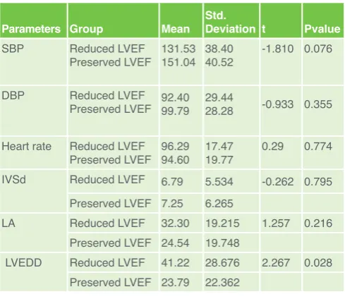
Patients with RLVEF had lower systolic and diastolic blood pressures, and higher pulse rate when compared with those with PLVEF, although not statistically significant. Also, patients with RLVEF had lower packed cell volume (PCV) when compared with PLEVEF, although not statistically significant (40.64 ± 11.47 vs 51.18 ± 75.56; t= -0.69; p= 0.49). The Clinical/ echocardiography parameters and LVEF distribution is shown in Table 2.
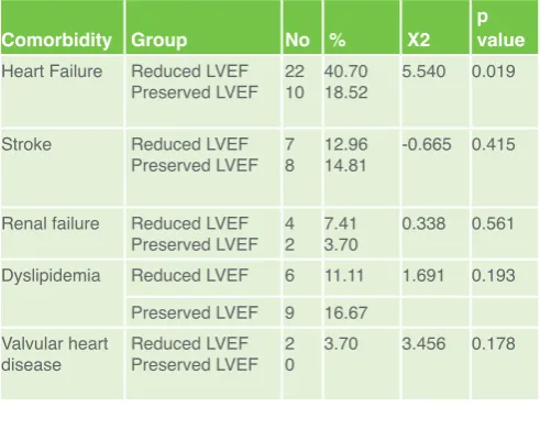
Heart failure, 32(59.3%) was the commonest comorbidity on admission followed by stroke,15(27.8%). RLVEF was significantly commoner in patients with heart failure compared with PLVEF (68.8% vs 31.3%; p- 0.019). Hypertension comorbidity/LVEF distribution is shown in Table 3. Mortality occurred in 1 (1.85%) patient, a man with heart failure, who had RLVEF.
The pattern of antihypertensive medication of the patients at the time of admission is shown in Table 4. There was no statistical difference in the pattern of use of the different medications among the two groups. Also there was no difference in the level of adherence to medication between the two groups (p=0.923).
Discussion
There is paucity of data on the prevalence, clinical characteristics, comorbidities and outcomes of hypertensive with preserved and reduced left ventricular ejection fraction, apart from cases of heart failure where this has been well studied. This multicenter Table 1 Age group, sex and LVEF distribution
LVEFgroup Sex
Total Male Female
Reduced
LVEF age group <45 26.7% 1136.7% 1343.3%
45-64 4 3 7
13.3% 10.0% 23.3%
>64 5 5 10
16.7% 16.7% 33.3%
Total 11 19 30
36.7% 63.3% 100.0% Preserved
LVEF age group <45 14.2% 416.7% 520.8%
45-64 3 8 11
12.5% 33.3% 45.8%
>64 2 6 8
8.3% 25.0% 33.3% Total
% of Total Count 625.0% 75.0%18 24100.0%
Table 2 shows clinical and echocardiography parameters and LVEF
Parameters Group Mean Std. Deviation t Pvalue
SBP Reduced LVEF
Preserved LVEF 131.53151.04 38.4040.52 -1.810 0.076
DBP Reduced LVEF
Preserved LVEF 92.4099.79 29.4428.28 -0.933 0.355
Heart rate Reduced LVEF
Preserved LVEF 96.2994.60 17.4719.77 0.29 0.774 IVSd Reduced LVEF 6.79 5.534 -0.262 0.795
Preserved LVEF 7.25 6.265
LA Reduced LVEF 32.30 19.215 1.257 0.216 Preserved LVEF 24.54 19.748
study was borne out of the need to assess the prevalence, clinical characteristics, comorbidities, and outcomes of hospitalized hypertensives with preserved and reduced ejection fraction from three teaching hospitals in Nigeria.
Our study revealed that 55.6% of the patients had RLVEF. This is very high when compared with studies by Verdecchia et al [9] and Ojji et al [14] where the prevalence were 3.6% and 6.7% respectively. The very low prevalence in the two studies when compared with our findings is probably because while they recruited uncomplicated hypertensive patients, ours is a study of very ill patients who were on admission as a result of hypertension complications already. Also, in the Genetic Epidemiology Network (HyperGEN) study [15], a population based study that recruited 2086 hypertensive subjects, RLVEF defined as Ejection fraction ≤ 54% was reported in 14% of the subjects. The inclusion of subjects with previous myocardial infarction or overt CHF in this study may explain the greater prevalence of systolic dysfunction when compared with the result by Verdecchia and Ojji et al. The prevalence in the HyperGEN study, however, is still very much lower when compared with our result since majority of their subjects were healthy people from the community. When we compare the three studies and ours there seem to be a narrative: reduced LVEF is associated with increased cardiovascular events and morbidity. Indeed studies have identified RLEVF as a marker of disease progression in hypertensive patients [11,16].
The present study reveals that 43% of those with systolic LV dysfunction were young people, less than 45 years old. This is at variance with the community based study by Wang etal [16] where LV systolic dysfunction is associated with increasing age. This may be due to the fact that hypertension develops quite early in blacks and progresses rapidly, during which the majority are unaware of it [17-19]. Moreover, a number of young black patients find it hard to accept that they are hypertensive and so do not take their drugs or resort to detrimental home remedies [17]. Furthermore, studies have shown that young black patients are more likely to have a variable medication-taking routine than older patients, with lower rate of medication adherence, predisposing them to hypertension complications very fast [20]. The attendant, subtle disease progression with
early systolicdysfunction is a major risk and prognostic factor in hypertension comorbidities. Happening at a young age puts more burden, in terms of morbidity, on the patients who are supposed to be in their most productive years. The resultant reduction in the quality of life of these patients as the disease progresses can have very dire consequences on them in every way: emotionally, financially, socially and other aspects.
The study also revealed that RLVEF had lower SBP, DBP and higher pulse rate compared with patients with PLVEF, though not statistically significant, probably due to the small sample size. This is similar to the observation in the hyperGen study [15]. The reason for this trend, though not totally clear, may be due to better cardiac contractility and ultimately higher blood pressure in the PLVEF group [21,22]. On the other hand, however, this may be an early pointer to future challenge of progressive pump failure that tends to develop with disease progression in those with RLVEF [16,22]. Aggressive management to prevent or delay this malady that often results in cardiogenic shock with very bleak prognosis should be the goal of management of such patients [22].
Our study also revealed that patients with RLVEF had significantly larger left ventricular end diastolic diameter (LVEDD) when compared with those with PLVEF, as well as larger left atrial diameter, though not statistically significant. This is similar to findings by other researchers [14,16]. Left ventricular remodeling and dilatation may develop in the absence of recurrent ischemic injury, as a result of neurohormonal pathway activation, with attendant changes at the cellular and molecular level in cardiac myocytes and the extracellular matrix, with resultant fibrosis, apoptosis and other deleterious cascade [16,22,23]. This has been posited to be a herald of early dilated cardiomyopathy especially, with disease progression [23].
Diastolic dysfunction was found in 77% of those with RLVEF, and 62.5% with PLVEF. This result shows that diastolic dysfunction is still a very common dysfunction in hypertensive patients, more common than systolic dysfunction. This is in keeping with previous reports [8, 9]. Our result also confirm the observation by other workers that diastolic dysfunction, though often an early and first dysfunction in hypertensive patients does not always precede systolic dysfunction [9,11]. Systolic dysfunction can develop in some hypertensive patients without prior diastolic dysfunction. This scenario is commonly seen in the setting of Ischemic heart disease and myocardial infarction [9, 16]. However, this has also been observed in non-ischemic scenario where there is apoptosis with interstitial fibrosis and myocyte loss, all of which are long term sequelae of neurohormonal pathway activation [22-24].
Table 3 shows hypertension comorbidities and LVEF distribution.
Comorbidity Group No % X2 p value
Heart Failure Reduced LVEF
Preserved LVEF 2210 40.7018.52 5.540 0.019
Stroke Reduced LVEF
Preserved LVEF 78 12.9614.81 -0.665 0.415
Renal failure Reduced LVEF
Preserved LVEF 42 7.413.70 0.338 0.561 Dyslipidemia Reduced LVEF 6 11.11 1.691 0.193
Preserved LVEF 9 16.67 Valvular heart
disease Reduced LVEFPreserved LVEF 20 3.70 3.456 0.178
Table 4. Antihypertensive use among the patients Antihypertensive RLVEF % PLVEF % X2 P
ACEI 60.0 43.5 2.254 0.521
ARB 86.4 75.0 0.796 0.372
CCB 86.4 68.8 1.729 0.189
Heart failure was the commonest comorbidity of patients on admission, followed by Stroke and dyslipidemia. While RLVEF was significantly commoner than PLVEF in heart failure, there was no statistical difference between these two groups in stroke, renal failure, valvular heart disease, dyslipidemia. It is well documented that hypertensive patients can develop and progress from asymptomatic left ventricular dysfunction to overt heart failure [23,25,26]. Previous studies reveal that RLVEF is associated with a 5-9 fold increased risk of CHF [9,16]. Proactive and aggressive management of preclinical stages of heart failure, as well as treating known risk factors will go a long way to reduce the overall societal burden of this syndrome. Adequate management of hypertension has been found to decrease risk of progression to heart failure by 50% [27].
Finally, our study revealed a relatively low in-hospital mortality of 1.85% in the RLVEF group. No mortality was recorded in the PLVEF group. Although the mortality in our study was so low that no major conclusions can be made from it, studies by other workers revealed higher mortality in patients with RLVEF [10, 16]. Indeed, the study by Wang et al indicate that even mild, asymptomatic left ventricular dysfunction have very important prognostic implications, with rates of CHF and death that are 2- to 4-fold higher than those of individuals with normal LV systolic function [16]. With this in mind, the need for early, proactive and aggressive management to prevent and delay disease progression cannot be overemphasized.
Limitations
This study has a number of limitations that need to be highlighted. The first and major limitation is incomplete data collection due to inadequate record keeping, resulting in the small sample size. A larger sample size, with complete data, may help to reveal more relevant findings. Furthermore, we were unable to assess the possible contributions of duration of hypertension, cigarette smoking, alcohol and diabetes which are major risk factors that can impact on left ventricular function, cardiovascular events and outcomes. Also, the contribution of adherence to medications in the natural history of these patients could not be assessed.
Conclusion
RLVEF is common in hypertensive patients admitted in hospital and this is associated with comorbidities, although in-hospital mortality is low. Early, proactive and aggressive management of hypertension, right from preclinical stages of target organ involvement is important to prevent and delay disease progression.
Declarations of interest
The authors declare no conflict of interest.
Acknowledgements
We thank the personnel in the Medical records department, and cardiovascular laboratory for their assistance during data collection. The authors state that they abide by the authors’ responsibilities and ethical publishing guidelines of the International Cardiovascular Forum Journal [28].
References
1. World Health Organization. A global brief on Hypertension. Silent killer, global public health crisis. World Health Day 2013. Geneva, World Health Organization, 2013.
2. Global health risks: Mortality and burden of disease attributable to selected major risks. Geneva, World Health Organization, 2009.
3. Lawes CMM, Vander Hoorn S, Rodgers A, for the International Society of Hypertension. Global burden of blood pressure related disease, 2001. Lancet 2008; 371: 1513–1518. DOI: http://dx.doi.org/10.1016/S0140- 6736(08)60655-8.
4. Lim SS, Vos T, Flaxman AD, Danaei G, et al A comparative risk assessment of burden of disease and injury attributable to 67 risk factors and risk factor clusters in 21 regions, 1990-2010 : a systematic analysis for the Global Burden of Disease Study 2010. Lancet. 2012; 380 (9859): 2224-60. 5. Kenchaiah S, Pfeffer MA. Cardiac remodeling in systemic hypertension.
Med Clin North Am. 2004; 88(1):115-30.
6. Bountioukos M, Schinkel AFL, Bax JJ, MD, Lampropoulos S, Poldermans D. The impact of hypertension on systolic and diastolic left ventricular function. A tissue Doppler echocardiographic study. Am Heart J. 2006;151(6):1323.e7-1323.e1.
7. Lawler PR, Hiremath P, Cheng S. Cardiac Target Organ Damage in Hypertension: Insights from Epidemiology. Curr Hypertens Rep. 2014 Jul; 16(7): 446. doi: 10.1007/s11906-014-0446-8.
8. Anguita SM. Arterial hypertension and systolic left ventricular dysfunction: therapeutic approach. Rev Esp Cardiol. 1999;52 Suppl 3:34-8.
9. Verdecchia P, Angeli F, Gattobigio R, Sardone M, Porcellati C. Asymptomatic Left Ventricular Systolic Dysfunction in Essential Hypertension: Prevalence, Determinants, and Prognostic Value. Hypertension. 2005;45:412-418. https:// doi.org/ 10. 1161/01.HYP.0000154822.37141.f6.
10. Gottdiener JS, McClelland RL, Marshall R, Shemanski L, Furberg CD, Kitzman DW, Cushman M, Polak J, Gardin JM, Gersh BJ, Aurigemma GP, Manolio TA. Outcome of congestive heart failure in elderly persons: influence of left ventricular systolic function. The Cardiovascular Health Study. Ann Intern Med. 2002; 137: 631–639.
11. Yeboah J., Rodriguez C.J., Stacey B., et al. Prognosis of individuals with asymptomatic left ventricular systolic dysfunction in the Multi-Ethnic Study of Atherosclerosis (MESA). Circulation 2012; 126:2713–2719.
12. Picard MH, Adams D, Bierig SM, Dent JM, Douglas PS, Gillam LD, Keller AM, Malenka DJ, Masoudi FA etal. American Society of Echocardiography Recommendations for Quality Echocardiography Laboratory Operations. J Am Soc Echocardiogr. 2011 Jan;24(1):1-10. doi: 10.1016/j. echo.2010.11.006.
13. McKee PA, Castelli WP, McNamara PM, Kannel WB. The natural history of congestive heart failure: the Framingham study. N Engl J Med. 1971 Dec 23;285(26):1441-6
14. Dike Ojji, John Atherton, Karen Sliwa, Jacob Alfa, Murtala Ngabea, Lionel Opie; Left Ventricular Systolic Dysfunction in Asymptomatic Black Hypertensive Subjects, American Journal of Hypertension. 2015; 28 (7): 924–929, https://doi.org/10.1093/ajh/hpu247.
15. Devereux RB, Bella JN, Palmieri V, Oberman A, Kitzman DW, Hopkins PN, Rao DC, Morgan D, Paranicas M, Fishman D, Arnett DK; Hypertension Genetic Epidemiology Network Study Group. Left ventricular systolic dysfunction in a biracial sample of hypertensive adults: The Hypertension Genetic Epidemiology Network (HyperGEN) Study. Hypertension. 2001; 38: 417–423.
16. Wang TJ, Evans JC, Benjamin EJ, Levy D, Leroy EC, Vasan RS. Natural History of Asymptomatic Left Ventricular Systolic Dysfunction in the Community. Circulation. 2003;108:977-982. doi: 10.1161/01. CIR.0000085166.44904.79.
17. Pettey CM, McSweeney JC, Stewart KE. African Americans’ Perceptions of Adherence to Medications and Lifestyle Changes Prescribed to Treat Hypertension. Sage. 2016;6. https://doi.org/10.1177/2158244015623595 18. Ergul A: Hypertension in black patients: An emerging role of the endothelin
system in salt-sensitive hypertension. Hypertension. 2000; 36: 62–67. 19. Victor RG, Leonard D, Hess P, Bhat DG, Jones J, Vaeth PAC, etal. Factors
Associated With Hypertension Awareness, Treatment, and Control in Dallas County, Texas. Arch Intern Med. 2008;168(12):1285-1293. doi:10.1001/ archinte.168. 12.1285.
20. Medication Routines and Adherence Among Hypertensive African Americans Abida Solomon, PhD;1 Antoinette Schoenthaler, EdD;1,2 Azizi Seixas, PhD;2 Gbenga Ogedegbe, MD;2 Girardin Jean-Louis, PhD;2 Dejian Lai, PhD2
21. Hollenberg SM, Kavinsky CJ, Parrillo JE. Cardiogenic Shock. Ann Intern Med. 1999;131:47-59.
22. Little WC,Applegate RJ. Congestive heart failure: systolic and diastolic function. J Cardiothorac Vasc Anesth. 1993;7(4):2-5.
23. Drazner MH. The Progression of Hypertensive Heart Disease. Circulation. 2011;123:327-334. https://doi.org/10.1161/ CIRCULATIONAHA.108.845792.
Chronic Heart Failure. Rev Esp Cardiol. 2004;57:834-41 - Vol. 57 Num.09 DOI: 10.1016/S1885-5857(06)60648-8.
25. Mann DL. Mechanisms and models in heart failure: a combinatorial approach. Circulation. 1999;100:999–1008.
26. McMurray JV, McDonagh TA, Davie AP, et al. Should we screen for asymptomatic left ventricular dysfunction to prevent heart failure? Eur Heart J. 1998;19:842–846.
27. Moser M, Herbert PR. Prevention of disease progression, left ventricular hypertrophy and congestive heart failure in hypertension treatment trials. J Am Coll Cardiol. 1996; 27: 1214–1218.