ARTIGO ORIGINAL
RESUMO
Introdução: A prematuridade e o baixo peso ao nascer têm sido associados a maior morbilidade e mortalidade neonatais. Este estudo teve como objetivo avaliar possíveis fatores de risco para a prematuridade associada a restrição do crescimento fetal e a recém --nascidos leves para a idade gestacional e determinar a incidência da morbilidade nestes dois grupos de recém--nascidos.
Material e Métodos: Estudo caso-controlo retrospetivo dos recém-nascidos com idade gestacional inferior a 32 semanas, com o diagnóstico obstétrico de restrição do crescimento fetal e com o diagnóstico clínico de leves para a idade gestacional, internados na Unidade de Cuidados Intensivos Neonatais de um hospital terciário, durante um período de seis anos.
Resultados: Foram estudados 356 recém-nascidos, observando-se uma incidência de 11% de restrição do crescimento fetal e 18% de leves para a idade gestacional. A pré-eclâmpsia foi o fator de risco da gestação com maior significado estatístico (47% vs 16%, p < 0,001) nos recém-nascidos leves para a idade gestacional. Observou-se também, nestes recém-nascidos, maior incidência de displa -sia broncopulmonar ligeira (66% vs 38%, p = 0,005), de sépsis tardia (59% vs 37%, p = 0,003), de retinopatia da prematuridade (58% vs 26%, p = 0,003) e de enterocolite necrotizante (20% vs 9%, p = 0,005). A mortalidade foi idêntica nos três grupos.
Discussão: Encontraram-se menos recém-nascidos do sexo masculino diagnosticados com restrição do crescimento fetal durante a gravidez comparativamente ao sexo feminino. Observaram-se diferenças significativas no grupo destes recém-nascidos, quanto à ocorrência de corioamnionite e de pré-eclâmpsia, face ao grupo controlo. Tanto os recém-nascidos com restrição do crescimento fetal como os leves para a idade gestacional apresentaram uma pontuação mais elevada nos índices de risco clínico comparativamente ao grupo controlo. De forma global, os recém-nascidos leves para a idade gestacional tiveram maior incidência de morbilidade que os recém-nascidos com restrição do crescimento fetal e que o grupo controlo.
Conclusão: Os avanços nos cuidados intensivos neonatais diminuíram a mortalidade nos recém-nascidos prematuros. Contudo, observam-se ainda diferenças significativas na incidência da morbilidade nos recém-nascidos com compromisso do crescimento. A colaboração entre obstetras e neonatalogistas constitui a base para uma correta avaliação clínica, sinalização precoce e intervenção global sobre estes recém-nascidos, com impacto significativo no prognóstico a curto e longo prazo.
Morbidity in Prematurity Associated with Fetal Growth
Restriction: Experience of a Tertiary Care Center
Morbilidade na Prematuridade Associada a Restrição do
Crescimento Fetal e nos Prematuros Leves para a Idade
Gestacional: Experiência de um Centro de Referência
1. Serviço de Pediatria. Unidade de Faro. Centro Hospitalar do Algarve. Faro. Portugal. 2. Serviço de Pediatria. Hospital de Santa Maria. Centro Hospitalar Lisboa Norte. Lisboa. Portugal.
3. Serviço de Neonatologia. Departamento de Pediatria. Hospital Universitário de Santa Maria. Centro Hospitalar Lisboa Norte. Lisboa. Portugal. 4. Clínica Universitária de Pediatria. Faculdade de Medicina. Universidade de Lisboa. Lisboa. Portugal.
Autor correspondente: Noémia Rosado da Silva. [email protected]
Recebido: 26 de agosto de 2017 - Aceite: 17 de setembro de 2018 | Copyright © Ordem dos Médicos 2018
Noémia Rosado da SILVA1, Joana OLIVEIRA2, Alberto BERENGUER3,4, André M. GRAÇA3,4, Margarida ABRANTES3,4, Carlos MONIZ3,4
Acta Med Port 2018 Nov;31(11):648-655 ▪ https://doi.org/10.20344/amp.9599 ABSTRACT
Introduction: Prematurity and low birth weight have been associated with increased neonatal morbidity and mortality. This study aimed to evaluate possible risk factors for prematurity associated with fetal growth restriction and being small for gestational age and to determine the incidence of morbidity in these two groups of infants.
Material and Methods: Retrospective case-control study of newborns with gestational age of less than 32 weeks, with obstetric diagnosis of fetal growth restriction and with the clinical diagnosis of small for gestational age, admitted to the Neonatal Intensive Care Unit of a tertiary hospital for a period of six years.
Results: A total of 356 newborns were studied, with an incidence of 11% of fetal growth restriction and 18% of small for gestational age. Pre-eclampsia was the risk factor for gestation with higher statistical significance (47% vs 16%, p < 0.001) in small for gestational age newborns. There was also a higher incidence of mild bronchopulmonary dysplasia (66% vs 38%, p = 0.005), late sepsis (59% vs 37%, p = 0.003), retinopathy of prematurity (58% vs 26%, p = 0.003) and necrotizing enterocolitis (20% vs 9%, p = 0.005). Mortality was similar in all three groups.
Discussion: There were fewer newborn males diagnosed with fetal growth restriction during pregnancy compared to women. Significant differences were observed in the group of these infants regarding the occurrence of chorioamnionitis and pre-eclampsia in comparison to the control group. Newborns with fetal growth restriction and small for age had higher scores on clinical risk indices compared to the control group. In general, small for gestational age newborns had a higher incidence of morbidity than infants with fetal growth restriction and the control group.
Conclusion: Advances in neonatal intensive care decreased mortality in preterm infants. However, there are still significant differences in the incidence of morbidity in newborns with growth compromise. The collaboration between obstetricians and neonatologists provides the basis for a correct clinical evaluation, early signaling and global intervention on these newborns, with a significant impact on short and long-term prognosis.
ARTIGO ORIGINAL INTRODUCTION
An increased prevalence of caesarean section births and induction of labour with a medical indication have been found, with an impact on prematurity rates.1
Prematurity is a consequence of foetal growth restric
-tion (FGR) and an increase in the incidence of small for gestational age (SGA) newborns (NB) has been found with a lower gestational age (GA).2 Preterm birth and growth re -striction represent an increased risk for NB as both are as
-sociated with neonatal complications.3
FGR (or intrauterine growth restriction) refers to a condi
-tion in which NB failed to reach genetic growth potential,4,5 even though there is no international consensus on its defi
-nition.6 Diagnosis is established by foetal growth rate ob -tained through serial obstetric examinations7 and 5-7% of pregnancies are estimated to be affected.8 Placental insuf -ficiency is the major cause of this pathology.8 In addition, the diagnosis of SGA NB is a clinical diagnosis and is de
-fined as growth below the 10th percentile for GA. However, after birth, a birth weight below the 10th percentile for GA is widely used as a measure of growth restriction.9 There -fore, the strengths and weaknesses of both measures, as well as the selection of patterns and percentiles are under continuous debate.9 It is known that not every SGA NB pre -sent with FGR and can only be constitutionally small.10 In addition, not all the foetuses having failed to reach genetic growth potential are below the 10th percentile for the ex
-pected weight.2,4 The causes may be maternal, placental or foetal and FGR severity and SGA NB outcome depend on the aetiology.11 Secular trends in birth weight mediated by inter-generational factors have also been described by some authors.12
Advanced maternal age, maternal pre-pregnancy over
-weight or low -weight, maternal toxic habits, occupational, social and economic level and medically-assisted reproduc
-tion have been described as maternal risk factors associ
-ated with FGR.6,13-15 Other conditions, including pre-eclamp -sia, pregnancy-induced hypertension, autoimmune dis
-eases, diabetes mellitus and essential hypertension have been considered.13,16 The prevalence of SGA NB is a good indicator of maternal health and nutritional status, according to some authors.17
As regards placental factors, placental size and function abnormalities have been described, producing dysfunction
-al matern-al-foet-al nutrient exchange, endocrine and growth factors, as well as cytokines.18 As regards foetal factors, birth defects, TORCH group (toxoplasmosis, rubella, syphi
-lis, cytomegalovirus and herpes) congenital infections and genetic disorders have been identified.11
Together with prematurity, FGR and/or SGA NB have been associated with higher morbidity and mortality throughout the first year of life8,19 and with an increased risk of different pathologies in adulthood.1,20 A higher inci -dence of neonatal asphyxia, hypothermia, metabolic aci
-dosis, polycythaemia, hypoglycaemia and hypocalcaemia
has been found.11 Psychomotor developmental disorders in the first year of live8,15 and impaired somatometric recovery have also been described.21 Higher predisposition to meta -bolic syndrome, stroke and recurrence of low birth weight in the offspring have also been found in adulthood.1,22-24 Even though preterm births represent only 5-10% of births, these correspond to over two-thirds of neonatal deaths (35% - 60% with gestational age less than 32 weeks) and are an important challenge to healthcare systems due to the need for highly specialised medical-surgical care.19 This study aimed at the assessment of the incidence of preterm births with antenatal diagnosis of FGR as well SGA preterm births in our unit, the assessment of pregnancy and delivery-related risk factors and the characterisation of neonatal morbidity associated with these situations, namely regarding the incidence of patent arterial duct, necrotising enterocolitis, late-onset sepsis, retinopathy of prematurity, neurological complications and bronchopulmonary dyspla
-sia.
MATERIAL AND METHODS Study type
This was a retrospective case-control study with data from the Registo Nacional de RN de Muito Baixo Peso
na-tional database (Nana-tional Registry of Very Low Birth Weight Newborns).
Participants
NB admitted to the Neonatal Intensive Care Unit (NICU) of the Hospital de Santa Maria with a diagnosis of preterm, FGR and SGA NB, throughout a six-year period between 1 Jan 2010 and 31 Dec 2015 were included in the study. Study inclusion criteria included GA of less than 31 weeks and six days, with an obstetric diagnosis of FGR (birth weight below the 10th percentile and abnormal umbili -cal artery Doppler)25 and/or clinical diagnosis of SGA (birth weight below the 10th percentile for GA according to 2013 gender-specific Fenton charts).26
Data collection
Data regarding our unit and from the Registo Nacional
de RN de Muito Baixo Peso database were analysed.
The presence of the following pregnancy-related risk factors was assessed: pre-eclampsia, diabetes mellitus, chorioamnionitis and early-onset intraventricular haemor
-rhage. The following gestation characteristics were also considered - spontaneous pregnancy or in-vitro fertilisation (IVF), singleton or twin pregnancy and vaginal or caesar
-ean-section delivery.
As regards NB, the following parameters were as
-sessed: GA, gender, birth weight (BW), induction of foe
-tal lung maturity, Apgar score, CRIB (clinical risk index for babies), SNAPPE (score for neonatal acute physiology – perinatal extension), need for endotracheal intubation (ETI),
ARTIGO ORIGINAL need for invasive (IV) or non-invasive ventilation (NIV), patent arterial duct (PAD) with an indication for medical or sur- -gical treatment, late-onset sepsis, necrotizing enterocolitis, need for supplemental oxygen at 28 days and at 26 weeks of life,27 retinopathy of prematurity (ROP), length of stay in hospital and abnormal transfontanellar ultrasound [periven
-tricular leukomalacia, peri/intraven-tricular haemorrhage (P/ IVH) and periventricular haemorrhagic infarction (PVHI)]. The following medical complications were also assessed: hydrocephalus with the need for ventriculoperitoneal (VP) shunt and for supplemental oxygen at discharge. NB pre
-senting with birth defects were excluded from the study.
Data analysis
Both groups of patients (NB antenatally diagnosed with
FGR and SGA NB) were compared to a control group in
-cluding NB with the same GA, although with a somatometry adequate to the gestational age and not diagnosed with FGR, admitted to the same NICU over the study period. Microsoft Excel® 2013 and SPSS® 20 software were used for data analysis. Mann-Whitney’s test was used for mean comparison; chi-square test and Fisher’s exact test were used for the comparison of dichotomous variables; median value was used as measure of central tendency of GA and BW variables and interquartile range as measure of dispersion. A 5% significance level was considered.
RESULTS
A total of 356 NB at less than 32 weeks’ GA (median GA of 29 weeks) admitted to the NICU over the 2010-2015 peri
-od. From these, 40 NB diagnosed with FGR were included, corresponding to an 11% incidence and 63 as SGA (51 with BW below P10 and 12 with BW below P3), corresponding to an 18% incidence. A median BW of 900 g was found in NB with FGR, 1,150 g in SGA NB and 1,155 g in the control group. Only 22 SGA NB were diagnosed with FGR. Mortality distribution per gestational age and birth weight in the group of FGR/SGA NB and in the control group is shown in Table 1.
A slight female predominance regarding the diagnoses of FGR (52%) and SGA (57%) compared to the control group (44%) was found.
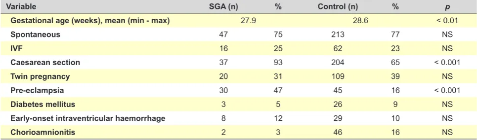
Pathology was presented by 44% of the mothers of NB with FGR and by 67% of the mothers of SGA NB, in contrast with 51% in the control group. Pre-eclampsia was found in 47% of the mothers of SGA NB vs. 16% in the control group, corresponding to a significant difference (p < 0.001). Twin pregnancy was found in around 30% of the FGR/SGA NB. Spontaneous pregnancy was mostly found in SGA NB group (75%) vs. only 50% in the group of NB with FGR. C-section delivery was mostly found in both groups (>80%). These numbers were not significantly different when com
-pared to the control group and GA, maternal pathology and delivery type comparisons are shown in Tables 1, 2 and 3.
Table 1 – Mortality per gestational age and birth weight in the group of FGR/SGA NB and in the control group
GA (weeks) FGR/SGA (n) BW (g) Min - Max Deaths (n) Control (n) BW (g) Mín - Máx Deaths (n)
23 0 NA NA 2 545 - 550 2
24 4 470 - 500 3 9 510 - 799 8
25 4 430 - 560 3 16 600 - 930 6
26 14 530 - 649 1 22 639 - 1100 4
27 12 300 - 680 2 28 720 - 1230 2
28 25 549 - 810 2 43 750 - 1409 5
29 7 568 - 924 0 48 950 - 1640 4
30 21 640 - 1043 0 47 930 - 1790 2
31 16 909 - 1200 0 60 1110 - 2240 2
Total 103 11 (11%) 275 35 (13%)
GA: gestational age; FGR: foetal growth restriction; SGA: small for gestational age; BW: birth weight; Min: minimum; Max: maximum; NA: not applicable
Table 2 – Pregnancy and delivery-related risk factors – comparison between SGA NB and the control group
Variable SGA (n) % Control (n) % p
Gestational age (weeks), mean (min - max) 27.9 28.6 < 0.01
Spontaneous 47 75 213 77 NS
IVF 16 25 62 23 NS
Caesarean section 37 93 204 65 < 0.001
Twin pregnancy 20 31 109 39 NS
Pre-eclampsia 30 47 45 16 < 0.001
Diabetes mellitus 3 5 26 9 NS
Early-onset intraventricular haemorrhage 8 12 29 10 NS
Chorioamnionitis 2 3 46 16 NS
ARTIGO ORIGINAL
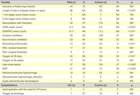
The need for IV was required by 60% of SGA NB and by 50% of NB with FGR, compared to 47% in the control group while NIV was required by 84% of SGA NB and by 87% of NB with FGR, compared to 53% in controls. The need for supplemental oxygen at 28 days was required by 66% of SGA NB and by 55% of NB with FGR vs. 38% in the control group (significant difference vs. SGA NB, p = 0.005). Other variables with significant differences in SGA NB group vs.
control group included: late-onset sepsis (59% vs. 37%, p =
0.003), ROP (58% vs. 26%, p = 0.003) and necrotizing en
-terocolitis (20% vs. 9%, p = 0.005). Late-onset sepsis was
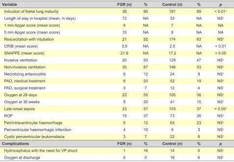
the only pathology with a statistical significance (p = 0.04) in NB with FGR.
The need for supplemental oxygen at discharge was the major complication found in SGA NB (27%). Data regarding morbidity are shown in Tables 4 and 5.
DISCUSSION
The presence of possible pregnancy or delivery-relat
-ed risk factors for prematurity associat-ed with FGR and/or SGA NB was assessed in this study, in addition to the as
-sessment of morbidity and mortality. Birth weight estimate
Table 3 – Pregnancy and delivery-related risk factors – comparison between NB with FGR and the control group
Variable FGR (n) % Control (n) % p
Gestational age (weeks), mean (min - max) 28.9 28.4 NS
Spontaneous 20 50 213 77 NS
IVF 20 50 62 23 NS
Caesarean section 53 84 188 64 < 0.005
Twin pregnancy 13 32 109 39 NS
Pre-eclampsia 13 32 45 16 NS
Diabetes mellitus 2 5 26 9 NS
Early-onset intraventricular haemorrhage 2 5 29 10 NS
Chorioamnionitis 1 2 46 16 < 0.005
FGR: foetal growth restriction. IVF: in vitro fertilization; NS: non-significant
Table 4 – Neonatal Morbidity – comparison between SGA NB and the control group
Variable SGA (n) % Control (n) % p
Induction of foetal lung maturity 44 70 191 69 NSa
Length of stay in hospital (mean, in days) 86 NA 53 NA < 0.001b
1 min Apgar score (mean score) 7 NA 7 NA NA
5 min Apgar score (mean score) 9 NA 9 NA NA
Resuscitation with intubation 44 70 174 63 NSa
CRIB (mean score) 5.3 NA 2.5 NA < 0.001b
SNAPPE (mean score) 31.7 NA 17.2 NA < 0.01b
Invasive ventilation 38 60 129 47 NSa
Non-invasive ventilation 53 84 146 53 NSa
Necrotizing enterocolitis 13 20 24 9 < 0.01a
PAD, medical treatment 17 27 52 19 NSa
PAD, surgical treatment 6 9 12 4 NSa
Oxygen at 28 days 34 66 105 38 < 0.01a
Oxygen at 36 weeks 17 34 41 15 NSa
Late-onset sepsis 37 59 103 37 < 0.005a
ROP 27 58 73 26 < 0.005a
Peri/intraventricular haemorrhage 10 16 63 23 NSa
Periventricular haemorrhagic infarction 3 5 9 3 NSc
Cystic periventricular leukomalacia 4 9 22 8 NSc
Complications LIG (n) % Control (n) % p
Hydrocephalus with the need for VP shunt 1 4 14 5 NSc
Oxygen at discharge 7 27 16 6 NSc
ARTIGO ORIGINAL and antenatal detection of FGR play an important role in obstetric decisions regarding the indications for induction of labour and parental counselling. In contrast, NB growth percentile, calculated by birth weight, is the key information used by paediatricians for decision making at the neonatal
unit.9 An 11% incidence of FGR and 18% of SGA NB has
been found in our group of preterm NB, probably due to the fact that the hospital is a neonatal and obstetric national ref
-erence centre. Two thirds of the SGA NB in our study were not diagnosed with FGR during pregnancy. Even though ul
-trasound imaging is the gold standard test for the diagnosis of FGR, a 20% error associated to the weight estimate has been usually described by different authors.28,29 The use of growth curves in clinical practice not adapted to severe pre
-maturity has been described as one of the reasons for this result.30,31
Pregnancy and delivery-related risk factors
A strong association between birth weight and preg
-nancy complications associated with prematurity has been described by Zeitlin et al.32 Some of the risk factors probably responsible by low birth weight, that were described in lit
-erature, were assessed in our study within the retrospective analysis of pregnancy and delivery.6 No differences between the groups regarding the type of gestation (spontaneous or with IVF) were found in our group of patients. In addition, a
third of FGR/SGA NB have resulted from a twin gestation, in line with the control group and that is why this variable was not considered as a significant factor. Significant differences regarding the presence of pregnancy pathology were found in FGR NB, namely regarding the presence of chorioamnio
-nitis. A high percentage of patients with pre-eclampsia has also been found in the SGA NB group, when compared to the control group (47% vs. 16%, p < 0.001), clearly corre
-sponding to the pregnancy pathology more closely associ
-ated with neonatal morbidity.
Gender differences
Less male than female NB diagnosed with FGR dur
-ing pregnancy were found in our group of patients, in line with Monier et al. and Magalhães et al., probably explained by the use of NB growth references non-adjusted to gen
-der, which can also be adapted to what was found in SGA NB.30,33
The absence of a template of somatometry at birth in the Portuguese population is worth mentioning and there
-fore leading to the use of international graphic patterns, with subsequent non-adjustment between these and the characteristics of each population. The population in our hospital usually shows a great socio-cultural heterogeneity, including European immigrants, as well as of African and Asian origin, which is worth mentioning. This mismatch in
Table 5 – Neonatal morbidity – comparison between FGR NB and the control group
Variable FGR (n) % Control (n) % p
Induction of foetal lung maturity 36 90 191 69 < 0.01a
Length of stay in hospital (mean, in days) 72 NA 53 NA NSb
1 min Apgar score (mean score) 8 NA 7 NA NA
5 min Apgar score (mean score) 10 NA 9 NA NA
Resuscitation with intubation 21 52 174 63 NSa
CRIB (mean score) 3.9 NA 2.5 NA < 0.01
SNAPPE (mean score) 21.8 NA 17.2 NA < 0.05
Invasive ventilation 20 50 129 47 NSa
Non-invasive ventilation 35 87 146 53 NSa
Necrotizing enterocolitis 5 12 24 9 NSa
PAD, medical treatment 8 20 52 19 NSa
PAD, surgical treatment 3 7 12 4 NSc
Oxygen at 28 days 22 55 105 38 NSa
Oxygen at 36 weeks 8 20 41 15 NSa
Late-onset sepsis 23 57 103 37 < 0.05a
ROP 15 37 73 26 NSa
Peri/intraventricular haemorrhage 5 12 63 23 NSa
Periventricular haemorrhagic infarction 4 10 9 3 NSc
Cystic periventricular leukomalacia 3 7 22 8 NSc
Complications FGR (n) % Control (n) % p
Hydrocephalus with the need for VP shunt 1 16 14 5 NSc
Oxygen at discharge 0 0 16 6 NSc
ARTIGO ORIGINAL
the classification of NB also has an impact on the assess
-ment of the individual diagnosis and subsequently on any subsequent medical approach.34
The fact that the height of the parents of SGA NB was not obtained is a limitation of the study that is worth men
-tioning.
CRIB and SNAPPE score assessment
The initial clinical severity in preterm NB is assessed by CRIB (clinical risk index for babies) score based on birth weight, gestational age, presence of birth defect, base ex
-cess and fraction of inspired oxygen, which was developed in the United Kingdom by the International Neonatal Net
-workin 1993 and is used for the measurement of the risk of hospital death.35 A 5.3 average maximum score in SGA NB (p < 0.001) and 3.9 in NB with FGR (p < 0.01) have been found in our study. A significantly lower score of severity was found in the control group, with a 2.5 average score. Even though the three groups correspond to a CRIB level I in medical practice, differences are significant, showing a trend towards a higher clinical risk in both groups, mostly in SGA NB.
The SNAPPE (score for neonatal acute physiology – perinatal extension) score is an extension of the SNAP (score for neonatal acute physiology) scale, which was de
-veloped by Richardson et al.36 aimed at the assessment of the clinical severity in NB at admission to a NICU, includ
-ing all the physiological parameters of the SNAP, in addition to the assessment of birth weight, perinatal history includ
-ing the Apgar score and the classification of SGA NB.37,38 A higher score in the SNAP-PE when compared to the control group has been found in NB with FGR/SGA, in line with was found when the CRIB application was used (31.7 in SGA and 21.8 in FGR vs. 17.2 in the control group). Again, sig
-nificantly different scores were found in both groups and a higher risk of a poorer outcome in these NB was confirmed.
Bronchopulmonary dysplasia
An increased prevalence of bronchopulmonary dyspla
-sia (BPD) was found in SGA NB in a recent study, regard
-less of an antenatal diagnosis of FGR.30 The risk of BPD was also strongly associated with SGA NB in another study, with a decline in prevalence as birth weight increased.32 The need for supplemental oxygen at 28 days of life was found in 66% of SGA NB (p = 0.005) and the double of what was found in the control group at 36 weeks (34% vs. 15%), even though this was not a significant difference. The depend
-ence on supplemental oxygen was also the main complica
-tion in SGA NB at discharge (27%), which was not found in NB with FGR in our study, in line with literature, even though it can be due to the presence of lung maturity in only 70% of the SGA NB, compared to 90% in NB with FGR.
Neurological outcomes
Brain volume abnormalities that are sensitive to multiple perinatal risk factors and neonatal morbidities/interventions have been found in extreme preterm NB.39,40 An association
between smaller regional brain volume and neurological impairment in prematurity has been found in different stud
-ies,39-41 including one study developed by our group.40 These large volume decline was associated with an increased cer
-ebrospinal fluid (CSF) volume and smaller total brain size, suggesting cerebral atrophy.39,40
Significantly reduced thalamus and basal ganglia vol
-umes have been found in another study with NB with FGR when compared to NB with an appropriate weight for ges
-tational age.42 These abnormalities have probably induced clinical outcomes regarding learning and memory, language function and visuospatial function.42
Periventricular haemorrhage (PVH > 2) in preterm NB is an acquired injury with a direct impact on morbidity, mortal
-ity and long-term neurodevelopment.43 However, SGA was not associated with PVH or cystic periventricular leukoma
-lacia in the study by Zeitlin et al.32 A 16% and 12% rates of PVH were found in SGA NB and NB with FGR in our study, respectively, while a 9% rate of cystic periventricular leu
-komalacia was found in SGA NB and 7% in NB with FGR, with no significant differences when compared to the control group.
Mortality and morbidity
A higher risk of mortality and morbidity associated with birth weight below the 10th percentile was found in the Mod
-els of organising access to intensive care for very preterm births in Europe (MOSAIC Research Group study),44 which was a population cohort study in children born before the 32 weeks of gestation. Nevertheless, other studies have found a significant decline (around 20%) in perinatal morbidity and mortality with the early detection and orientation of foetuses with FGR.45 An adequate obstetric and perinatal approach can explain the absence of a significant difference in mortal
-ity between NB with FGR and the control group.
A higher prevalence of late-onset sepsis has been found in NB with FGR (p < 0.05) and in SGA NB (p < 0.005) in our study, regarding the analysis of the incidence of morbidity, when compared to the control group. The rate of late-onset sepsis in these NB can be explained by a delayed enteral feeding, which is associated with an increased duration of parenteral nutrition and the need for central catheter. Higher incidence of ROP (58% vs. 26%, p < 0.005) and necrotizing enterocolitis (20% vs. 9%, p < 0.01) has been found in SGA NB when compared to the control group, with significant dif
-ferences.
Globally, a higher morbidity has been found in SGA NB when compared to both the NB with FGR and the control group in our study, giving support to the hypothesis that low weight percentile at birth reflects a real deficit in organ growth and development.
Limitations of the study
ARTIGO ORIGINAL 2. A great social and cultural heterogeneity has been found in the population attended by our hospital, with a possible mismatch regarding the classification of NB, with an impact on the assessment of the indi
-vidual outcome and on the subsequent medical ap
-proaches.
3. The height of the parents of SGA NB was not ob
-tained. Strengths:
1. This study was developed based on the experience of a reference centre in Neonatal Intensive Care and including a group of 356 NB with GA less than 32 weeks.
2. Seventeen clinical parameters for the assessment of morbidity were analysed, as well as the character
-istics of gestation and the major pregnancy-related risk factors described in literature.
What does this study add to national medical literature? This study was aimed at the re-assessment of the pos
-sible pregnancy and delivery-related risk factors of prema
-turity associated with FGR and/or SGA NB and found that pre-eclampsia was the pathology most strongly associated with neonatal morbidity.
A higher incidence of morbidity has been found in SGA NB when compared to NB with FGR and the control group, giving support to the hypothesis that low weight percentile at birth reflects a real deficit in organ growth and develop
-ment.
CONCLUSIONS
Significantly increased survival and reduced morbidity and mortality in preterm NB have been produced by the
advances in neonatal intensive care. However, significant differences have still been found as regards the prevalence of BPD, necrotizing enterocolitis, late-onset sepsis and retinopathy of prematurity in SGA NB and late-onset sepsis in NB with FGR. A strong association between SGA NB and maternal pre-eclampsia has also been found in our study. An impaired foetal growth corresponding to low birth weight for GA and gender has shown higher impact on NB morbidity when compared to patients antenatally diagnosed with FGR and no impaired growth.
Active communication and collaboration between obste
-tricians and neonatologists are crucial for a correct clinical assessment, signalling and global action on these NB, re
-ducing early morbidity and late outcomes.
HUMAN AND ANIMAL PROTECTION
The authors declare that the followed procedures were according to regulations established by the Ethics and Clini
-cal Research Committee and according to the Helsinki Dec
-laration of the World Medical Association.
DATA CONFIDENTIALITY
The authors declare that they have followed the proto
-cols of their work centre on the publication of patient data.
CONFLICTS OF INTEREST
The authors declare that there were no conflicts of inter
-est in writing this manuscript.
FINANCIAL SUPPORT
The authors declare that there was no financial support in writing this manuscript.
REFERENCES
1. Coutinho P, Cecatti J, Surita F, Costa M, Morais S. Perinatal outcomes associated with low birthweight in a historical cohort. Reprod Health. 2011;8:1-6.
2. Alexandrino A. Período neonatal - restrição de crescimento intra-uterino: decisões obstétricas com repercussão no RN – mesa redonda, XXIV Reunião Anual de Pediatria do Centro Hospitalar do Porto. Nascer Crescer. 2012;21:167-8.
3. Doyle L, Victorian Infant Collaborative Study Group. Outcome at 5 years of age of children 23 to 27 weeks’ gestation: refining the prognosis. Pediatrics. 2001;108:134-41.
4. Ross M. Fetal growth restriction: overview, causes of intrauterine growth restriction, perinatal implications. New York: Medscape; 2015. 5. Grandi C, Tapia J, Marshall G, Grupo Colaborativo NEOCOSUR.
Evaluación de la severidad, proporcionalidad y riesgo de muerte de recién nacidos de muy bajo peso con restricción del crecimiento fetal. Análisis multicéntrico sudamericano. J Pediatr. 2005;81:198-204. 6. Gaudineau A. Prévalence, facteurs de risque et morbi-mortalité
materno-foetale des troubles de la croissance foetale. J Gynecol Obstet Biol Reprod. 2013;42:895-910.
7. Magalhães J, Resende C, Braga A, Alexandrino A. Muito baixo peso e restrição de crescimento intrauterino – uma associação de mau prognóstico? Acta Pediatr Port. 2014;45:107-15.
8. Levine T, Grunau R, McAuliffe F, Pinnamaneni R, Foran A, Alderdice F. Early childhood neurodevelopment after intrauterine growth restriction: a systematic review. Pediatrics. 2015;135:126-41.
9. El Ayoubi M, Jarreau P, Van Reempts P, Cuttini M, Kaminski M, Zeitlin J et al. Does the antenatal detection of fetal growth restriction (FGR) have a prognostic value for mortality and short-term morbidity for very preterm infants? Results from the MOSAIC cohort. J Matern Fetal Neonatal Med. 2016;29:596-601.
10. Royal College of Obstetricians and Gynaecologists. The investigation and management of the small–for–gestational–age fetus. Green–top Guideline. 2013;31:1-34.
11. Saldanha M, Machado M, Matos C, Pinto F, Barroso R, Carreiro H. Recém-nascidos leves para a idade gestacional numa população suburbana. Incidência e factores de risco. Acta Pediatr Port. 2003;1:25-32.
12. Drake A, Walker B. The intergenerational effects of fetal programming: non-genomic mechanisms for the inheritance of low birth weight and cardiovascular risk. J Endocrinol. 2004;180:1–16.
13. Sehested L, Pedersen P. Prognosis and risk factors for intrauterine growth retardation. Danish Med J. 2014;61:1-4.
14. Aghamolaei T, Eftekhar H, Zare S. Risk factors associated with intrauterine growth retardation (IUGR) in Bandar Abbas. J Med Sci. 2007;7:665-9.
15. Conley D, Bennett N. Birth weight and income: interactions across generations. J Health Soc Behav. 2001;42:450–65.
ARTIGO ORIGINAL 17. Budhathoki S, Poudel P, Bhatta N, Singh R, Shrivastava M, Niraula S,
et al. Clinico-epidemiological study of low birth weight newborns in the Eastern part of Nepal. Nepal Med Coll J. 2014;16:190-3.
18. Forbes K, Westwood M. Maternal growth factor regulation of human placental development and fetal growth. J Endocrinol. 2010;207:1–16. 19. Buitendijka S, Zeitlinb J, Cuttinic M, Langhoff-Roosd J, Bottu J. Indicators
of fetal and infant health outcomes. Eur J Obstet Gynecol Reprod Biol. 2003;111:66–77.
20. Wilcox J. On the importance – and the unimportance – of birthweight. Int J Epidemiol. 2001;30:1233-41.
21. Gill A, Yu V, Bajuk B, Astbury J. Postnatal growth in infants born before 30 weeks’ gestation. Arch Dis Child. 1986;61:549-53.
22. Jong F, Monuteaux M, Elburg R, Gillman, Belfort M. Systematic review and meta-analysis of preterm birth and later systolic blood pressure. Hypertension. 2012;59:226-34.
23. Barker DJ. Adult consequences of fetal growth restriction. Clin Obstet Gynecol. 2006;49:270-83.
24. Ross M, Beall M. Adult sequelae of intrauterine growth restriction. Semin Perinatol. 2008;32:213–8.
25. Dias C. Vigilância pré-natal e decisão do parto. Nascer Crescer. 2012;21:159-66.
26. Fenton T, Kim J. A systematic review and meta-analysis to revise the Fenton growth chart for preterm infants. BMC Pediatr. 2013;13:59. 27. Proença E, Vasconcellos G, Rocha G, Carreira M, Mateus M, Santos I,
et al.Displasia broncopulmonar. Consensos em Neonatologia. Lisboa: Sociedade Portuguesa de Neonatologia; 2009.
28. Copel J, Bahtiyar M. A practical approach to fetal growth restriction. Obstet Gynecol. 2014;123:1057-69.
29. Dudley N. A systematic review of the ultrasound estimation of fetal weight. Ultrasound Obstet Gynecol. 2005;25:80-9.
30. Monier I, Ancel P, Ego A, Jarreau P, Lebeaux C, Kaminski M, et al. Fetal and neonatal outcomes of preterm infants born before 32 weeks of gestation according to antenatal vs postnatal assessments of restricted growth. Am J Obstet Gynecol. 2017;216:516.e1-10.
31. Marsál K, Persson P, Larsen T, Lilja H, Selbing A, Sultan B. Intrauterine growth curves based on ultrasonically estimated foetal weights. Acta Paediatr. 1996;85:843-8.
32. Zeitlin J, El Ayoubi M, Jarreau P, Draper E, Blondel B, Kunzel W, et al. Impact of fetal growth restriction on mortality and morbidity in a very preterm birth cohort. J Pediatr. 2010;157:733-9.
33. Monier I, Blondel B, Ego A, Kaminski M, Goffinet F, Zeitlin J. Does the presence of risk factors for fetal growth restriction increase the
probability of antenatal detection? A French national study. Paediatr Perinat Epidemiol. 2016;30:46-55.
34. Rodrigues T, Teles T, Miguel C, Pereira A, Barros H. Recém-nascidos leves para a idade gestacional. Influência das curvas padrão de peso ao nascimento no cálculo da sua prevalência e dos factores de risco. Acta Med Port. 1996;9:335-40.
35. Sarquis A, Miyaki M, Cat M. Aplicação do escore CRIB para avaliar o risco de mortalidade neonatal. J Pediatr. 2002;78:225-9.
36. Richardson DK, Corcoran JD, Escobar GJ, Lee SK. SNAP-II and SNAPPE-II: simplified newborn illness severity and mortality risk scores. J Pediatr. 2001;138:92-100.
37. Harsha S, Archana B. SNAPPE-II (Score for Neonatal Acute Physiology with Perinatal Extension-II) in predicting mortality and morbidity in NICU. J Clin Diagn Res. 2015;9:10-2.
38. Silveira R, Schlabendorff M, Procianoy R. Valor preditivo dos escores de SNAP e SNAP-PE na mortalidade neonatal. J Pediatr. 2001;77:455-60. 39. Parikh N, Lasky R, Kennedy A, McDavid G, Tyson J. Perinatal factors
and regional brain volume abnormalities at term in a cohort of extremely low birth weight infants. PLoS One. 2013;8:1-11.
40. Graça AM, Cardoso K, Costa J, Cowan FM. Cerebral volume at term age: comparison between preterm and term-born infants using cranial ultrasound. Early Hum Dev. 2013;89:643–8.
41. Peterson B, Anderson A, Ehrenkranz R, Staib L, Tageldin M, Colson E, et al. Regional brain volumes and their later neurodevelopmental correlates in term and preterm infants. Pediatrics. 2003;111:939-48. 42. Bruno C, Bengani S, Gomes W, Brewer M, Vega M, Xie X, et al. MRI
differences associated with intrauterine growth restriction in preterm infants. Neonatology. 2017;111:317–23.
43. Luque M, Tapia J, Villarroel L, Marshall G, Musante G, Carlo W, et al. A risk prediction model for severe intraventricular hemorrhage in very low birth weight infants and the effect of prophylactic Indomethacin. J Perinatol. 2014;34:43-8.
44. Zeitlin J, Papiernik E, Bréart G, Draper E, Kollée L, MOSAIC Research Group. Presentation of the European project models of organising access to intensive care for very preterm births in Europe (MOSAIC) using European diversity to explore models for the care of very preterm babies. Eur J Obstet Gynecol Reprod Biol. 2005;118:272-4.