Address for correspondence Dr. Mohammad Amin Almasi, Young Researchers and Elites Club,
North Tehran Branch, Islamic Azad University, Tehran, Iran.
Email: [email protected]
Original Article
Design and application of a loop-mediated isothermal
amplification
for
detection
of
molluscum
contagiosum virus
Introduction
Molluscum contagiosum virus (MCV) lesions, known as Molluscipox virus from the Poxviridae family, were first described by Bateman as papules of umbilicated centers in humans and other vertebrates.1,2 The infection commonly
involves children of 1-10 years, immunocompromised individuals, and sexually active adults.3 In children, the lesions are seen in
the face, arms, hands, neck, soles, armpits, and
mucous membrane, i.e. lips, buccal mucosa, and tongue. In adults, they commonly appear in the genital area, abdomen, and inner parts of thighs.4,5
Immunohistochemical methods using a polyclonal antibody allow MCV recognition in fixed tissues. In-situ hybridization of MCV DNA has been also utilized.6,7 Polymerase chain
reaction (PCR)-based assays are the best alternative to MCV definitive diagnosis.8-10 On
the other hand, molecular diagnosis can provide additional information about Molluscum strain type; however, its usefulness may be limited by the need for trained staff working with the reagents and equipment in a professional operating space.11
Mohammad Amin Almasi, Rozhin Esmaili*
Young Researchers and Elites Club, North Tehran Branch, Islamic Azad University, Tehran, Iran. * Oncology and Hematology Department, Rassoul Akram Hospital, Iran University of Medical Sciences, Tehran, Iran.
Abstract Objective Molluscum contagiosum virus (MCV) is a poxvirus that causes localized papules in healthy persons. A loop-mediated isothermal amplification (LAMP) assay was developed and compared to polymerase chain reaction (PCR) assay for detection of MCV.
Methods The p43K protein gene of virus is basically used for designing the primers. These assays were employed to examine the lesion samples taken from 50 patients, who were aged between 1 and 60 years and diagnosed with clinical MCV lesions in different parts of their bodies.
Results 44 MCV-positive patients (88%), children aged between 1 and 10 years were a higher percentage. The results show LAMP is an advantageous method because it is highly sensitive (1000-fold), quite cheap, user-friendly, and safe; in addition, it is performed quickly by visual detection using hydroxynaphthol blue (HNB) Dye in a water bath.
Conclusion LAMP technique can be simply and reliably applied with basic instruments through visual inspection in laboratory studies.
Key words
Recently, isothermal amplification methods providing simple and cost-effective molecular tests in low resource settings have been developed with an increasing demand.12
Loop-mediated isothermal amplification (LAMP) is one of these methods, which has been most widely adopted today. In a single-step reaction, it can provide amplification of 109 copies out of a few copies of the target in less than an hour even in the presence of large amounts of non-target DNA.11 LAMP can be thwarted by the Bst
DNA polymerases commonly present in the clinical samples and insects as they are more tolerant to the inhibitors.13 Nevertheless,
amplification based on LAMP assay can be simply done via the visual detections of turbidity made by magnesium pyrophosphate precipitation, intercalating dye fluorescence, or color variations of metal-sensitive indicators by setting a simple electric device like a water bath or heat block at a constant temperature.14
In the present study, we developed a LAMP assay based on the p43K gene of MCV via a visualized system of detection. We also evaluated the efficiency, speed and sensitivity of LAMP for MCV and we compared its performance with PCR.
Methods
This cross-section study was conducted on all the patients attending the Outpatient Clinic of Dermatology of Emam Reza Hospital in Iran during 2016-2017. Collection of the patients’ samples was done in the dermatology unit following the dermatologists’ diagnoses of the cases. Then, the samples were submitted for diagnosis, while an MCV isolate of subtype 1, i.e., Keyvan Lab MCV214, was applied as the positive control. The patients’ demographic information included age, sex, and address. After curetting each patient’s lesion and placing it in 5 ml of Phosphate-Buffered Saline (PBS)
with a pH of 7.1, it was immediately transferred to the laboratory. The specimens were stored at -37 °C until DNA extraction. 50 samples were selected depending on the sizes of the patient’s lesions (≥30mg) according to the method described by Geneaid Company (http://www.geneaid.com). After neglecting the PBS, the DNA extraction and purification were performed as instructed by the Viral Nucleic Acid Extraction Kit II (Geneaid Co., Taiwan VR050/100/300).
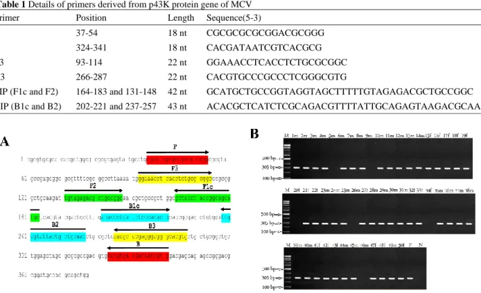
The Oligo7 and Primer Explorer V.4 software were utilized to design PCR (F and B) and LAMP (F3, B3, FIP and BIP) specific primers based on the p43K gene of MCV (GenBank: EF138623.1) (Table 1). Subsequently, they were examined based on sequence alignments using ClustalX 2.11 (Des Higgins). The positions of the designed primers on the sequence are displayed in Figure 1A.
PCR amplification was carried out in a thermocycler (iCycler, BIO RAD, CA, USA). The amplification was done in a 25 μl volume containing 10 × PCR buffer (10 mMTris-HCl, pH 8.3 and 50 mMKCl), 1.5 mM MgCl2
(CinnaGen Co., Iran), 0.5 μM of each F and B primers, 0.2 mM ofdNTPs (CinnaGen Co., Iran), 2 U of Taq DNA polymerase(CinnaGen Co., Iran) and 2 μl template DNA. Amplification was performed with the following PCR profile: 3 min at 94 °C (1 cycle); 35 cycles of 1 min at 94 °C, 1 min at 54 °C, 1 min at 72 °C and 10 min at 72 °C for final extension. PCR products were visualized by staining with ethidium bromide after electrophoresis on 1% agarose gel. Finally, using a UV transilluminator (GELDOC 2000, Bio-Rad, USA) equipped with a photo of each gel containing PCR fragments (expected size 305 bp).
Table 1 Details of primers derived from p43K protein gene of MCV Primer Position Length Sequence(5-3)
F 37-54 18 nt CGCGCGCGCGGACGCGGG
B 324-341 18 nt CACGATAATCGTCACGCG
F3 93-114 22 nt GGAAACCTCACCTCTGCGCGGC
B3 266-287 22 nt CACGTGCCCGCCCTCGGGCGTG
FIP (F1c and F2) 164-183 and 131-148 42 nt GCATGCTGCCGGTAGGTAGCTTTTTGTAGAGACGCTGCCGGC
BIP (B1c and B2) 202-221 and 237-257 43 nt ACACGCTCATCTCGCAGACGTTTTATTGCAGAGTAAGACGCAA
Figure 1 Oligonucleotide primers used for detection of the p43K protein gene of MCV (A). Results of electrophoresis of PCR
products (B). M,DNA size marker (100 bp); P, positive control; N, negative control; m, male and f, female
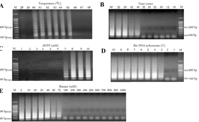
DNTP, time, betaine and Bst DNA polymerase concentrations in LAMP reaction. Afterwards, the isolated DNA was involved in the LAMP assay. DNA (2 μl) as the template in the LAMP total volume (25 μl) contained Tris–HCl (20 mM, pH 8.8), (NH4)2SO4 (10 mM), KCl (10
mM), dNTPs (10 mM each), betaine (5 mM), FIP and BIP primers (0.8 μM each), F3 and B3 (0.2 μM each), Bst DNA polymerase (2 U), and Triton X-100 (0.1%). After incubating the mixture in a water bath at 63 °C for 60 min, segregation of the products was conducted using electrophoresis on a 1.5% agarose gel. Also, before the amplification, 1 μl of hydroxynaphthol blue (HNB) (Lemongreen, Shanghai, China) was added to the LAMP master mix to provide a visual detection of the LAMP products. The color change occurring in the tubes (positive reaction) from violet (prior to amplification) to blue (post-amplification) was monitored by simply watching them.
Comparison of sensitivity of detection methods was carried out using DNA of positive control (a tenfold dilution from 1 × 10¹⁰ to 1 × 101).
Results
MCV was detected in samples and positive control by PCR and the predicted DNA fragment (304 bp) was observed on agarose gel (Figure 1B). 44 and 6 patients (88 and 12%) out of 50 patients with skin lesions demonstrated positive and negative results for MCV, respectively.
Figure 2 Results of optimization of LAMP reaction. Effects of temperature (A), Effects of the reaction time (B), Effects of dNTPs concentration (C), Effects of Bst DNA polymerase concentrations (D) and Effects of Betaine concentrations (E). M, DNA size marker (100 bp)
Figure 3 Results of LAMP reaction by agarose gel electrophoresis (A), with HNB (B) and comparison of sensitivity using a ten
Table 2 Comparison of PCR and LAMP assays
Samples Age (year) Male
samples Female samples Male positive results Male negative results Female positive results Female negative results
1-22 1-10 14 8 12 2 7 1
23-27 11-20 4 1 3 1 1 0
28-34 21-30 4 3 4 0 2 1
35-43 31-40 6 3 6 0 3 0
44-48 41-50 3 2 2 1 2 0
49-50 51-60 1 1 1 0 1 0
Total 32 18 28 4 16 2
Table 3 Distribution of MCV according to the age and sexes of patients
Assay amplification time
Detection
method Safety
Need to UV ray
Need to detect instruments Cost
User
Friendly Sensitivity
PCR 180 min Gel
electrophoresis No Yes Yes High High 10
5
LAMP 30 min Visual/Gel
electrophoresis Yes/No No/Yes No/Yes Low Very high 10
2
To test the effects of dNTPs concentration on LAMP reaction, final concentration of 1 mM to 10 mM was prepared. The results showed that at 7 mM to10 mM, ladder-like DNA fragments were clearly observed (Figure 2C). Different concentrations (1 U to 10 U) of Bst DNA polymerase were used to select the minimum concentrations with the good performance. With low concentration of the enzyme (3U), poor amplification of DNA was observed, but with increasing the enzyme concentration to 4 U to 10 U, the amplification considerably improved
(Figure 2D). A Betaine concentration from 5
mM to 1000 mM was examined. When the concentration of Betaine increased from 5 mM to 75 mM, the intensity of the amplified products increased but no visible products were detected when the concentration increased to 200 mM (Figure 2E).
After achieving the optimization conditions and testing 50 samples via LAMP assay, a large number of the DNA fragments was observed in a ladder-like pattern by electrophoresing the amplicons on the 1.5% agarose gel (Figure 3A). It was possible to see the amplicons with the naked eye by detecting their color changes in the solutions with the help of various visual dyes. All the positive and negative samples could be
clearly and successfully distinguished from each other by HNB.
LAMP assay yielded products even with a lower concentration of sap dilutions (1 × 10² or more),
whilst PCR required a higher concentration (1 × 10⁵ or more) (Figure 3B). LAMP assay had
higher sensitivity for detection of MCV in comparison with PCR (1000-fold).
Discussion
It is highly essential to develop easy, rapid, reliable, and cost-effective diagnostic protocols with great sensitivities to perform precise diagnoses. Therefore, we aimed at assessing MCV detection based on LAMP assay in this study. Even LAMP and PCR techniques had enough potential to make differentiation and detect infected samples accurately, LAMP proved to be much more useful as some factors including time, safety, cost and being user friendly are taken into account (Table 2).
Molluscum contagiosum, which has been most commonly seen to occur in the age group of 5-10 years and then 1-5 and 5-10-14 years, but less in the age group of 1 year. Our results are also in line with those of another study conducted in the USA reporting that approximately 80% of the patients have been younger than 8 years of age.16
In this research, MCV was found to be more prevalent in the males (28 patients, 63.6%) compared to the females (16 patients, 36.4%) though the difference was not significant. Moreover, the MCV percentages showed an agreement with those reported by other researchers arriving at no statistically significant differences,9,17 but there is a disagreement with
those of an Egyptian study, in which the males and females had represented 42.9 and 57.1% of MCV, respectively, being stil statistically insignificant.18 These differences may be due to
the lack of adequate education and attention to health issues, particularly when the emergence of such diseases is not of special interest to the elites with little knowledge of health matters.
In our study, the products amplified via LAMP could be readily visualized with the help of different color indicators without any additional staining systems involving toxic materials. Contrary to the proposed approach displaying the advantages of simplicity, user-friendly, and cost-effectiveness, any other methods for MCV detection, including PCR and immunohistochemical assays, require professional personnel to work in labs equipped with costly molecular instruments.12
Furthermore, no thermocycler and gel electrophoresis were needed for accomplishing LAMP assay as it could be easily conducted in a water bath or through temperature block.11
Generally, the need for additional staining for pursuing post-amplification processes can be obviated by making easier and quicker visual detections via LAMP in-tube or in-plate color indicators.14.
Conclusion
In this research, the LAMP positive amplicons were observed with naked eye by adding fluorescent dye to the reaction tubes. Conclusively, cross-contamination risks would be reduced by using and adding HNB dye to the reaction mixture before amplification without the need for opening the assayed samples. Hence, LAMP assay has several remarkable advantages over any other colorimetric-based methods and can serve as a suitable approach not only to the laboratory detection of MCV, but also to the field diagnoses of other viruses.
Acknowledgement
The author is grateful to the Young Researchers and Elites Club, North Tehran Branch, Islamic Azad University, Tehran, Iran for financial support for this study. Also would like to thank Dr. Hossein Ahmadi from the Outpatient Clinic of Dermatology of Emam Reza Hospital in Iran for providing patients samples and positive control.
References
1. Zhang Q, Davis JC, Lamborn IT, Freeman AF, Jing H, Favreau AJ et al. Combined immunodeficiency associated with DOCK8 mutations. N Engl J Med 2009; 361: 2046-2055.
2. Hourihane J, Hodges E, Smith J, Keefe M, Jones A, Connett G. Interferon a treatment of molluscumcontagiosum in immunodeficiency. Arch Dis Child. 1999; 80: 77-79.
3. Hanson D, Diven DG.
MolluscumContagiosum. Dermatol online J. 2003; 9: 2.
4. Penneys NS, Matsuo S, Mogollon R. The identification of molluscum infection of immunohistochemical means. J Cutan Pathol 1986; 13: 97-101.
6. Bikowski JB Jr. Molluscumcontagiosum: The Need for physician intervention and new treatment options. Cutis 2004; 73: 202-206.
7. Stulberg DL, Hutchinson AG. Molluscumcontagiosum and and warts. Am Fam Physician 2003; 67: 1233-1240. 8. Eleftheriou LI, Kerr SC, Stratman EJ.
Diagnosis of atypical
Molluscumcontagiosum: The utility of a squash preparation. Clin Med Res 2001; 9: 50-51.
9. Nunez A, Funes JM, Agromayor M, Moratilla M, Varas AJ, Lopez-Estebaranz JL et al. Detection and typing of Molluscumcontagiosum virus in skin lesions by using a simple lysis method and polymerase chain reaction. J Med Virol 1996; 50: 342-349.
10. Thompson CH. Identification and typing of Molluscumcontagiosum virus in clinical specimens by polymerase chain reaction. J Med Virol 1997; 53: 205-211.
11. Notomi T, Okayama H, Masubuchi H, Yonekawa T, Watanabe K, Amino N et al. Loop-mediated isothermal amplification of DNA. Nucleic Acids Res 2000; 28: e63. 12. Nagamine K, Hase T, Notomi T.
Accelerated reaction by loop mediated
isothermal amplification using loop primers. Mol Cell Probes 2002; 16: 223-229.
13. Tomita N, Mori Y, Kanda H, Notomi T. Loop-mediated isothermal amplification (LAMP) of gene sequences and simple visual detection of products. Nat Protoc 2008; 3: 877-882.
14. Dukes JP, King DP, Alexandersen S. Novels reverse transcription loop-mediated isothermal amplification for rapid detection of Foot-and-mouth disease virus. Arch Virol 2006; 151: 1093-1106.
15. Laxmisha C1, Thappa DM, Jaisankar TJ. Clinical profile of molluscumcontagiosum in children versus adults. Dermatol Online J 2002; 9: 1.
16. Dohil MA, Lin P, Lee J, Lucky AW, Paller AS, Eichenfield LF. The epidemiology of molluscumcontagiosum in children. J Am Acad Dermatol 2006; 54: 47-54.
17. Zandi F, Shamsaddini S, Kambin N. Prevalence of Molluscum Contagiosum in students of elementary schools of Kerman. Iranian J Dermatol 1999; 2: 25-30.