TOXICOLOGICAL CHEMISTRY
The attached two chapters are taken from the book Environmental Chemistry, Stanley Manahan, Taylor & Francis/CRC Press, 2009. The first of the chapters, “Chapter 23,” defines toxicological chemistry and describes the basic principles of the science including exposure, metabolism of toxicants, biochemical mechanisms of toxicant action, and toxic effects. The second chapter, “Chapter 24,” discusses specific toxic substances. PowerPoint presentations for each of these two chapters can be downloaded from: http://sites.google.com/site/environmentalchemistry1/
Toxicological chemistry is covered in detail in the book Toxicological Chemistry and Biochemistry, Third Edition, Stanley Manahan, Taylor & Francis/CRC Press, 2002.
Distribution of free, bound, or metabolite form
Liver Bile
Feces (excretion)
Blood and lymph system
Metabolism Protein
binding
Kidney
Bladder Cell membrane
Receptor cells
Urine (excretion) Gastrointestinal tract
Ingestion (entry site)
Inhaled air (entry site)
Exhaled air (excretion)
Skin
Toxicant storage Bone
Fat
Dermal exposure (entry site) Pulmonary system
(lung and alveoli)
Portal vein
23 TOXICOLOGICAL CHEMISTRY
23.1 INTRODUCTION TO TOXICOLOGY AND
TOXICOLOGICAL CHEMISTRY
Ultimately, most pollutants and hazardous substances are of concern because of their toxic effects. The general aspects of these effects are addressed in this chapter under the heading of toxicological chemistry; the toxicological chemistry of specific classes of chemical substances is addressed in Chapter 23. In order to understand toxicological chemistry, it is essential to have some understanding of biochemistry, the science that deals with chemical processes and materials in living systems. Biochemistry was summarized in Chapter 22.
Toxicology
A poison, or toxicant, is a substance that is harmful to living organisms because of its detrimental effects on tissues, organs, or biological processes. Common end point effects of toxic substances are destruction of cells, mutation of DNA leading to cancer, and disruption of the signaling mechanisms by which cell development and function are controlled. Most toxicants are foreign to the bodies of individuals that are affected (see xenobiotics in Section 23.5) and tend to have an affinity for lipids. Therefore, they have a tendency to cross the lipid membranes of cells and to build up to toxic levels. In many cases toxicants undergo metabolism to produce an active species that causes poisoning.
Toxicology is the science of poisons. Whether a substance is poisonous depends upon the type of organism exposed, the amount of the substance, and the route of exposure. In the case of human exposure, the degree of harm done by a poison can depend strongly upon whether the exposure is to the skin, by inhalation, or through ingestion.
Toxicants to which subjects are exposed in the environment or occupationally may be in several different physical forms. This may be illustrated for toxicants that are inhaled. Gases are substances such as carbon monoxide in air that are normally in the gaseous state under ambient conditions of temperature and pressure. Vapors are gas-phase materials that have evaporated or sublimed from liquids or solids. Dusts are respirable solid particles produced by grinding bulk solids, whereas fumes are solid particles from the condensation of vapors, often metals or metal oxides. Mists are liquid droplets.
There are numerous variables related to the ways in which organisms are exposed to toxic substances. One of the most crucial of these, dose, is discussed in Section 23.2. Another important factor is the toxicant concentration, which may range from the pure substance (100%) down to a very dilute solution of a highly potent poison. Both the duration of exposure per exposure incident and the frequency of exposure are important. The rate of exposure and the total time period over which the organism is exposed are both important situational variables. The exposure site and route also affect toxicity.
It is possible to classify exposures on the basis of acute vs. chronic and local vs. systemic exposure, giving four general categories. Acute local exposure occurs at a specific location over a time period of a few seconds to a few hours and may affect the exposure site, particularly the skin, eyes, or mucous membranes. The same parts of the body can be affected by chronic local exposure, for which the time span may be as long as several years. Acute systemic exposure is a brief exposure or exposure to a single dose and occurs with toxicants that can enter the body, such as by inhalation or ingestion, and affect organs, such as the liver, that are remote from the entry site. Chronic systemic exposure differs in that the exposure occurs over a prolonged time period.
In discussing exposure sites for toxicants it is useful to consider the major routes and sites of exposure, distribution, and elimination of toxicants in the body as shown in Figure 23.1. The major routes of accidental or intentional exposure to toxicants by humans and other animals are the skin (percutaneous or dermal route), the lungs (inhalation, respiration, pulmonary route), and the mouth (oral route); minor routes of exposure are rectal, vaginal, and parenteral (intravenous or intramuscular, a common means for the administration of drugs or toxic substances in test subjects). Of these, the dermal route is the most difficult to quantify. It is particularly important in children, whose activities bring them into contact with contaminated dirt, pesticides, household chemicals, and other environmental pollutants. Furthermore, the skin of children is relatively more permeable to toxic substances, increasing their risk of dermal exposure.
The way that a toxic substance is introduced into the complex system of an organism is strongly dependent upon the physical and chemical properties of the substance. The pulmonary system is most likely to take in toxic gases or very fine, respirable solid or liquid particles. In other than a respirable form, a solid usually enters the body orally. Absorption through the skin is most likely for liquids, solutes in solution, and semisolids, such as sludges.
The defensive barriers that a toxicant may encounter vary with the route of exposure. Toxic substances ingested orally are absorbed through the intestinal epilthelium which has detoxification systems that help reduce the effects of the substances. Toxic elemental mercury is absorbed through the alveoli in the lungs much more readily than through the skin or gastrointestinal tract. Most test exposures to animals are through ingestion or gavage (introduction into the stomach through a tube). Pulmonary exposure is often favored with subjects that may exhibit refractory behavior when noxious chemicals are administered by means requiring a degree of cooperation from the subject. Intravenous injection may be chosen for deliberate exposure when it is necessary to know the concentration and effect of a xenobiotic substance in the blood. However, pathways used experimentally that are almost certain not to be significant in accidental exposures can give misleading results when they avoid the body’s natural defense mechanisms.
order soles and palms > abdomen, back, legs, arms > genital (perineal) area. Evidence of the susceptibility of the genital area to absorption of toxic substances is to be found in accounts of the high incidence of cancer of the scrotum among chimney sweeps in London described by Sir Percival Pott, Surgeon General of Britain during the reign of King George III. The cancer-causing agent was coal tar condensed in chimneys. This material was more readily absorbed through the skin in the genital areas than elsewhere, leading to a high incidence of scrotal cancer. (The chimney sweeps’ conditions were aggravated by their lack of appreciation of basic hygienic practices, such as bathing and regular changes of underclothing.)
Figure 23.1. Major sites of exposure, metabolism, and storage, routes of distribution and elimination of toxic substances in the body.
Organisms can serve as indicators of various kinds of pollutants. In this application, organisms are known as biomonitors. For example, higher plants, fungi, lichens, and mosses can be important biomonitors for heavy metal pollutants in the environment.
Synergism, Potentiation, and Antagonism
The biological effects of two or more toxic substances can be different in kind and degree from those of one of the substances alone. One of the ways in which this can occur is when one substance affects the way in which another undergoes any of the steps in the kinetic phase as discussed in Section 23.7 and illustrated in Figure 23.9. Chemical interaction between substances may affect their toxicities. Both substances may act upon the same physiologic function, or two substances may compete for binding to the same receptor (molecule or other
Distribution of free, bound, or metabolite form
Liver Bile
Feces (excretion)
Blood and lymph system Metabolism
Protein binding
Kidney Bladder Cell membrane
Receptor cells
Urine (excretion) Gastrointestinal tract
Ingestion (entry site)
Inhaled air (entry site)
Exhaled air (excretion)
Skin
Toxicant storage Bone
Fat
Portal vein
Dermal exposure (entry site) Pulmonary system
entity acted upon by a toxicant). When both substances have the same physiologic function, their effects may be simply additive or they may be synergistic (the total effect is greater than the sum of the effects of each separately). Potentiation occurs when an inactive substance enhances the action of an active one, and antagonism when an active substance decreases the effect of another active one.
23.2 DOSE-RESPONSE RELATIONSHIPS
Toxicants have widely varying effects upon organisms. Quantitatively, these variations include minimum levels at which the onset of an effect is observed, the sensitivity of the organism to small increments of toxicant, and levels at which the ultimate effect (particularly death) occurs in most exposed organisms. Some essential substances, such as nutrient minerals, have optimum ranges above and below which detrimental effects are observed (see Section 23.5 and Figure 23.4).
Factors such as those just outlined are taken into account by the dose-response relationship, which is one of the key concepts of toxicology. Dose is the amount, usually per unit body mass, of a toxicant to which an organism is exposed. Response is the effect upon an organism resulting from exposure to a toxicant. In order to define a dose-response relationship, it is necessary to specify a particular response, such as death of the organism, as well as the conditions under which the response is obtained, such as the length of time from administration of the dose. Consider a specific response of a population of the same kinds of organisms. At relatively low doses, none of the organisms exhibits the response (for example, all live), whereas at higher doses all of the organisms exhibit the response (for example, all die). In between, there is a range of doses over which some of the organisms respond in the specified manner and others do not, thereby defining a dose-response curve. Dose-response relationships differ among different kinds and strains of organisms, types of tissues, and populations of cells.
Figure 23.2 shows a generalized dose-response curve. Such a plot may be obtained, for example, by administering different doses of a poison in a uniform manner to a homogeneous population of test animals and plotting the cumulative percentage of deaths as a function of the log of the dose. The dose corresponding to the mid-point (inflection point) of the resulting S-shaped curve is the statistical estimate of the dose that would kill 50 percent of the subjects and is designated as LD50. The estimated doses at which 5 percent (LD5) and 95 percent (LD95) of
Figure 23.2. Illustration of a dose-response curve in which the response is the death of the organism. The cumulative percentage of deaths of organisms is plotted on the Y axis.
100
50
0
Log dose
Percent deaths
the test subjects die are obtained from the graph by reading the dose levels for 5 percent and 95 percent fatalities, respectively. A relatively small difference between LD5 and LD95 is reflected by a steeper S-shaped curve, and vice versa. Statistically, 68 percent of all values on a dose-response curve fall within ±1 standard deviation of the mean at LD50 and encompass the range from LD16 to LD84.
23.3 RELATIVE TOXICITIES
Table 23.1 illustrates standard toxicity ratings that are used to describe estimated toxicities of various substances to humans. In termsof fatal doses to an adult human of average size, a “taste” of a supertoxic substances (just a few drops or less) is fatal. A teaspoonful of a very toxic substance could have the same effect. However, as much as a quart of a slightly toxic substance might be required to kill an adult human.
Table 23.1. Toxicity Scale with Example Substances1
_____________________________________________________________________________ Substance Approximate LD50 Toxicity rating
_____________________________________________________________________________
_____________________________________________________________________________ 1 Doses are in units of mg of toxicant per kg of body mass. Toxicity ratings on the right are given as numbers ranging from 1 (practically nontoxic) through 6 (supertoxic) along with estimated lethal oral doses for humans in mg/kg. Estimated LD50 values for substances on the left have been measured in test animals, usually rats, and apply to oral doses.
2 Bis(2-ethylhexyl)phthalate 3 Tetraethylpyrophosphate 4 Toxin from pufferfish
5 TCDD represents 2,3,7,8,-tetrachlorodibenzodioxin, commonly called “dioxin.” – – – – – – – – – – – – – – – – – – – – – 105 104 103 102 10 1
1. Practically nontoxic DEHP2 Ethanol Sodium chloride Malathion Chlordane Heptachlor Parathion TEPP3 Tetrodotoxin4 TCDD5 Botulinus toxin
3. Moderately toxic, 500 to 5000 mg/kg
6. Supertoxic, <5 mg/kg 5. Extremely toxic, 5 to 50 mg/kg 4. Very toxic, 50 to 500 mg/kg 10-1 10-2 10-3 10-4 10-5
> 1.5 x 10⁴ mg/kg 2. Slightly toxic, 5 • 103
When there is a substantial difference between LD50 values of two different substances, the one with the lower value is said to be the more potent. Such a comparison must assume that the dose-response curves for the two substances being compared have similar slopes.
Nonlethal Effects
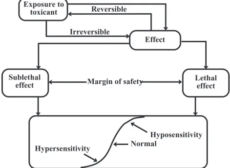
So far, toxicities have been described primarily in terms of the ultimate effect—deaths of organisms or lethality. This is obviously an irreversible consequence of exposure. In many, and perhaps most, cases, sublethal and reversible effects are of greater importance. This is obviously true of drugs, where death from exposure to a registered therapeutic agent is rare, but other effects, both detrimental and beneficial, are usually observed. By their very nature, drugs alter biological processes; therefore, the potential for harm is almost always present. The major consideration in establishing drug dose is to find a dose that has an adequate therapeutic effect without undesirable side effects. A dose-response curve can be established for a drug that progresses from noneffective levels through effective, harmful, and even lethal levels. A low slope for this curve indicates a wide range of effective dose and a wide margin of safety (see Figure 23.3). This term applies to other substances, such as pesticides, for which it is desirable to have a large difference between the dose that kills a target species and that which harms a desirable species.
Figure 23.3. Effects of and responses to toxic substances.
23.4 REVERSIBILITY AND SENSITIVITY
Sublethal doses of most toxic substances are eventually eliminated from an organism’s system. If there is no lasting effect from the exposure, it is said to be reversible. However, if the effect is permanent, it is termed irreversible. Irreversible effects of exposure remain after the toxic substance is eliminated from the organism. Figure 23.3 illustrates these two kinds of effects. For various chemicals and different subjects, toxic effects may range from the totally reversible to the totally irreversible.
Hypersensitivity
Hyposensitivity Normal
Margin of safety Exposure to
toxicant Reversible
Lethal effect Sublethal
effect
Hypersensitivity and Hyposensitivity
Examination of the dose-response curve shown in Figure 23.2 reveals that some subjects are very sensitive to a particular poison (for example, those killed at a dose corresponding to LD5), whereas others are very resistant to the same substance (for example, those surviving a dose corresponding to LD95). These two kinds of responses illustrate hypersensitivity and hyposensitivity, respectively; subjects in the mid-range of the dose-response curve are termed normals. These variations in response tend to complicate toxicology in that there is no specific dose guaranteed to yield a particular response, even in a homogeneous population.
In some cases hypersensitivity is induced. After one or more doses of a chemical, a subject may develop an extreme reaction to it. This occurs with penicillin, for example, in cases where people develop such a severe allergic response to the antibiotic that exposure is fatal if countermeasures are not taken.
23.5 XENOBIOTIC AND ENDOGENOUS SUBSTANCES
Xenobiotic substances are those that are foreign to a living system, whereas those that occur naturally in a biologic system are termed endogenous. Xenobiotic substances that cause harm in organisms are commonly metabolized by them. The levels of endogenous substances must usually fall within a particular concentration range in order for metabolic processes to occur normally. Levels below a normal range may result in a deficiency response or even death, and the same effects may occur above the normal range. This kind of response is illustrated in Figure 23.4.
Figure 23.4. Biological effect of an endogenous substance in an organism showing optimum level, deficiency, and excess.
Examples of endogenous substances in organisms include various hormones, glucose (blood sugar), and some essential metal ions, including Ca2+, K+, and Na+. The optimum level of calcium in human blood serum occurs over a rather narrow range of 9 – 9.5 milligrams per deciliter (mg/dL). Below these values a deficiency response known as hypocalcemia occurs, manifested by muscle cramping. At serum levels above about 10.5 mg/dL hypercalcemia occurs, the major effect of which is kidney malfunction.
Dose
Deficiency Excess, toxicity
Normal physiological effect
Detrimental effect
23.6 TOXICOLOGICAL CHEMISTRY
Toxicological Chemistry
Toxicological chemistry is the science that deals with the chemical nature and reactions of toxic substances, including their origins, uses, and chemical aspects of exposure, fates, and disposal.1 Toxicological chemistry addresses the relationships between the chemical properties and molecular structures of molecules and their toxicological effects. Figure 23.5 outlines the terms discussed above and the relationships among them.
Figure 23.5. Toxicology is the science of poisons. Toxicological chemistry relates toxicology to the chemical nature of toxicants.
Much of what is known about xenobiotic substances in living systems is based upon intensive research upon pharmaceutical compounds in organisms. Pharmacodynamics deals with what a drug does to a body including the dose/response relationship, sites and mechanisms of pharmaceutical actions, therapeutic effects, and side effects. What the body does to a drug is addressed by pharmacokinetics, including uptake, distribution, metabolism, retention, and excretion.
Toxicants in the Body
The processes by which organisms metabolize xenobiotic species are enzyme-catalyzed Phase I and Phase II reactions, which are described briefly here. The substances that undergo these reactions are treated theoretically by the science of quantitative structure-activity relationships, (QSAR) which relates the chemical nature of substances to their biochemical reactions.
Phase I Reactions
Lipophilic xenobiotic species in the body tend to undergo Phase I reactions that make them more water-soluble and reactive by the attachment of polar functional groups, such as –OH (Figure 23.6). Most Phase I processes are “microsomal mixed-function oxidase” reactions catalyzed by the cytochrome P-450 enzyme system. associated with the endoplasmic reticulum of the cell and occurring most abundantly in the liver of vertebrates.2 An example of a Phase I reaction of a xenobiotic is epoxide formation of chloroprene, used to make solvent-resistant synthetic rubbers:
Toxicant Organism Toxic
effect
. . . + . . . . .
Figure 23.6. Illustration of Phase I reactions.
Phase II Reactions
A Phase II reaction occurs when an endogenous species is attached by enzyme action to a polar functional group which often, though not always, is the result of aPhase I reaction on a xenobiotic species. Phase II reactions are called conjugation reactions in which enzymes attach conjugating agents to xenobiotics, their Phase I reaction products, and nonxenobiotic compounds (Figure 23.7). The conjugation product of such a reaction is usually less toxic than the original xenobiotic compound, less lipid-soluble, more water-soluble, and more readily eliminated from the body. The major conjugating agents and the enzymes that catalyze their Phase II reactions are glucuronide (UDP glucuronyltransferase enzyme), glutathione (glutathionetransferase enzyme), sulfate (sulfotransferase enzyme), and acetyl (acetylation by acetyltransferase enzymes). The most abundant conjugation products are glucuronides. A glucuronide conjugate is illustrated in Figure 23.8, where -X-R represents a xenobiotic species conjugated to glucuronide, and R is an organic moiety. For example, if the xenobiotic compound conjugated is phenol, HXR is HOC6H5, X is the O atom, and R represents the phenyl group, C6H5.
C C C C H H Cl H H
H C C
C C H H Cl H H O C C C C O H Cl H H H H C C C C H H Cl H OH H OH H Chloroprene {O} P450
H2O
C C C C H H H O H H Cl C C C C H H H H H Cl O {O} P450 Rearrange (23.6.1)
Product that is more water-soluble and reactive Lipophilic, poorly
Figure 23.8. Glucuronide conjugate formed from a xenobiotic, HX-R.
Figure 23.7. Illustration of Phase II reactions.
23.7 KINETIC PHASE AND DYNAMIC PHASE
Kinetic Phase
The major routes and sites of absorption, metabolism, binding, and excretion of toxic substances in the body are illustrated in Figure 23.1. Toxicants in the body are metabolized, transported, and excreted; they have adverse biochemical effects; and they cause manifestations of poisoning. It is convenient to divide these processes into two major phases, a kinetic phase and a dynamic phase.
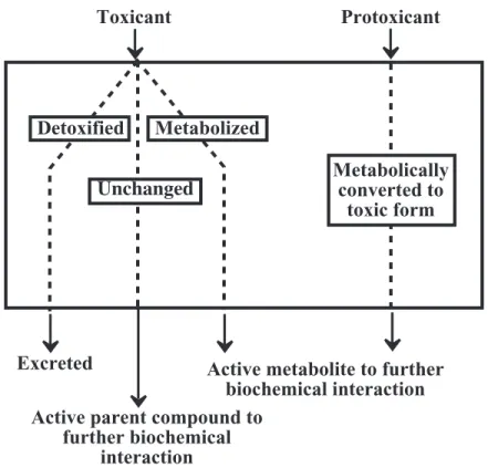
In the kinetic phase, a toxicant or the metabolic precursor of a toxic substance (protoxicant) may undergo absorption, metabolism, temporary storage, distribution, and excretion, as illustrated in Figure 23.9. A toxicant that is absorbed may be passed through the kinetic phase unchanged as an active parent compound, metabolized to a detoxified metabolite that is excreted, or converted to a toxic active metabolite. These processes occur through Phase I and Phase II reactions discussed above.
Xenobiotic
Glucuronide HO
O O OH OH
OH
X R C
C O
OH Carboxyl:
OH Hydroxyl:
F,Cl,Br,I Halogen:
N H
H Amino:
: Epoxide C
C O
Functional groups that react with a conjugating agent
+
• More easily eliminated • Greater water solubility • Higher polarity Xenobiotic
compound, often Phase I reaction product
Conjugation product Endogenous conjugating
Figure 23.9. Processes involving toxicants or protoxicants in the kinetic phase.
Dynamic Phase
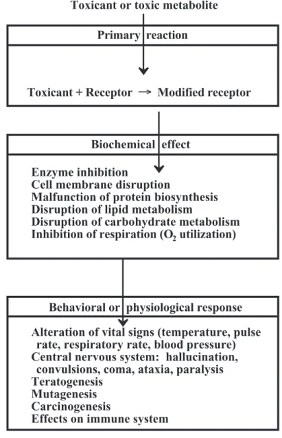
In the dynamic phase (Figure 23.10) a toxicant or toxic metabolite interacts with cells, tissues, or organs in the body to cause some toxic response. The three major subdivisions of the dynamic phase are the following:
• Primary reaction with a receptor or target organ • A biochemical response
• Observable effects
Primary Reaction in the Dynamic Phase
A toxicant or an active metabolite reacts with a receptor. The process leading to a toxic response is initiated whensuch a reaction occurs. A typical example is when benzene epoxide, the initial product of the Phase I reaction of benzene (see Chapter 23, Figure 23.1), forms an adduct with a nucleic acid unit in DNA (receptor) resulting in alteration of the DNA. (Many species that cause a toxic response are reactive intermediates, such as benzene epoxide, which have a brief lifetime but a strong tendency to undergo reactions leading to a toxic response while they are around.) DNA adduct formation with benzene epoxide is an irreversible reaction between a toxicant and a receptor. A reversible reaction that can result in a toxic response is illustrated by the binding between carbon monoxide and oxygen-transporting hemoglobin (Hb) in blood:
O2Hb + CO ←→ COHb + O2 (23.7.1)
Toxicant Protoxicant
Unchanged Metabolicallyconverted to toxic form
Excreted Active metabolite to further biochemical interaction Active parent compound to
further biochemical interaction
Figure 23.10. The dynamic phase of toxicant action.
Biochemical Effects in the Dynamic Phase
The binding of a toxicant to a receptor may result in some kind of biochemical effect. The major ones are the following:
• Impairment of enzyme function by binding to the enzyme, coenzymes, metal activators of enzymes, or enzyme substrates
• Alteration of cell membrane or carriers in cell membranes • Interference with carbohydrate metabolism
• Interference with lipid metabolism resulting in excess lipid accumulation (“fatty liver”)
• Interference with respiration, the overall process by which electrons are transferred to molecular oxygen in the biological oxidation of energy-yielding substrates
• Stopping or interfering with protein biosynthesis by their action on DNA Primary reaction
Toxicant + Receptor ! Modified receptor
Biochemical effect
Enzyme inhibition
Cell membrane disruption
Malfunction of protein biosynthesis Disruption of lipid metabolism
Disruption of carbohydrate metabolism Inhibition of respiration (O2 utilization)
Toxicant or toxic metabolite
Behavioral or physiological response
Alteration of vital signs (temperature, pulse rate, respiratory rate, blood pressure) Central nervous system: hallucination, convulsions, coma, ataxia, paralysis Teratogenesis
Mutagenesis Carcinogenesis
• Interference with regulatory processes mediated by hormones or enzymes.
Responses to Toxicants
Among the more immediate and readily observed manifestations of poisoning are alterations in the vital signs of temperature, pulse rate, respiratory rate,and blood pressure. Poisoning by some substances may cause an abnormal skin color (jaundiced, yellow skin from CCl4 poisoning) or excessively moist or dry skin. Toxic levels of some materials or their metabolites cause the body to have unnatural odors, such as the bitter almond odor of HCN in tissues of victims of cyanide poisoning. Symptoms of poisoning manifested in the eye include miosis (excessive or prolonged contraction of the eye pupil), mydriasis (excessive pupil dilation), conjunctivitis (inflammation of the mucus membrane that covers the front part of the eyeball and the inner lining of the eyelids) and nystagmus (involuntary movement of the eyeballs). Some poisons cause a moist condition of the mouth, whereas others cause a dry mouth. Gastrointestinal tract effects including pain, vomiting, or paralytic ileus (stoppage of the normal peristalsis movement of the intestines) occur as a result of poisoning by a number of toxic substances.
Central nervous system poisoning may be manifested by convulsions, paralysis, hallucinations, and ataxia (lack of coordination of voluntary movements of the body), as well as abnormal behavior, including agitation, hyperactivity, disorientation, and delirium. Severe poisoning by some substances, including organophosphates and carbamates, causes coma, the term used to describe a lowered level of consciousness.
Prominent among the more chronic responses to toxicant exposure are mutations, cancer, and birth defects and effects on the immune system. Other observable effects, some of which may occur soon after exposure, include gastrointestinal illness, cardiovascular disease, hepatic (liver) disease, renal (kidney) malfunction, neurologic symptoms (central and peripheral nervous systems), and skin abnormalities (rash, dermatitis).
Often the effects of toxicant exposure are subclinical in nature. The most common of these are some kinds of damage to the immune system, chromosomal abnormalities, modification of functions of liver enzymes, and slowing of conduction of nerve impulses.
23.8 TERATOGENESIS, MUTAGENESIS, CARCINOGENESIS, AND
EFFECTS ON THE IMMUNE AND REPRODUCTIVE SYSTEMS
Teratogenesis
Teratogens are chemical species that cause birth defects. These usually arise from damage to embryonic or fetal cells. However, mutations in germ cells (egg or sperm cells) may cause birth defects, such as Down’s syndrome.
The biochemical mechanisms of teratogenesis are varied. They include enzyme inhibition by xenobiotics; deprivation of the fetus of essential substrates, such as vitamins; interference with energy supply; or alteration of the permeability of the placental membrane.
Mutagenesis
Mutagens alter DNA to produce inheritable traits. Although mutation is a natural process that occurs even in the absence of xenobiotic substances, most mutations are harmful. The mechanisms of mutagenicity are similar to those of carcinogenicity, and mutagens often cause birth defects as well. Therefore, mutagenic hazardous substances are of major toxicological concern.
Biochemistry of Mutagenesis
To understand the biochemistry of mutagenesis, it is important to recall from Chapter 22 that DNA contains the nitrogenous bases adenine, guanine, cytosine, and thymine. The order in which these bases occur in DNA determines the nature and structure of newly produced RNA, a substance generated as a step in the synthesis of new proteins and enzymes in cells. Exchange, addition, or deletion of any of the nitrogenous bases in DNA alters the nature of RNA produced and can change vital life processes, such as the synthesis of an important enzyme. This phenomenon, which can be caused by xenobiotic compounds, is a mutation that can be passed on to progeny, usually with detrimental results.
There are several ways in which xenobiotic species may cause mutations. It is beyond the scope of this work to discuss these mechanisms in detail. For the most part, however, mutations due to xenobiotic substances are the result of chemical alterations of DNA, such as those discussed in the two examples below.
Nitrous acid, HNO2, is an example of a chemical mutagen that is often used to cause mutations in bacteria. To understand the mutagenic activity of nitrous acid it should be noted that three of the nitrogenous bases—adenine, guanine, and cytosine—contain the amino group, –NH2. The action of nitrous acid is to replace amino groups with a hydroxy group. When this occurs, the DNA may not function in the intended manner, causing a mutation to occur.
Alkylation, the attachment of a small alkyl group, such as –CH3or –C2H5, to an N atom on one of the nitrogenous bases in DNA is one of the most common mechanisms leading to mutation. The methylation of “7” nitrogen in guanine in DNA to form N_Methylguanine is shown in Figure 23.11. O-alkylation may also occur by attachment of a methyl or other alkyl group to the oxygen atom in guanine.
Figure 23.11. Alkylation of guanine in DNA.
A number of mutagenic substances act as alkylating agents. Prominent among these are the compounds shown in Figure 23.12.
N C
C C N C
N C N H
O
H2N
H
N C
C C N C
N C N H
O
H2N
H CH3
Guanine bound to DNA Methylated guanine in DNA +
Figure 23.12. Examples of simple alkylating agents capable of causing mutations.
Alkylation occurs by way of generation of positively charged electrophilic species that bond to electron-rich nitrogen or oxygen atoms on the nitrogenous bases in DNA. The generation of such species usually occurs by way of biochemical and chemical processes. For example, dimethylnitrosamine (structural formula in Figure 23.12) is activated by oxidation through cellular NADPH to produce the following highly reactive intermediate:
This product undergoes several nonenzymatic transitions, losing formaldehyde and generating a methyl carbocation, +CH
3, that can methylate nitrogenous bases on DNA:
(23.7.1)
One of the more notable mutagens is tris(2,3-dibromopropyl)phosphate, commonly called “tris,” that was used as a flame retardant in children’s sleepwear. Tris was found to be mutagenic in experimental animals and metabolites of it were found in children wearing the treated sleepwear. This strongly suggested that tris is absorbed through the skin and its uses were discontinued.
Carcinogenesis
Cancer is a condition characterized by the uncontrolled replication and growth of the body’s own (somatic) cells. Carcinogenic agents may be categorized as follows:
• Chemical agents, such as nitrosamines and polycyclic aromatic hydrocarbons • Biological agents, such as hepadnaviruses or retroviruses
• Ionizing radiation, such as X-rays
• Genetic factors, such as selective breeding.
Clearly, in some cases, cancer is the result of the action of synthetic and naturally occurring chemicals. The role of xenobiotic chemicals in causing cancer is called chemical
N CH3 CH3
O N N H
CH3 H3C
H N
O H3CO S CH3
O N N CH3
CH3 N
Dimethylnitros-amine 3,3-Dimethyl-1-phenyltriazine 1,2-Dimethyl-hydrazine Methylmethane-sulfonate
HO C N N O
H
H
CH
3O N N C H
H
CH3
OH H C H O
O N N H
CH3 HO
-+N N H
CH3
Other products +
carcinogenesis. It is often regarded as the single most important facet of toxicology and is clearly the one that receives the most publicity.
Chemical carcinogenesis has a long history. As noted earlier in this chapter, in 1775 Sir Percival Pott, Surgeon General serving under King George III of England, observed that chimney sweeps in London had a very high incidence of cancer of the scrotum, which he related to their exposure to soot and tar from the burning of bituminous coal. Around 1900 a German surgeon, Ludwig Rehn, reported elevated incidences of bladder cancer in dye workers exposed to chemicals extracted from coal tar; 2-naphthylamine,
was shown to be largely responsible. Other historical examples of carcinogenesis include observations of cancer from tobacco juice (1915), oral exposure to radium from painting luminescent watch dials (1929), tobacco smoke (1939), and asbestos (1960).
An important and unresolved question regarding carcinogens is the existence of thresholds above which carcinogenesis occurs and below which there is no effect.4 Rodent tests for carcinogens are normally conducted at doses 1,000-10,000 times those to which humans are exposed and the probabilities of substances causing cancer in humans are extrapolated linearly to the low doses at which humans are exposed under the assumption that there is no threshold below which the carcinogen has no effect. Some authorities believe that such extrapolations are unrealistic and overstate cancer risk in part because they do not take into account mechanisms possessed by humans to combat genotoxic and carcinogenic insults leading to cancer. Such mechanisms include detoxication and error-free DNA repair.
Biochemistry of Carcinogenesis
Large expenditures of time and money on the subject in recent years have yielded a much better understanding of the biochemical bases of chemical carcinogenesis. The overall processes for the induction of cancer may be quite complex, involving numerous steps. However, it is generally recognized that there are two major steps in carcinogenesis: an initiation stage followed by a promotional stage. These steps are further subdivided as shown in Figure 23.13.
Figure 23.13. Outline of the process by which a carcinogen or procarcinogen may cause cancer.
NH2
2-Naphthylamine
Chemical carcinogen or precursor (procarcinogen)
Metabolic
action carcinogenUltimate Binding to DNA or other(epigenetic) effect
Elimination of compound or its metabolite without adverse effect
Detoxication
(no effect) Altered
DNA DNA repaired,
no adverse effect
Neoplastic cells Promotion of
tumor cell growth Tumor
tissue
Progression of tumor
tissue growth Malignant tumor(neoplasm) Metastasis
Initiation of carcinogenesis may occur by reaction of a DNA-reactive species with DNA, or by the action of an epigenetic carcinogen that does not react with DNA and is carcinogenic by some other mechanism. Most DNA-reactive species are genotoxic carcinogens because they are also mutagens. These substances react irreversibly with DNA. They are either electrophilic or, more commonly, metabolically activated to form electrophilic species, as is the case with electrophilic +CH3 generated from dimethylnitrosamine, discussed under mutagenesis above. Most cancer-causing substances require metabolic activation to produce electrophilic species capable of forming adducts with DNA thereby causing gene mutations. The substances that are activated are called procarcinogens. The metabolic species actually responsible for carcinogenesis is termed an ultimate carcinogen. Some species that are intermediate metabolites between precarcinogens and ultimate carcinogens are called proximate carcinogens. Carcinogens that do not require biochemical activation are categorized as primary or direct-acting carcinogens. Some procarcinogens and primary carcinogens are shown in Figure 23.14.
Most substances classified as epigenetic carcinogens are promoters that act after initiation. Manifestations of promotion include increased numbers of tumor cells and decreased length of time for tumors to develop (shortened latency period). Promoters do not initiate cancer, are not electrophilic, and do not bind with DNA. The classic example of a promoter is decanoyl phorbol acetate or phorbol myristate acetate, which is extracted from croton oil.
Alkylating Agents in Carcinogenesis
Chemical carcinogens usually have the ability to form covalent bonds with macromolecular life molecules. Such covalent bonds can form with proteins, peptides, RNA, and DNA. Although most binding is with other kinds of molecules, which are more abundant, the DNA adducts are the significant ones in initiating cancer. Prominent among the species that bond to DNA in carcinogenesis are the alkylating agents which attach alkyl groups—methyl (CH3) or ethyl (C2H5)—to DNA. A similar type of compound, arylating agents, act to attach aryl moieties, such as the phenyl group,
to DNA. As shown by the examples in Figure 23.15, the alkyl and aryl groups become attached to N and O atoms in the nitrogenous bases that compose DNA. This alteration in DNA can trigger initiation of the sequence of events that results in the growth and replication of neoplastic (cancerous) cells. The reactive species that donate alkyl groups in alkylation are usually formed by metabolic activation through the action of enzymes. This process was shown for conversion of dimethylnitrosamine to a methylating metabolic intermediate in the discussion of mutagenesis earlier in this section.
Testing for Carcinogens
Only a few chemicals have definitely been established as human carcinogens. A well documented example is vinyl chloride, CH2=CHCl, which is known to have caused a rare form of liver cancer (angiosarcoma) in individuals who cleaned autoclaves in the polyvinylchloride fabrication industry. In some cases chemicals are known to be carcinogens from epidemiological studies of exposed humans. Animals are used to test for carcinogenicity, and the results can be extrapolated, although with much uncertainty, to humans.
Figure 23.14. Examples of the major classes of naturally occurring and synthetic carcinogens, some of which require bioactivation, and others which act directly.
Figure 23.15. Alkylated (methylated) forms of the nitrogenous base guanine.
Bruce Ames Test
Mutagenicity used to infer carcinogenicity is the basis of the Bruce Ames test, in which observations are made of the reversion of mutant histidine-requiring Salmonella bacteria back to a form that can synthesize its own histidine. The test makes use of enzymes in homogenized liver tissue to convert potential procarcinogens to ultimate carcinogens. Histidine-requiring
Salmonella bacteria are inoculated onto a medium that does not contain histidine, and those that O
O
H3C H3CO
OCH3 OCH 3 O Cl CH H H C O O C H H H H N N CH3 C H O C C Cl H H H
Cl C O C Cl H
H
H
H C C
N H H H H H O C C H C H H H O Naturally occurring carcinogens that require bioactivation
Bis(chloromethyl)ether Ethyleneimine !-Propioacetone Benzo(a)pyrene
Primary carcinogens that do not require bioactivation Synthetic carcinogens that require bioactivation
N CH3 CH3 N
N
Griseofulvin (produced by
Penicillium griseofulvum)
Saffrole (from sassafras)
N-methyl-N-formylhydrazine (from edible false morel mushroom)
4-Dimethylaminoazobenzene Vinyl chloride
N N CH3 N N OH
H2N
N
N N N OCH3
H2N
Attachment to the remainder of the DNA molecule Methyl groups attached to
mutate back to a form that can synthesize histidine establish visible colonies that are assayed to indicate mutagenicity.
According to Bruce Ames, the pioneer developer of the test which bears his name, animal tests for carcinogens that make use of massive doses of chemicals have a misleading tendency to give results that cannot be accurately extrapolated to assess cancer risks from smaller doses of chemicals. This is because the huge doses of chemicals used kill large numbers of cells, which the organism’s body attempts to replace with new cells. Rapidly dividing cells greatly increase the likelihood of mutations that result in cancer simply as the result of rapid cell proliferation, not genotoxicity.
Immune System Response
The immune system acts as the body’s natural defense system to protect it from xenobiotic chemicals; infectious agents, such as viruses or bacteria; and neoplastic cells, which give rise to cancerous tissue. Adverse effects on the body’s immune system are being increasingly recognized as important consequences of exposure to hazardous substances. Toxicants can cause immunosuppression, which is the impairment of the body’s natural defense mechanisms. Xenobiotics can also cause the immune system to lose its ability to control cell proliferation, resulting in leukemia or lymphoma.
Another major toxic response of the immune system is allergy or hypersensitivity. This kind of condition results when the immune system overreacts to the presence of a foreign agent or its metabolites in a self-destructive manner. Among the xenobiotic materials that can cause such reactions are beryllium, chromium, nickel, formaldehyde, some kinds of pesticides, resins, and plasticizers.
Endocrine Disruption
Some pollutants are of particular concern because of their potential to disrupt the crucial endocrine gland activities that regulate the metabolism and reproductive functions of organisms.5 Because they live in water, fish, frogs, and reptiles such as alligators are particularly susceptible to such substances present as water pollutants. Exposed fish have exhibited reproductive dysfunction, alterations in secondary sex characteristics, and abnormal serum steroid levels. Substances that exhibit hormone-like activity in test subjects are called hormonally active agents. Of particular concern are the substances that act like estrogen, the female sex hormone, that survive wastewater treatment and get into waterways receiving treated wastewater. Examples of such compounds are shown in Figure 23.16. Among such substances are estrogen, an endogenous sex hormone; 17a-ethinylestradiol, an ingredient of oral contraceptives, and chemicals from industrial and consumer sources, such as the last two examples in Figure 23.16. Estrogenic substances from artificial sources are called xenoestrogens and include antioxidants, bisphenol A, dioxins, PCBs, phytoestrogens (from plants), some pesticides (chlordecone, dieldrin, DDT and its metabolites, methoxychlor, toxaphene), preservatives, and phthalic esters (butylbenzyl phthalate).
22.9 HEALTH HAZARDS
substantial, their assessment is very difficult because of factors such as uncertainties in exposure, low occurrence above background levels of disease, and long latency periods.
Figure 23.16. Endocrine disruptors that may be discharged into the environment and affect organisms. Estrone (a natural estrogen), 17α-ethinylestradiol (a constituent of oral contraceptives), 17β-estradiol, and estriol are steroid estrogens. Both 4-tert-octylphenol and p-nonylphenol are breakdown products of nonionic surfactants, bisphenol A is an ingredient of epoxy resins and other polymers, genistein is synthesized by trees to make the wood disease-resistant and is present in pulp mill discharges, and dibutylphthalate is a plasticizer in plastics.
Assessment of Potential Exposure
A critical step in assessing exposure to toxic substances, such as those from hazardous waste sites is evaluation of potentially exposed populations. The most direct approach to this is
O
HO
H3C
Estrone
H3C
HO C C C CH3
CH3
CH3 H
H H3C
4-tert-Octylphenol
C CH3
CH3
HO OH
Bisphenol A
17!!-Ethinylestradiol
O
O HO
OH
OH
Genistein 17""-Estradiol
HO
H3C OH
HO
H3C OH OH
Estriol
p-Nonylphenol
HO C C C C
H
H H
H H
H
C C C C C H
H H
H H
H H
H H
H
H H
H
O O O
O
Dibutylphthalate
HO
C C H OH
to determine chemicals or their metabolic products in organisms. For inorganic species this is most readily done for heavy metals, radionuclides, and some minerals, such as asbestos. Symptoms associated with exposure to particular chemicals may also be evaluated. Examples of such effects include readily apparent effects, for example, skin rashes, or subclinical effects, such as chromosomal damage.
Epidemiological Evidence
Epidemiological studies applied to toxic environmental pollutants, such as those from hazardous wastes, attempt to correlate observations of particular illnesses with probable exposure to such wastes. There are two major approaches to such studies. One approach is to look for diseases known to be caused by particular agents in areas where exposure is likely from such agents in hazardous wastes. A second approach is to look for clusters consisting of an abnormally large number of cases of a particular disease in a limited geographic area, then attempt to locate sources of exposure to hazardous wastes that may be responsible. The most common types of maladies observed in clusters are spontaneous abortions, birth defects, and particular types of cancer.
Epidemiologic studies are complicated by long latency periods from exposure to onset of disease, lack of specificity in the correlation between exposure to a particular waste and the occurrence of a disease, and background levels of a disease in the absence of exposure to a hazardous waste capable of causing the disease.
E
stimation of Health Effects Risks
An important part of estimating the risks of adverse health effects from exposure to toxicants involves extrapolation from experimentally observable data. Usually the end result needed is an estimate of a low occurrence of a disease in humans after a long latency period resulting from low-level exposure to a toxicant for a long period of time. The data available are almost always taken from animals exposed to high levels of the substance for a relatively short period of time. Extrapolation is then made using linear or curvilinear projections to estimate the risk to human populations. There are, of course, very substantial uncertainties in this kind of approach.
Risk Assessment
Toxicological considerations are very important in estimating potential dangers of pollutants and hazardous waste chemicals. One of the major ways in which toxicology interfaces with the area of hazardous wastes is in health risk assessment, providing guidance for risk management, cleanup, or regulation needed at a hazardous waste site based upon knowledge about the site and the chemical and toxicological properties of wastes in it. Risk assessment includes the factors of site characteristics; substances present, including indicator species; potential receptors; potential exposure pathways; and uncertainty analysis. It may be divided into the following major components:
LITERATURE CITED
1. Manahan, Stanley E., Toxicological Chemistry and Biochemistry , 3rd ed., Lewis Publishers/CRC Press, Boca Raton, FL, 2002.
2. Myasoedova, K. N, “New Findings in Studies of Cytochromes P450,” Biochemistry
(Moscow), 73, 965-969, (2008).
3. Barr, Dana B.; Bishop, Amanda; Needham, Larry L., “Concentrations of Xenobiotic Chemicals in the Maternal-Fetal Unit,” Reproductive Toxicology, 23, 260-266 (2007).
4. Nohmi, Takehiko, Naomi Toyoda-Hokaiwado, Masami Yamada, Kenichi Masumura, Masamitsu Honma, and Shoji Fukushima, "Meeting Report on the International Symposium on Genotoxic and Carcinogenic Thresholds," Genes and Environment, 30, 101-107 (2008). 5. Norris, David O., and James A. Carr, Endocrine Disruption: Biological Basis for Health
Effects in Wildlife and Humans, Oxford University Press, New York, 2006.
SUPPLEMENTARY REFERENCES
Baselt, Randall C., Disposition of Toxic Drugs and Chemicals in Man, 6th ed., Biomedical Publications, Foster City, CA, 2002.
Benigni, Romualdo , Ed., Quantitative Structure-Activity Relationship (QSAR) Models of Mutagens and Carcinogens, CRC Press, Boca Raton, FL, 2003.
Bingham, Eula, Barbara Cohrssen, and Charles H. Powell, Patty’s Toxicology, 5th ed., Wiley, New York, 2001.
Boelsterli, Uhrs A., Mechanistic Toxicology: The Molecular Basis of How Chemicals Disrupt Biological Targets, 2nd ed., CRC Press, Boca Raton, FL, 2007.
Dart, Richard C., Medical Toxicology, 3rd ed., Lippincott, Williams & Wilkins, Philadelphia, 2003.
Fenton, John, Toxicology: A Case-Oriented Approach, CRC Press, Boca Raton, FL, 2002. Greenberg, Michael I., Richard J. Hamilton, Scott D. Phillips, and Gayla McCluskey, Eds.,
Occupational, Industrial, and Environmental Toxicology, 2nd ed., Mosby, St. Louis, 2003. Hoffman, David J., Barnett A. Rattner, G. Allen Burton, Jr., and John Cairns, Jr., Handbook of Ecotoxicology, 2nd ed., Lewis Publishers/CRC Press, Boca Raton, FL, 2002.
Joshi, B. D. P. C. Joshi, and Namita Joshi, and Namita Joshi, Eds., Environmental Pollution and Toxicology, A.P.H. Publishing Corporation, 2008.
Smart, Robert C., and Ernest Hodgson, Eds., Molecular and Biochemical Toxicology, 4th ed., Wiley, Hoboken, NJ, 2008.
Ioannides, Costas, Ed., Cytochromes P450: Role in the Metabolism and Toxicity of Drugs and Other Xenobiotics, RSC Publications, Cambridge, U.K., 2008.
Klaassen, Curtis D., Ed., Casarett and Doull's Toxicology: The Basic Science of Poisons, 7th ed., McGraw-Hill Medical, New York, 2008.
Landis, Wayne G. and Ming-Ho Yu, Introduction to Environmental Toxicology: Impacts of Chemicals upon Ecological Systems, 3rd ed., CRC Press/Lewis Publishers, Boca Raton, FL, 2004.
Leonard, Barry, Leonard, Ed., Report on Carcinogens: Carcinogen Profiles,10th ed., Collingdale PA, 2002.
Lippmann, Morton, Ed., Environmental Toxicants: Human Exposures and their Health Effects, 3rd ed., Wiley, New York, 2009.
Newman, Michael C., and William H. Clements, Ecotoxicology: A Comprehensive Treatment, Taylor & Francis/CRC Press, Boca Raton, FL 2008.
Nichol, John, Bites and Stings. The World of Venomous Animals, Facts on File, New York, 1989.
Parvez, S. H., Ed., Molecular Responses to Xenobiotics, Elsevier, Amsterdam, 2001.
Pohanish, Richard P., and Marshall Sittig, Sittig's Handbook of Toxic and Hazardous Chemicals and Carcinogens, Knovel Corporation, Norwich NY, 2002.
Public Health Service, National Toxicology Program, 11th Report on Carcinogens, U.S. Department of Health and Human Services, Washington, DC, 2008, available from the following website: http://ntp.niehs.nih.gov/ntp/roc/toc11.html
Romano, James A., Brian J. Lukey, and Harry Salem, Eds., Chemical Warfare Agents: Chemistry, Pharmacology, Toxicology, and Therapeutics, 2nd ed., Taylor and Francis/CRC Press, Boca Raton, FL, 2008.
Rosenstock , Linda, Textbook of Clinical Occupational and Environmental Medicine, Saunders Health Sciences Division, Philadelphia, 2004.
Saferstein, Richard, Forensic Science: From the Crime Scene to the Crime Lab, Pearson Prentice Hall, Upper Saddle River, NJ, 2009.
Santos, Eduardo B., Ed., Ecotoxicology Research Developments, Nova Science Publishers, New York, 2009.
Stine, Karen E., Thomas M. Brown, Principles of Toxicology, 2nd ed. Taylor and Francis/CRC Press, Boca Raton, FL 2006.
Timbrell, John A., Principles of Biochemical Toxicology, 4th ed., Informa Healthcare, New York, 2009.
Ullmann’s Industrial Toxicology, Wiley-VCH, New York, 2005.
Walker, C. H., Principles of Ecotoxicology, 3rd ed., Taylor and Francis/CRC Press, Boca Raton, FL, 2006.
Ware, George, Reviews of Environmental Contamination and Toxicology, 190, Springer, New York, 2007.
Wexler, Philip, Ed., Encyclopedia of Toxicology, 2nd ed., Elsevier, New York, 2005.
Williamson, John A., Peter J. Fenner, Joseph W. Burnett, and Jacqueline F. Rifkin, Eds,
Venomous and Poisonous Marine Animals: A Medical and Biological Handbook, University of New South Wales Press, Sydney, Australia , 1996.
Wilson, Samuel H., and William A. Suk, Biomarkers of Environmentally Associated Disease, Boca Raton, FL, 2002.
QUESTIONS AND PROBLEMS
1. How are conjugating agents and Phase II reactions involved with some toxicants?
2. What is the toxicological importance of proteins, particularly as related to protein structure? 3. What is the toxicological importance of lipids? How are lipids related to hydrophobic
pollutants and toxicants?
4. What are Phase I reactions? What enzyme system carries them out? Where is this enzyme system located in the cell?
5. Name and describe the science that deals with the chemical nature and reactions of toxic substances, including their origins, uses, and chemical aspects of exposure, fates, and disposal.
6. What is a dose-response curve?
7. What is meant by a toxicity rating of 6?
8. What are the three major subdivisions of the dynamic phase of toxicity, and what happens in each?
9. Characterize the toxic effect of carbon monoxide in the body. Is its effect reversible or irreversible? Does it act on an enzyme system?
10. Of the following, choose the one that is not a biochemical effect of a toxic substance (explain): (A) impairment of enzyme function by binding to the enzyme, (B) alteration of cell membrane or carriers in cell membranes, (C) change in vital signs, (D) interference with lipid metabolism, (E) interference with respiration.
11. Distinguish among teratogenesis, mutagenesis, carcinogenesis, and immune system effects. Are there ways in which they are related?
12. As far as environmental toxicants are concerned, compare the relative importance of acute and chronic toxic effects and discuss the difficulties and uncertainties involved in studying each.
13. What are some of the factors that complicate epidemiologic studies of toxicants?
14. Alkylating agents do not or are not (explain): (A) formed by metabolic activation, (B) attach groups such as CH3 to DNA, (C) include some species that cause cancer, (D) alter DNA, (E) noted for being electron-pair donors or nucleophiles.
24 TOXICOLOGICAL CHEMISTRY OF CHEMICAL
SUBSTANCES
24.1 INTRODUCTION
Toxicological chemistry
, defined and discussed in Chapter 22, centers on the relationship between the chemical nature of toxicants and their toxicological effects. This chapter discusses this relationship with regard to some of the major pollutants and hazardous substances. The first section deals with toxicological aspects of elements (particularly heavy metals) the presence of which in a compound frequently means that the compound is toxic. It also discusses the toxicities of some commonly used elemental forms, such as the chemically uncombined elemental halogens. The following section discusses the toxicological chemistry of inorganic compounds, many of which are produced from industrial processes. It also provides a brief discussion of organometallic compounds. The next section deals with the toxicology of organic compounds. The toxicological properties of hydrocarbons and oxygen-containing organic compounds are discussed as well as other organic substances oxygen-containing functional groups, such as alcohols and ketones. This section also discusses the toxicities of organic nitrogen, halide, sulfur, and phosphorous compounds, some of which are used as pesticides or military poisons. Finally, toxic natural products are discussed.ATSDR Toxicological Profiles
A very useful source of information about the toxicological chemistry of various kinds of toxic substances is published by the U. S. Department of Health and Human Services, Public Health Service Agency for Toxic Substances and Disease Registry ASTDR’s Toxicological Profiles. The detailed documents pertaining to these substances, which are listed in Table 24.1, can be accessed through the website cited in the table.
24.2. TOXIC ELEMENTS AND ELEMENTAL FORMS
Ozone
Table 24.1. Materials Listed by ATSDR1
_______________________________________________________________
Acetone Acrolein Acrylonitrile Aldrin/Dieldrin Aluminum Americium Ammonia Aniline Antimony Arsenic Asbestos Atrazine Barium Benzene Benzidine 2,3-Benzofuran Beryllium Bis(2-chloroethyl)ether Bis(chloromethyl)ether Blister Agents (Lewisite) Boron Bromodichloromethane Bromoform and Dibromochloromethane Bromomethane 1,3-Butadiene 2-Butanone 2-Butoxyethanol CadmiumCalcium or sodium hypochlorite Carbon Disulfide Carbon Tetrachloride Cesium Chlordane Chlorfenvinphos Clofenvinfos Chlorinated dibenzo-p-dioxins (CDDs) Chlorodibenzofurans (CDFs) | Dibenzofuranos Chloroethane Chloroform Chloromethane Chlorophenols Chlorpyrifos Chromium Cobalt Copper
Crankcase oil, used Creosote
Cresols
Crotonaldehyde Cyanide
DDT, DDE, DDD DEHP, Di(2-ethyl-hexyl)phthalate Diazinon Diborane 1,2-Dibromo-3-chloropropane 1,2-Dibromoethane Dichlorobenzenes Dichlorobenzidine 1,1-Dichloroethane 1,2-Dichloroethane 1,2-Dicloroetano 1,1-Dichloroethene 1,2-Dichloroethene 1,2-Dichloropropane 1,3-Dichloropropene Dichlorvos Diethyl phthalate Diisopropyl Methylphosphonate (DIMP) Di-n-butylphtalate Di-1,3-dinitrobenzene and 1,3,5-Trinitrobenzene Dinitrocresols Dinitrophenols 2,4- and 2,6-Dinitrotoluene (HCCPD)
Di-n-octylphthalate (DNOP)
Dioxins (CDDs) | Dibenzo-p-Dioxinas Diphenylhydrazine Disulfoton Endosulfan Endrin Ethion Ethylbenzene Ethylene Glycol Ethylene Oxide Fluorides, hydrogen fluoride, and Fluorine Fuel Oils Gasoline, Automotive Heptachlor Heptachlor epoxide Hexachlorobutadiene Hexachlorocyclohexane Hexachlorocyclopenta-diene Hexachloroethane Hexamethylene diisocyanate (HDI) Hydrogen Peroxide Hydrogen Sulfide Iodine Ionizing Radiation Isophorone Jet Fuels
Kerosene (Fuel Oils) Lead Malathion Manganese 4,4'-Methylenebis (2-Chloroaniline) Mercury Methoxychlor Methyl Isocyanate Methyl Mercaptan Methyl Parathion | Methyl t-Butyl Ether (MTBE)
Methylene Chloride Methylenedianiline Mirex and Chlordecone Naphthalene,
___________________________________________________________________________
1. U. S. Agency for Toxic Substances and Disease Registry: http://www.atsdr.cdc.gov/toxfaq.html#bookmark05
GD, VX) Nickel Nitrobenzene Nitrogen Oxides Nitrophenols
n-Nitrosodimethylamine
n-Nitrosodi-n-propylamine
Nitrosodiphenylamine Otto Fuel II
Pentachlorophenol Perchlorates Phenol Phosgene
Phosgene Oxime Phosphine Phosphorus, Plutonium Polybrominated Biphenyls (PBBs) Polybrominated Diphenyl Ethers (PBDEs) Polychlorinated Biphenyls (PCBs) Polycyclic Aromatic Hydrocarbons (PAHs) Propylene Glycol | Pyrethroids
Pyridine Radium RDX Selenium
Selenium Hexafluoride Silver
Sodium Hydroxide Stoddard Solvent Strontium
Styrene Sulfur Dioxide Sulfur Trioxide | Sulfuric Acid Synthetic Vitreous Fibers
1,1,2,2-Tetrachloroethane
Tetrachloroethylene Thallium
Thorium Tin
Titanium Tetrachloride Toluene
Total Petroleum Hydrocarbons Toxaphene Trichloroethane
Trichloroethylene (TCE) Trichloropropane
Trinitrotoluene (TNT) | 2,4,6-Trinitrotolueno (TNT)
Tungsten Uranium Vanadium Vinyl Acetate Vinyl Chloride Xylene
Ozone generates free radicals in tissue. These reactive species can cause lipid peroxidation, oxidation of sulfhydryl (–SH) groups, and other destructive oxidation processes. Compounds that protect organisms from the effects of ozone include radical scavengers, antioxidants, and compounds containing sulfhydryl groups.
White Phosphorus
Elemental white phosphorus can enter the body by inhalation, by skin contact, or orally. It is a systemic poison, that is, one that is transported through the body to sites remote from its entry site. White phosphorus causes anemia, gastrointestinal system dysfunction, bone brittleness, and eye damage. Exposure also causes phossy jaw, a condition in which the jawbone deteriorates and becomes fractured.
Elemental Halogens
Elemental fluorine (F2) is a pale yellow, highly reactive gas that is a strong oxidant. It is a toxic irritant and attacks skin, eye tissue, and the mucous membranes of the nose and respiratory tract. Chlorine (Cl2) gas reacts in water to produce a strongly oxidizing solution. This reaction is responsible for some of the damage caused to the moist tissue lining the respiratory tract when the tissue is exposed to chlorine. The respiratory tract is rapidly irritated by exposure to 10-20 ppm of chlorine gas in air, causing acute discomfort that warns of the presence of the toxicant. Even brief exposure to 1,000 ppm of Cl2 can be fatal.
Bromine (Br2) is a volatile, dark red liquid that is toxic when inhaled or ingested. Like chlorine and fluorine, it is strongly irritating to the mucous tissue of the respiratory tract and eyes and may cause pulmonary edema. The toxicological hazard of bromine is limited somewhat because its irritating odor elicits a withdrawal response.
Elemental solid iodine (I2) is irritating to the lungs much like Cl2 or Br2. However, the relatively low vapor pressure of iodine limits exposure to I2 vapor.
Heavy Metals
Heavy metals (Section 7.3) are toxic in their chemically combined forms and some, notably mercury, are toxic in the elemental form. The toxic properties of some of the most hazardous heavy metals and metalloids are discussed here.
Although not truly a heavy metal, beryllium (atomic mass 9.01) is one of the more hazardous toxic elements. Its most serious toxic effect is berylliosis, a condition manifested by lung fibrosis and pneumonitis, which may develop after a latency period of 5-20 years. Beryllium is a hypersensitizing agent and exposure to it causes skin granulomas and ulcerated skin. Beryllium was used in the nuclear weapons program in the U. S., and it is believed that 500 to 1000 cases of beryllium poisoning have occurred or will occur in the future as a result of exposure to workers. In July 1999, the U. S. Department of Energy acknowledged these cases of beryllium poisoning and proposed legislation to compensate the victims. This resulted in the Energy Employees Occupational Illness Compensation Program of 2000 under which qualified workers with beryllium disease can receive a lump sum payment of $150,000 as reimbursement for disability and medical care resulting from the disease.1