Address for correspondence Dr. Sabyasachi Banerjee,
Associate Professor, Department of
Dermatology, Malda Medical College, Malda, West Bengal.
E-mail: [email protected]
Case Report
Porokeratosis palmaris et plantaris disseminata - a
rare entity
Kar Chinmay, Sarkar Prodip, Banerjee Sabyasachi, Biswas Rabindranath, Dutta Pallab*
Department of Dermatology, Malda Medical College and Hospital, West Bengal, India * Centre for Healthcare Science and Technology, Indian Institute of Engineering Science and Technology, Shibpur, Howrah, West Bengal, India.
Abstract
Porokeratosis is a well-known keratinization disorder where cornoid lamella is characteristically seen. There are seven clinical variants of which porokeratosis palmaris et plantaris disseminata (PPPD) is a very rare entity. Our PPPD case was sporadic and diagnosed early at 35 years of age. The lesions started on trunk and disseminated to extremities, face, oral cavity, palms and soles. Involvement of oral mucosa made easier to differentiate our case from disseminated superficial porokeratosis. The response was good with isotretinoin therapy.Key words
Porokeratosis, porokeratosis palmaris et plantaris disseminata, cornoid lamella.
Introduction
Porokeratosis is a morphologically distinct disorder of keratinisation. It is characterised clinically by hyperkeratotic papules or plaques with raised and advancing edge, which histologically corresponds to a column of parakeratotic cells. There are seven clinical
variants.1 Among those, porokeratosis
palmaris et plantaris disseminata (PPPD) is a very rare variety, originally described by Guss et al.2 in 1971. This variety is usually inherited
in an autosomal dominant manner, though some sporadic cases have been reported. More than one type of porokeratosis may be found
in same patient.3 Different types of
porokeratosis may also be found in multiple
members of an affected family.4 Malignant
transformation has been reported in all forms except punctate variety. The incidence of malignant transformation is 6.8-11% of all
porokeratosis patients.5 Older and linear
variants have higher risk and actinic variant has lowest risk of malignant transformation.6
Here we report a case of PPPD because of its atypicality and rarity.
Case Report
A 35-year-old male presented with numerous blackish brown papules and plaques all over the body including oral mucosa since last fifteen years. The lesions of trunk appeared first, followed by lesions on extremities, face, oral cavity, palms and soles, respectively. All the lesions appeared within one-year duration. The patient was farmer by profession. There was no history of long-term drug intake. None of his family was affected by similar disease.
On clinical examination, the lesions of trunk, extremities and face 2-7mm in diameter
in diameter (Figure 1b, 1d). A few of these lesions had characteristic border with furrow.
Figure 1 a) Porokeratosis of trunk (front) and upper limbs.
Figure 1 b) Porokeratosis of palms. Figure 1 c) Porokeratosis of palate.
Figure 1 d) Porokeratosis of soles. Figure 2 a) HPE of PPPD (back) showed basket weave orthokeratosis, the cornoid lamella with hypogranulosis or agranulosis in subcornoid lamella region in epidermis (X10). Inset: HPE focussing cornoid lamella, H&E stain (X20). (Nikon Eclipse Ti microscope).
Figure 2 b) HPE of PPPD (palm) showed compact orthokeratosis with cornoid lamella in epidermis (X10). Inset: HPE focussing cornoid lamella (X20).
Figure 3 a) HPE of back focussing cornoid lamella, H & E stain, X80
magnification. magnification.
Oral lesions with characteristic border were clearly seen over hard palate (Figure 1c).
All basic blood investigations were within normal limit. Estimation of arsenic level from hair and nail were also normal. Punch biopsy was taken from back and histopathological examination (HPE) of hematoxylin and eosin-stained (H&E) section revealed basket weave orthokeratosis with a column of parakeratotic cells, the cornoid lamella, characteristic of porokeratosis. There was hypogranulosis or agranulosis in subcornoid lamella region along with mild dyskeratosis and a few vacuolar cells. Dermis showed chronic inflammatory infiltrates in perivascular regions (Figure 2a,
3a). Punch biopsy from palm showed marked
compact orthokeratosis with characteristic
cornoid lamella (Figure 2b, 3b). Other
findings were similar to trunk lesion. So clinical and histopathological examination of our case revealed rare variant of porokeratosis palmaris et plantaris disseminata There was no malignant change in our case.
Our case was treated with isotretinoin as 30 milligram per day and about 30% lesional improvement was observed within four weeks.
Some lesions became hyperpigmented
macules following treatment. Mostly the lesions of trunk, extremities and face improved
without any visible improvement of
palmoplantar lesions. Regular follow-up is needed to note whether there is any complete clearance of disease, the duration of treatment and further recurrence.
Discussion
In 1893, Mibelli first described the classic form of porokeratosis. After that, various types of porokeratosis were discovered. After introduction of PPPD by Guss et al.2 a few
cases were reported. In PPPD, it is usually observed that porokeratosis starts from palms and soles and then disseminates to other areas
including mucous membrane. It is mostly autosomal dominant with a few sporadic occurrences. Males are affected more than females and it usually starts in late teens to
early twenties.2 But due to very slow progress
of the disease, diagnosis of PPPD is usually confirmed at a much later age. It also has malignant potential due to abnormal DNA ploidy in lesional epidermis like other porokeratosis.7 The lesions of our case started
typically at the age of twenty but appeared first on trunk. Then the lesions disseminated to other areas including oral mucosa within one year. The palms and soles were the last sites to be involved. Though it was not typical, a few
reports were found with this sequence.8
Clinically and histopathologically, our case had no abnormal change.
Lesions of trunk, extremities and face are similar to lesions of disseminated superficial porokeratosis (DSP) and disseminated actinic superficial porokeratosis (DSAP) clinically,
morphologically and even
histopathologically.9 But palms, soles and
mucosae are usually spared in DSP and
DSAP.10 Palmoplantar lesions of PPPD are
close to palmoplantar porokeratosis (PP).11 So
PPPD is difficult to differentiate from DSP or DSAP with PP. But with involvement of any mucosa, PPPD can easily be differentiated from DSP or DSAP with PP. So our case is a classical case of PPPD. This PPPD should also
be differentiated from systematized,
widespread form of porokeratotic eccrine ostial and dermal ductal nevus (PEODDN). This PEODDN is a rare, benign hamartoma of eccrine sweat gland with porokeratotic histology and clinically presents as keratotic
papules or plaques with central plugged pits.12
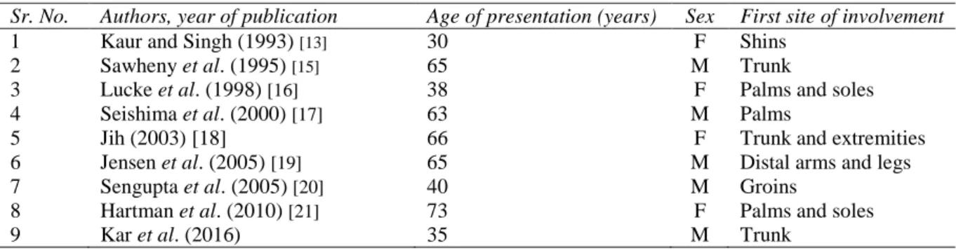
The sporadic occurrence of PPPD is also rare. We have found only eight previous sporadic
cases of PPPD in English literature (Table 1).
Our sporadic case was diagnosed at the age of thirty five, which is, most probably the second
of PPPD by isotretinoin has been reported
earlier.14 Our case also showed visible
improvement with isotretinoin.
Table 1 Sporadic cases of PPPD
Sr. No. Authors, year of publication Age of presentation (years) Sex First site of involvement
1 Kaur and Singh (1993) [13] 30 F Shins
2 Sawheny et al. (1995) [15] 65 M Trunk
3 Lucke et al. (1998) [16] 38 F Palms and soles
4 Seishima et al. (2000) [17] 63 M Palms
5 Jih (2003) [18] 66 F Trunk and extremities
6 Jensen et al. (2005) [19] 65 M Distal arms and legs
7 Sengupta et al. (2005) [20] 40 M Groins
8 Hartman et al. (2010) [21] 73 F Palms and soles
9 Kar et al. (2016) 35 M Trunk
References
1. O’Regan GM, Irvine AD. Porokeratosis. In: Goldsmith AL, Karz IS, Gilchrest AB, Paller SA, Leffell JD, Wolff K, editors. Fitzpatrick's Dermatology in General Medicine. 8th ed. New York: McGraw-Hill; 2012. P. 563-8.
2. Guss SB, Osbourn RA, Lutzner MA. Porokeratosis palmaris et plantaris disseminata: a third type of porokeratosis. Arch Dermatol. 1971;104:366-73.
3. Dover JS, Phillips TJ, Burns DA. Disseminated superficial actinic porokeratosis: coexistence with other porokeratotic variants. Arch Dermatol. 1986;122:887-9.
4. Lucker GP, Steiflen PM. The coexistence of linear and giant porokeratosis associated with Bowen's disease. Dermatology. 1994;189:78-80.
5. Bhaskar S, Jaiswal AK, Raj N, Reddy D. Porokeratosis – Head to toe: An unusual presentation. Indian Dermatol Online J. 2015;6:101-4.
6. Sasson M, Krain A. Porokeratosis and cutaneous malignancy: a review. Dermatol Surg. 1996;22:339-42.
7. Beers B, Jaszcz W, Sheetz K, Hogan DJ, Lynch PJ. Porokeratosis palmaris et plantaris disseminata - Report of a case with abnormal DNA ploidy in lesional epidermis. Arch Dermatol. 1992;128:236-9.
8. Irisawa R, Yamazaki M, Yamamoto T, Tsuboi R. A case of porokeratosis palmaris plantaris et disseminata and literature review. Dermatol Online J. 2012;18(8):5. 9. Vasudevan B, Chatterjee M, Grewal R, Rana
V, Lodha N. A case of disseminated superficial porokeratosis associated with giant porokeratosis in pregnancy. Indian J Dermatol. 2014;59:492-4.
10. Requena L, Requena C, Cockerell JC. Benign epidermal tumours and proliferations. In: Bolognia LJ, Jorizzo LJ, Schaffer VJ, editors. Dermatology. 3rd ed.
Delhi, India: Reed Elsevier; 2014. P. 1795-815.
11. Lanka P, Lanka LR, Manivachagam D. Punctate porokeratosis palmaris et plantaris. Indian J Dermatol. 2015;60:284-6.
12. Kroumpouzos G, Stefanato CM, Wilkel CS, Bogaars H, Bhawan J. Systematized porokeratotic eccrine and hair follicle nevus: report of a case and review of the literature. Br J Dermatol. 1999;141:1092-6.
13. Kaur V, Singh G. Porokeratosis palmaris et plantaris disseminata. Indian J Dermatol Venereol Leprol. 1993;59:130-1.
14. McCallister RE, Estes SA, Yarbrough CL. Porokeratosis palmaris et plantaris disseminata. Report of a case and treatment with isotretinoin. J Am Acad Dermatol. 1985;13:598-603.
15. Sawheny MP, Mahatachar V, Bisht YS. Porokeratosis plantaris palmaris et disseminata. Indian J Dermatol Venereol Leprol. 1995;61:48-9.
16. Lucke TW, Fallowfield M, Kemmett D. A sporadic case of porokeratosis plantaris palmaris et disseminata. Br J Dermatol. 1998;138:556-7.
17. Seishima M, Izumi T, Oyama Z, Maeda M. Squamous cell carcinoma arising from lesions of porokeratosis palmaris et plantaris disseminata. Eur J Dermatol. 2000;10:478-80.
18. Jih MH. Porokeratosis plantaris, palmaris, et disseminata. Dermatol Online J. 2003;9(4):34.
19. Jensen JM, Egberts F, Proksch E, Hauschild A. Disseminated porokeratosis palmaris and plantaris treated with imiquimod cream to prevent malignancy. Acta Derm Venereol. 2005;85:550-1.
20. Sengupta S, Das JK, Gangopadhyay A. Porokeratosis confined to the genital area: a report of three cases. Indian J Dermatol Venereol Leprol. 2008;74:80.