INTRAVENOUS VITAMIN D TREATMENT IN HEMODIALYSIS PATIENTS: PATTERNS OF USE AND ASSOCIATION WITH FRACTURE RISK
Anne Christine Beaubrun
A dissertation submitted to the faculty of the University of North Carolina at Chapel Hill in partial fulfillment of the requirements for the degree of Doctor of Philosophy in the
Eshelman School of Pharmacy (Division of Pharmaceutical Outcomes and Policy).
Chapel Hill 2013
Approved by:
M. Alan Brookhart, PhD Gang Fang, PharmD, PhD Abhijit V. Kshirsagar, MD
Christine U. Oramasionwu, PharmD, PhD Betsy L. Sleath, PhD
© 2013
ABSTRACT
ANNE CHRISTINE BEAUBRUN: Intravenous Vitamin D Treatment in Hemodialysis Patients: Patterns of Use and Association with Fracture Risk
(Under the direction of Dr. M. Alan Brookhart and Dr. Betsy L. Sleath)
The administration of intravenous vitamin D therapy is central to the treatment of secondary hyperparathyroidism. Yet, there is little data documenting the variations in the use of these agents in large, representative samples and vitamin D’s clinical benefits are not clear. The objectives of this dissertation were to describe patterns in the use of vitamin D and to examine the association between vitamin D exposure and fracture risk among hemodialysis patients.
Vitamin D use has increased sharply from 58.6% of patients treated in 1999 to 83.9% of patients treated in 2008. Paricalcitol was the preferred formulation during the study years. In 2008, the average dose among black patients was 84% greater than among white patients. No significant relation was observed between the proportion of vitamin D users or the average vitamin D dose per patient at the facility-level and fracture rates for all fracture types. Specifically, for any fracture, the hazard ratio (HR) in adjusted models for a facility’s proportion of vitamin D users was 1.10 (95% CI 0.86-1.42) while the HR for a facility’s average vitamin D dose per patient was 0.99 (95% CI 0.90-1.09).
In summary, vitamin D use has increased and parallels the rise in use of paricalcitol and doxercalciferol. Increasing vitamin D use and average vitamin D dose administered per patient within dialysis facilities did not have an observed beneficial association with
DEDICATION
ACKNOWLEDGEMENTS
It takes a power so much greater than me to have assembled in one time and in one place some of the most impressive, genuine, astute, and caring individuals that I will ever have the pleasure of knowing. How fortuitous an occasion! The completion of this dissertation was possible because individuals were strategically placed into my life. I am eternally grateful that God placed me in an environment where I was supported both academically and emotionally.
I am forever indebted to my co-advisors, Dr. M. Alan Brookhart and Dr. Betsy Sleath. I benefited tremendously from both of your complementary mentoring styles and masterful coaching throughout the dissertation process. Alan, you are the academic mind and mentor that every health services researcher should strive to emulate. It has been an honor to work with a professor at your stature with such a genuine interest in student professional
development and in promoting an exciting culture of intellectual curiosity. I was constantly in awe of your dedication to advancing patient outcomes and your steadfast calm under all the pressures that are associated with high quality productivity. Betsy, you are the epitome of organization and character. Thank you for the countless number of hours you invested into ensuring that I was an efficient written communicator and for allowing me the opportunity to work under your distinguished scholarship.
Gang, a brilliant methodologist, and Christine for your meticulous review and valuable insights. Abhi, your clinical perspectives were instrumental to the completion of this undertaking and it benefited me remarkably to have access to your tutelage.
My deepest appreciation goes to my mother, Marie Quetly Beaubrun. The
culmination of all your sacrifices has made me the woman I am today. You amaze me and I love you!
To Dr. Lily Wang, a true angel is disguise. Your mental acuity and abundance of statistical knowledge are only matched by your devotion to serve humanity. Lily, it is an understatement to simply say that it was a blessing that our paths crossed. It is with absolute certainty that I am a better researcher and a better human being because I had the privilege of knowing you.
I would like to thank Dr. Timothy Carey, Dr. Mark Holmes, and the National Research Service Award (NRSA) team of collaborators for their valuable critiques.
Thank you to a host of friends and colleagues who have provided statistical guidance, encouraged and sustained me along the way. I am especially fortunate for Amica Yon, Raphael Yon, Stacie Dusetzina, Adrian Stephens, Chris Wiesen, Melissa Butler, Stedman Stevens, Mackenzi Pergolotti, Monica Jolles, Carla White, Alan Ellis, Chris Beadles, and Loren Robinson.
TABLE OF CONTENTS
LIST OF TABLES ... xii
LIST OF FIGURES ... xiii
LIST OF ABBREVIATIONS ... xiv
CHAPTER I: INTRODUCTION ... 15
1.1 Overview ... 15
1.2 Aims and Hypotheses ... 16
1.3 Significance of the Study ... 21
1.4 Summary ... 23
CHAPTER II: LITERATURE REVIEW ... 25
2.1 Secondary hyperparathyroidism in end-stage renal disease ... 26
2.1.1 Pathogenesis, epidemiology, and consequences ... 26
2.1.2 Disparities ... 28
2.2 Vitamin D therapy ... 31
2.2.1 Role of vitamin D therapy ... 32
2.2.2 Calcitriol ... 35
2.2.3 Paricalcitol ... 35
2.2.4 Doxercalciferol ... 36
2.2.5 Clinical and economic differences of vitamin D formulations ... 37
2.3 Adjunct therapies ... 40
2.4.3 Factors associated with fractures ... 45
2.5 Vitamin D therapy and non-skeletal and skeletal outcomes ... 47
2.5.1 Vitamin D therapy and non-skeletal outcomes ... 47
2.5.2 Vitamin D therapy and skeletal outcomes ... 50
2.5.3 Relationship between clinical parameters, secondary hyperparathyroidism treatment and skeletal outcomes ... 51
2.6 Conceptual Framework ... 55
2.6.1 Andersen’s Behavioral Model of Health Services Use ... 55
2.6.2 Proposed Conceptual Framework ... 57
CHAPTER III: RATIONALE FOR METHODS USED TO ASSESS FRACTURE RISK... 61
3.1 Justification for the use of observational studies ... 61
3.2 Justification for the grouped-treatment approach ... 62
CHAPTER IV: METHODS ... 69
4.1 Data source ... 69
4.2 Study design and cohort selection by aims ... 71
4.2.1 Aim 1 ... 71
4.2.2 Aim 2 ... 75
4.2.3 Sample size ... 79
4.3 Measurements ... 81
4.3.1 Vitamin D formulations and dose ... 81
4.3.2 Vitamin D exposure by aims ... 82
4.3.4 Covariates ... 88
4.4 Statistical analyses by aims ... 100
4.4.1 Analyses used to create case-mix adjusted measures of vitamin D exposure ... 100
4.4.2 Aim 1 ... 102
4.4.3 Aim 2 ... 103
4.5 Sensitivity Analyses ... 108
CHAPTER V: STUDY 1 RESULTS: TRENDS AND VARIATIONS IN INTRAVENOUS VITAMIN D USE AMONG HEMODIALYSIS PATIENTS IN THE UNITED STATES ... 110
5.1 Overview ... 110
5.2 Introduction ... 111
5.3 Methods ... 112
5.3.1 Data source ... 112
5.3.2 Study design and patient population ... 113
5.3.3 Patterns of vitamin D use assessment ... 113
5.3.4 Statistical analyses ... 114
5.4 Results ... 114
CHAPTER VI: STUDY 2 RESULTS: INCREASING USE OF INTRAVENOUS VITAMIN D MAY NOT REDUCE FRACTURE RISK AMONG HEMODIALYSIS PATIENTS ... 128
6.1 Overview ... 128
6.2 Introduction ... 129
6.3 Methods ... 130
6.3.3 Measurement of vitamin D exposure ... 132
6.3.4 Measurement of fracture outcomes ... 133
6.3.5 Measurement of Covariates ... 134
6.3.6 Statistical analyses to assess fracture risk ... 136
6.4 Results ... 136
6.5 Discussion ... 141
CHAPTER VII: DISCUSSION ... 145
7.1 Summary of findings ... 145
7.2 Implications ... 148
7.3 Study limitations ... 155
7.4 Study strengths ... 157
7.5 Recommendations for future research ... 160
7.6 Conclusion ... 164
APPENDICES ... 166
LIST OF TABLES
Table 1. Summary of Aim 1 eligibility criteria ... 71
Table 2. Summary of Aim 2 eligibility criteria ... 75
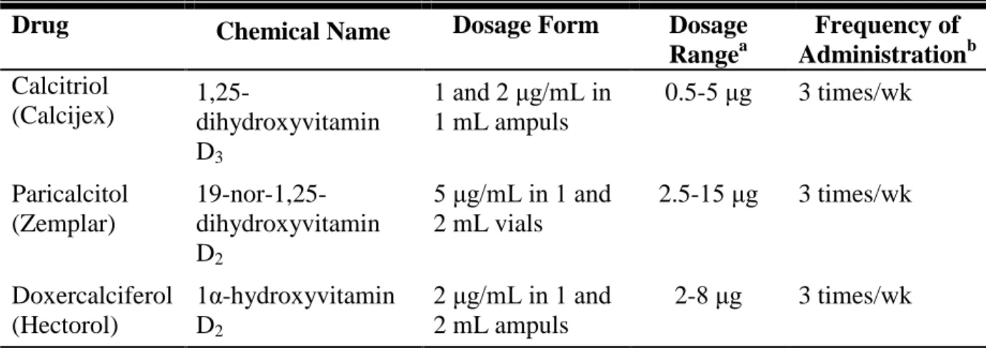
Table 3. Description of IV vitamin D formulations ... 81
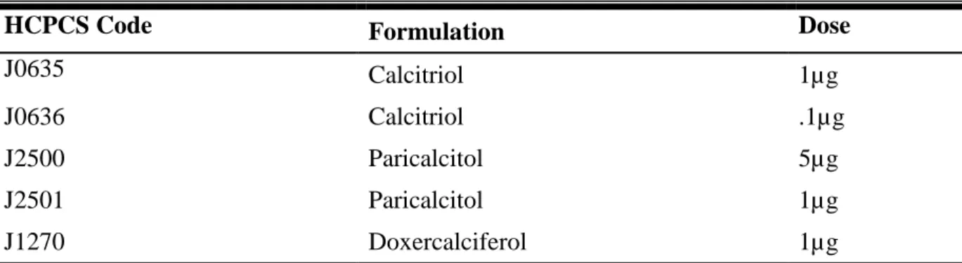
Table 4. HCPCS codes to identify IV vitamin D formulations ... 82
Table 5. Diagnostic codes used to identify fractures ... 86
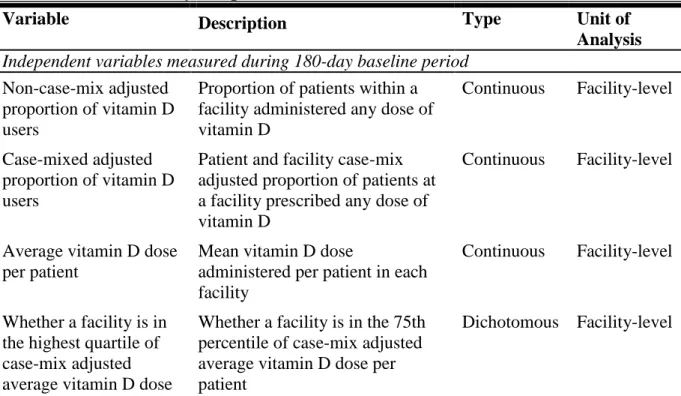
Table 6. Overview of key independent and dependent variables for Aim 2 ... 88
Table 7. Diagnostic codes used to identify comorbidities ... 91
Table 8. Diagnostic and procedural codes used to identify parathyroidectomies ... 95
Table 9. Procedural codes used to identify personal assistance aids ... 98
Table 10. Procedural codes used to identify fistula creation ... 99
Table 11. Baseline characteristics of patients between 1999-2008 ... 115
Table 12. Mean annual IV vitamin D dose (mcg) administered per patient by race ... 118
Table 13. Mean annual IV vitamin D dose (mcg) administered per patient by formulation ... 120
Table 14. Demographic and clinical characteristics of cohort by vitamin D user status ... 137
Table 15. Fracture rates per 100,000 person-years by vitamin D user status ... 139
Table 16. Cox models of the association between measures of vitamin D exposure and fracture risk ... 140
LIST OF FIGURES
Figure 1. Pathogenesis and consequences of secondary hyperparathyroidism ... 27
Figure 2. Structure of IV vitamin D formulations ... 33
Figure 3. Andersen’s behavioral model of health services use ... 55
Figure 4. Proposed conceptual framework ... 58
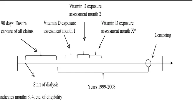
Figure 5. Study design for Aim 1 ... 74
Figure 6. Study design for Aim 2 ... 76
Figure 7. Sample size determination flow chart for Aim 2 ... 80
Figure 8. Levels of analysis when studying ESRD population ... 107
Figure 9. Annual percentage of patients treated with intravenous vitamin D by formulation ... 116
Figure 10. Annual percentage of intravenous vitamin D users by race ... 117
Figure 11. Annual percentage of intravenous vitamin D users by sex ... 119
Figure 12. Annual intravenous vitamin D dos per patient by state among both white and black patients, 1999-2008 ... 122
Figure 13. Annual intravenous vitamin D dos per patient by state among only black patients, 1999-2008 ... 123
LIST OF ABBREVIATIONS
AOR Adjusted odds ratio
BMI Body mass index
CI Confidence Interval
CKD Chronic kidney disease
CPT Current Procedural Terminology ESRD End-stage renal disease
FDA US Food and Drug Administration
HCPCS Healthcare Common Procedure Coding System
HR Hazard ratio
ICD-9 International Classification of Diseases, Ninth Revision
iPTH Intact PTH
IV Intravenous
PTH Parathyroid hormone
CHAPTER I
INTRODUCTION
1.1 Overview
Disordered bone mineral metabolism is rampant in end-stage renal disease (ESRD) patients and a considerable amount of time and resources are dedicated to its evaluation and treatment.1 Intravenous (IV) vitamin D has become a mainstay in bone-mineral disorder management and is used to treat secondary hyperparathyroidism (SHPT), a common complication among patients with ESRD.2 SHPT, characterized by increased parathyroid hormone (PTH) levels, has been associated with abnormalities in bone metabolism, soft tissue and vascular calcification and a range of other disorders.2, 3 Despite IV vitamin D’s widespread use and its proven effectiveness in decreasing PTH levels, there is a lack of evidence demonstrating that pharmacologically reducing PTH levels can actually result in improved fracture outcomes. There are a myriad of examples from various therapeutic areas documenting instances where medications were approved for their efficacy in manipulating a surrogate biomarker but were eventually found to confer no clinical benefit or even harm.1
It is important to evaluate whether vitamin D’s benefit extends beyond treating
SHPT. Patients with renal failure commonly experience fractures, associated with significant morbidity and mortality in this patient population.4 The age- and sex-adjusted risk of
morphologies that may be linked to an increased risk of fracture.5 Although it would be tempting for nephrologists to use vitamin D to ameliorate the high clinical burden of fractures observed among dialysis patients, it would be ill-advised given the general lack of valid, population-based studies or clinical trials documenting any benefits or harms of IV vitamin D use for this indication.
Also, studies exploring racial, gender, geographic secular variations, and patterns of vitamin D use are needed to document any secular trends in overuse of the drug, provide evidence in support of dialysis quality improvement initiatives, and alleviate any health disparities among patients with ESRD. There have been no large-scale population-based observational studies, thus far, examining the association between vitamin D exposure and fracture risk among dialysis patients. Vitamin D exposure refers to vitamin D-related treatment decisions regarding dialysis patients. To address these salient deficits in the nephrology literature, the aims and hypotheses that comprise this dissertation are described below.
1.2 Aims and Hypotheses
Data were derived from the United States Renal Data System (USRDS), a national registry of all renal disease patients. The aims of this study were:
Aim 1: To describe patient-level, facility-level, and state-level trends in the use and
dosage of three vitamin D analogs among prevalent hemodialysis patients.
of vitamin D formulation were presented in longitudinal graphs comparing secular trends in vitamin D use in each calendar year between 1999 and 2008.
Aim 2: To investigate the association between vitamin D exposure and fracture risk by
fracture type and among relevant subgroups among incident hemodialysis patients.
Null Hypotheses
H10: There is no association between the non-case-mix proportion of vitamin D users within a dialysis facility and fracture risk.
H20: There is no association between the case-mix adjusted proportion of vitamin D users within a dialysis facility and fracture risk.
H30: There is no association between the non-case-mix average vitamin D dose per patient within a dialysis facility and fracture risk.
H40: There is no association between the case-mix adjusted average vitamin D dose per patient within a dialysis facility and fracture risk.
H50: There is no association between high case-mix adjusted average vitamin D doses per patient at the facility-level (the 75th percentile) and fracture risk.
Alternative Hypotheses
H1a: The non-case-mix adjusted proportion of vitamin D users within a dialysis facility is negatively associated with fracture risk.
H2a: The case-mix adjusted proportion of vitamin D users within a dialysis facility is negatively associated with fracture risk.
H4a: The case-mix adjusted average vitamin D dose within a dialysis facility is negatively associated with fracture risk.
H5a: High case-mix adjusted average vitamin D doses per patient at the facility-level (the 75th percentile) are negatively associated with fracture risk.
We conducted a retrospective cohort, intention-to-treat analysis using data from 2000-2004 where vitamin D exposure variables were measured as ecological variables at the facility-level while covariates and fracture outcomes were measured at the individual-level. The measures of vitamin D exposure for Aim 2 were ecological variables measured at the facility-level during the 180-day baseline period: 1) the non-case-mix adjusted proportion of vitamin D users in each facility; 2) the case-mix adjusted proportion of vitamin D users in each facility; 3) the non-case-mix adjusted average vitamin D dose per patient in each facility; 4) the case-mix adjusted average vitamin D dose per patient in each facility; and 5) whether a facility was in the highest quartile of case-mix adjusted average vitamin D dose per patient in each facility. We focused the presentation of results on the case-mix adjusted measures of vitamin D exposure because they account for variations in patient characteristics at a dialysis facility that may have influenced how vitamin D was delivered.
end of the baseline period. In sum, fracture risk was used to describe our outcome and time to first fracture was used to describe the dependent variable in Cox proportional hazards models.
A number of statistical techniques were employed to address the high likelihood of confounding by indication in this analysis given that we did not have access to clinical variables that likely mediate the association between vitamin D use and fracture risk. We adopted a facility-practice-based, grouped-treatment approach whereby vitamin D exposure was measured ecologically while covariates and outcomes were measured at the individual-level. The main measures of vitamin D exposure (the case-mix adjusted proportion of vitamin D users and case-mix adjusted average vitamin D dose per patient) reflected the facility’s likelihood to prescribe vitamin D at certain doses based on the distribution of demographic and clinical characteristics of patients within the facility. There is empirical evidence in the nephrology literature suggesting that facility-level characteristics have a great influence on patient-level health outcomes. For instance, in a study of chronic hemodialysis patients within a non-profit dialysis provider, Chan and colleagues found evidence
suggesting that the most important determinant of achieving optimal anemia management may be at the dialysis facility-level.6 Even after adjusting for the use of facility treatment protocols, a patient’s dialysis center was strongly associated with a patient’s achievement of target hemoglobin values.6
analyses. Crude and covariate adjusted fracture rates were estimated. Cox proportional hazard models examined fracture risk in models with time to fracture as the dependent
variable. Analyses adjusted for baseline patient demographic and clinical characteristics. All analyses were conducted in the overall patient population within age, sex, and racial
subgroups, respectively.
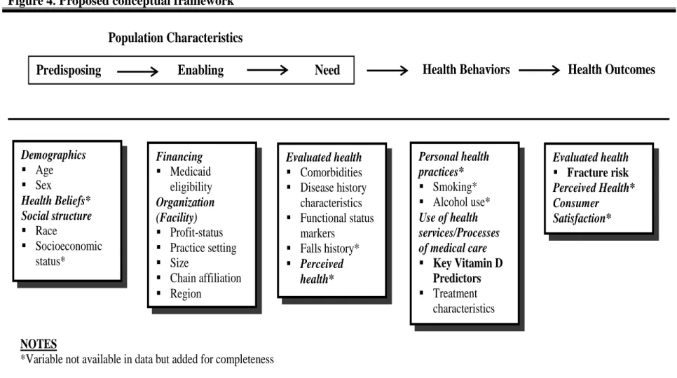
The choice of covariates and the hypothesized relationship between important
determinants of fracture risk was guided by Andersen’s Behavioral Model of Health Services Use.7 According to the model, predisposing, enabling and need factors comprise of
population characteristics that determine health behavior, health service use, and health outcomes. In all the Cox proportional hazard regression models performed, adjustments were made for predisposing characteristics such as age, sex, and race, attributes inherent to the individual prior to the onset of disease. We also controlled for enabling characteristics such as eligibility for Medicaid and organizational level factors like a dialysis facility’s profit-status to reflect the healthcare resources available to the patient. Comorbidities and functional status markers were included in our analysis to reflect need characteristics that compel individuals to seek health care services. These population characteristics
1.3 Significance of the Study
Surrogate endpoints are defined as symptoms, laboratory values (e.g., serum calcium levels), symptoms (e.g., inflammation), clinical markers (e.g., body mass index) , and other measures of treatment efficacy that are used as a proxy for clinical outcomes like morbidity and mortality.8 There are grave potential safety consequences, cost-inefficiencies, and potential for mismanagement of patient care when a surrogate endpoint is assumed to be an appropriate substitute for clinical endpoints. For instance, sodium fluoride was shown to effectively increase bone mineral density but it was proven to have no effect on fracture rates among postmenopausal women in clinical trials.9 In the nephrology community, there is a massive dearth in the literature regarding whether the metabolic changes in PTH levels induced by vitamin D administration actually correct the bone abnormalities and increased fracture risk observed among patients with ESRD. The prognostic value of altered PTH levels as a surrogate endpoint for changes in fracture risk must be validated with biochemical and epidemiological evidence from both randomized clinical trials and observational studies like that conducted herein. 9
Additionally, data generated from this analysis will most likely be relevant for
reducing the administration of vitamin D and injectable medications.1 Once contemporary data of the effect of the new bundled system becomes available, understanding the potential clinical benefit of vitamin D under the dosage practices of the old system can be used as evidence for task forces charged with evaluating the effect of reimbursement changes on dialysis patient care.12
Moreover, clinicians do have good reason to suspect an association between vitamin D and fracture even though the relation has yet to be proven. Due to its high prevalence and observable effects on bone structure, SHPT is believed to contribute meaningfully to the elevation in fracture risk observed in the dialysis population as a whole. SHPT, common among dialysis patients, has direct pathological effects on bone. Among dialysis patients, bone mineral disorders known as renal osteodystrophy has been associated with bone pain, muscle tendon ruptures and increased fracture risk.13 The action of PTH on bone is directly mediated through promoting osteoclast activity and bone resorption that can result in high-turnover bone disease as documented by bone histology.14, 15 These consequences are believed to increase the risk of fracture, which has been estimated to be 4.4 to 14 times higher among dialysis patients than in the general population.16
consequences for dialysis patients who fracture, the literature currently describes the effect of surrogate serum markers on fractures and no studies to date have examined the association between vitamin D dose and the risk of fracture among dialysis patients.
This was the first large, population-based study to examine the association between vitamin D exposure and bone outcomes by four fracture types and by age, sex and race. The burden of SHPT, bone diseases and fractures among a costly, and morbid ESRD population warrants the research conducted herein.
1.4 Summary
Vitamin D therapy helps to maintain appropriate mineral metabolism, prevents bone disease, and minimizes loss of bone strength by decreasing PTH levels.18 However, the increasing and perhaps excessive doses of vitamin D administered to dialysis patients may confer minimal clinical benefit with respect to fractures. The association between IV vitamin D exposure and fracture outcomes, to date, has not been investigated. In order to fill this gap, we first provide descriptive data of secular trends in IV vitamin D use among
hemodialysis patients in the United States to validate studies suggesting that the use of the drug has been increasing. Then, we examined the association between vitamin D exposure and various fracture outcomes by different subgroups and fracture type.
patients. Identifying disparities in vitamin D use may assist in providing evidence for
CHAPTER II
LITERATURE REVIEW
This section presents the epidemiology of secondary hyperparathyroidism (SHPT) and the adverse skeletal and extraskeletal health outcomes associated with the disease. Mechanisms of treating SHPT are explored with an emphasis on the three most commonly administered commercially available vitamin D formulations. The gaps in the evidence regarding the association between vitamin D, intermediate clinical markers, bone disease and fracture risk are presented to support the need for studies investigating the independent association between vitamin D and fracture risk among hemodialysis patients.
PubMed and Google Scholar were used to extract relevant articles published in English anytime before the 2013 calendar year. Google and Google Scholar were used to identify conference proceedings, academic presentations, websites and other sources with pertinent information. A free-text search strategy using a combination of Boolean operators was employed using search strings such as “vitamin D”, “fractures”, “paricalcitol”,
2.1 Secondary hyperparathyroidism in end-stage renal disease
2.1.1 Pathogenesis, epidemiology, and consequences
SHPT is an extremely common complication associated with chronic kidney disease (CKD) and ESRD. Approximately 78% of hemodialysis patients suffer from SHPT,19 a disease characterized by increased parathyroid hormone (PTH) levels.20 PTH is a polypeptide of 84 amino acids that plays a direct role in maintaining bone metabolism
homeostasis and regulating calcium levels including the release of calcium into the blood and intestinal absorption of calcium.21, 22 The primary role of PTH is to reduce the excretion of calcium from the kidneys, control the release of calcium and phosphorus from bone, increase urinary excretion of phosphorous, and direct the synthesis of active vitamin D in the
kidneys.23
To assess bone metabolism and disease, clinicians traditionally use the intact parathyroid hormone assay system which measures the full length PTH (1-84) but also has been found to react with large truncated fragments of non-1–84 PTH.24 Although there are newer generation assays that measure the full length 1-84 PTH24, current dialysis care
guidelines are based on iPTH levels, advising nephrologist to maintain the dialyzed patient at a range of 150 and 300 pg/mL.25 A full discussion of the differences between PTH assays and the implications of using one versus another is beyond the scope of this work. The central point is that different assays, even those from the same generation can produce highly different PTH levels thus affecting a patient’s SHPT and bone disorder classification.25
Calcium is the most important parameter dictating SHPT progression. PTH secretion is primarily regulated by calcium-sensing receptors located on the surface of parathyroid cells.23 In order to maintain homeostasis, calcium concentrations must be rigorously controlled and typically must not fluctuate above or below 2% of the normal level.23
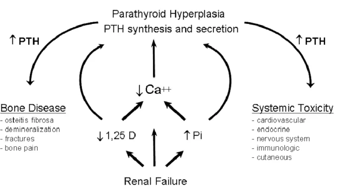
Figure 1 describes the pathogenesis and consequences associated with SHPT.
Figure 1. Pathogenesis and consequences of secondary hyperparathyroidism
Source: Brown Alex J., Slatopolsky Eduardo: Vitamin d analogs: Therapeutic applications and mechanisms for selectivity. Molecular Aspects of Medicine 29: 433-452, 2008
Progressive kidney decline is also associated with declines in vitamin D synthesis by the kidney.28 Reduced activation of parathyroid vitamin D receptors (VDRs) are a
consequence of vitamin D deficiency, fostering PTH mRNA transcription and inducing PTH synthesis.26 The mRNA transcription of PTH by the parathyroid gland is further increased because of the decreased ionized calcium available for binding to calcium sensing-receptors on the surface of the parathyroid glands.27
Increased PTH levels, a uremic toxin, are linked to a myriad of serious, adverse clinical skeletal and non-skeletal effects.29 Skeletal-related clinical consequences of elevated PTH levels include a series of bone abnormalities termed renal osteodystrophy while non-skeletal effects include hypertension, left ventricular hypertrophy, atherosclerosis, immune dysfunction and renal anemia.29 Renal function and PTH levels are inversely correlated as PTH levels continually increase with decreased renal function.30 SHPT-induced variations in bone histology and increased serum phosphorous and calcium, have all been implicated as factors in part responsible for the increased morbidity and mortality observed in hemodialysis patients.30
The hypocalcemia induced by the decrease in serum vitamin D levels and increased phosphorous retention leads to pathyroid gland hyperplasia, the effects of which are
clinically manifested with bone disease and system toxicity.28 The skeletal consequences of SHPT such as demineralization, bone pain and fractures are described in detail in later sections of this chapter.
2.1.2 Disparities
Compared to whites, the incidence of new black and Native American ESRD patients in 2009 was 3.5 and 1.9 times greater, respectively. The ESRD incidence rate among Hispanics was 1.5 times that found in non-Hispanic populations.3 Given the current racial disparities in ESRD incidence, it was important to consider possible racial variations in the manifestation of SHPT and other disorders clinically present in the dialysis population.
Race is a major determinant of SHPT.31 Black dialysis patients generally have higher iPTH levels in comparison to other races.19 Gupta and colleagues reported an average PTH level of 641.7 pg/mL in black dialysis patients and 346.0 pg/mL in white dialysis patients.31 In comparison with white patients, black patients were reported to have a higher mean PTH level in a cohort of 218 patients within an ambulatory nephrology. 32 Wolf and colleagues also reported that black patients are given the most vitamin D therapy when compared to other ethnicities33, presumably because black patients have these reportedly higher PTH levels.
In the general population, parathyroid gland mass is greater among blacks and there may be an increased risk of SHPT among black individuals when diagnosed with chronic kidney disease.29 Nearly all non-Hispanic blacks (97%) currently suffer from vitamin D deficiency in the general population.34 Additionally, some scholars have posited that because of their darker skin tones, black individuals synthesize less active vitamin D, 25(OH)D3, causing SHPT and greater parathyroid gland mass.29
Thus, compared to white patients, black dialysis patients are more likely to be vitamin D deficient and have more severe SHPT.35 However, black patients in the general
of fractures compared to other ethnicities. There is some evidence that the clinical
consequences of SHPT and renal osteodystrophy may vary by race, but the studies are scant and traditionally have focused on white subjects. Elevated PTH levels and SHPT of greater severity may actually be protective in blacks, serving as a physiologically adaptive
mechanism to maintain bone turnover.31 For instance, studies of predominantly white patients have concluded that a PTH level of 120 to 240 pg/mL is optimal for dialysis.31 However, treating black ESRD patients using these guidelines may led to over-suppression of parathyroid gland and a greater risk of adynamic bone disease.31
There is considerable debate regarding whether current therapeutic guidelines are applicable to black hemodialysis patients given documented differences in calcium balance and bone histomorphometry between blacks and non-blacks in the general population.13 Differences in iPTH level between blacks and non-blacks have been discussed in the literature but there is currently no consensus on the optimal level of iPTH and subsequent ideal vitamin D dosing for hemodialysis patients by patient ethnicity.13 Moore and
colleagues concluded that the published K/DOQI guideline iPTH threshold of less than 150 pg/mL may not accurately identify black hemodialysis patients with adynamic bonde disease because the authors identified many black patients with adynamic bone disease above this cutoff after performing transiliac bone biopsies.13
Adynamic bone disease, low born-turnover, affects approximately 30% of hemodialysis and 50% of peritoneal dialysis patients.31, 37 Patients with relative
unknown given that the relationship between PTH levels and bone turnover has been investigated predominantly among whites.31
Higher serum iPTH thresholds may be necessary among black hemodialysis patients to prevent adynamic bone disease, a disorder associated with fractures and increased
mortality.13 Black dialysis patients are therefore at risk for over-therapy with the PTH overestimation resulting in adynamic bone disease and subsequent fracture and death.38 This analysis was warranted because trends over time in vitamin D dosing among different
subgroups, including race, has not been documented to date. More importantly, the relation between vitamin D therapy and bone outcomes by race may contribute to our understanding of the association between facility-level vitamin D dosing practices and fractures among black dialysis patients.
2.2 Vitamin D therapy
SHPT therapy attempts to maintain mineral metabolism, prevent bone disease and minimize the skeletal complications that eventually induce loss of bone strength and fractures.18 Additionally, treatments for SHPT aim to prevent the numerous extraskeletal complications such as vascular calcification that are associated with the high cardiovascular morbidity observed in ESRD patients. SHPT is currently managed with the concurrent use of phosphate binders, phosphate diet restrictions, and vitamin D therapy.39 These therapeutic modalities aim to address the range of mineral metabolism disturbances found in SHPT.
These therapies are also instrumental in the prevention of hyperphosphatemia, phosphate retention, and the control of serum calcium levels. The consequences of
effects such as increased cardiac stroke index, vessel calcification and cardiac calcification.40 A 1mg/dL increase in phosphorous levels is associated with a 6% incremental increase in the relative mortality risk among hemodialysis patients.40, 41 The mortality rate among
hemodialysis patients has been shown to increase by 11% for every 10mg2/dL2 increase in calcium phosphorous product. 40, 41
The pathogenesis of SHPT and the confluence of factors that foster it illustrate the tremendous complexities associated with treating the disease. Vitamin D, phosphorous, calcium, and PTH levels must be simultaneously controlled, especially since the
manipulation of one parameter directly or indirectly elicits a profound influence on another. Treatment regimens must be evaluated often and tailored to the disparate needs of a growing ESRD population.
2.2.1 Role of vitamin D therapy
Vitamin D therapy suppresses PTH levels in both direct and indirect ways. Treatment with vitamin D directly reduces PTH levels by either inhibiting the enlargement of
parathyroid glands or decreasing PTH synthesis.42 When active vitamin D is administered, messenger RNA synthesis to induce PTH production by parathyroid glands is decreased.30 In addition to reducing PTH synthesis and secretion by the parathyroid glands, active vitamin D plays a role in the absorption of dietary calcium by the intestines and in skeletal bone
Figure 2. Structure of IV vitamin D formulations
Source: Martin KJ, González EA: Vitamin D analogues for the management of secondary hyperparathyroidism.
Am J Kidney Dis 38: S34-S40, 2001
Figure 2 depicts the structures of IV calcitriol (1α,25-dihydroxyvitamin D3; Calcijex, Abbott Laboratories, North Chicago, IL, USA), paricalcitol (19-nor-1α,25-dihydroxyvitamin D2; Zemplar, Abbott Laboratories) and doxercalciferol (1α-hydroxyvitamin D2; Hectorol, Genzyme). Calcitriol, paricalcitol and doxercalciferol are the three most commonly
Moreover, it is important to differentiate nutritional (inactive or native) from active (vitamin D3 or calcitriol) vitamin D medications. The generic term “vitamin D” refers to numerous substances and variants of vitamin D with very different effects and physical consequences. Nutritional vitamin D refers to compounds such as cholecalciferol and ergocalciferol found in foods high in vitamin D content.44 Active vitamin D compounds refer to agents with the ability to activate VDRs.44 In contrast to active vitamin D
compounds, nutritional vitamin D is less efficacious in the suppression of PTH levels and in improving or maintaining the status of bone histology in dialysis patients.46 Precursors to active vitamin D are found in food and ultraviolet light exposure.21 In healthy individuals, a series of enzymatic reactions convert these precursors to the calcitriol/active vitamin D3 molecule.21 The conversion of nutritional vitamin D (25-(OH)D3) to active vitamin D (1,25(OH)2D3) occurs due to the 1-α-hydrolase enzyme located in the mitochrondria of proximal tubular cells of the kidney.27 With declining renal function, the kidney becomes less able to perform 1α-hydroxylation, the final reaction response for the synthesis of active vitamin D, and PTH levels rise.21
IV rather than oral vitamin D formulations were the predictors of interest for the work presented herein. IV vitamin D is preferred for hemodialysis patients because these
patient intake of most oral medications, prescribers must consider the decrease in medication efficacy associated with patient non-adherence of oral vitamin D therapy.27
Evidence regarding optimal treatment of bone mineral disorders in dialysis patients is scant with guidance predominantly provided by the opinion-based National Kidney
Foundation’s Kidney Disease Outcomes Quality Initiative Clinical Practice Guidelines for Bone Metabolism and Disease in Chronic Kidney Disease (K/DOQI).47 Decisions to
administer vitamin D sterols are guided by PTH levels, with physicians urged to concurrently consider serum calcium and phosphorous levels.48 IV vitamin D should be given to dialysis patients with a PTH level greater than 300pg/mL in order to suppress PTH levels to the target range of 150pg/mL to 300pg/mL.47 The K/DOQI disseminates guidelines three opinion-based algorithms for the management of vitamin D sterols opinion-based on either serum calcium, phosphorous or intact PTH levels. Appendix 1 depicts the guideline based on dialysis patient intact PTH levels.47
2.2.2 Calcitriol
Calcitriol administration in dialysis patients has been associated with elevated serum calcium and phosphorous concentrations and also low bone turnover (hypodynamic bone disease).44 Nine chronic hemodialysis patients were administered 2µg of IV calcitriol three times a week for ten weeks.49 Following therapy, baseline PTH levels were reduced from 902 +/- 126 pg/mL to 466 +/- 152 pg/mL (p< 0.01).49
2.2.3 Paricalcitol
release and preventing PTH synthesis.39 Additionally, the drug promotes bone mineralization and intestinal calcium and phosphorous absorption.39 In multicenter, prospective trials of greater than 12 months, paricalcitol reduced PTH levels by approximately 59% to 82%.51 In hemodialysis patients, a 0.24mcg/kg bolus IV administration of paricalcitol has a mean elimination half-life of 19.9 hours. 39
The efficacy of paricalcitol has been evaluated in numerous clinical trials with the majority of trials comparing paricalcitol users to patients receiving placebo. Three double-blind, placebo-controlled, dose-escalating, randomized, multicenter trials of 78 hemodialysis patients treated for 12 weeks found a significant decrease in iPTH levels from 795 ± 86 to 406 ± 106 pg/mL (p<0.001).52 Long-term studies of paricalcitol have confirmed these findings. In an open-label, multicenter, 13-month study of 164 hemodialysis patients, IV paricalcitol administered at a dose of 0.04-0.394µg/kg 2-3 times per week rapidly and effectively suppressed iPTH levels.53 Mean iPTH levels reached designated target levels of 100-300 pg/mL, going from a baseline mean of 628.3 +/- 27.65 pg/mL to 295.3 +/- 25.69 pg/mL.53 Paricalcitol has been shown to suppress PTH levels even in in patients with protracted SHPT resistant to calcitriol therapy.54
2.2.4 Doxercalciferol
Both intermittent oral and IV doxercalciferol therapy effectively suppress iPTH levels but IV doxercalciferol does so with less instances of hypercalcemia and hypophosphatemia.56 2.2.5 Clinical and economic differences of vitamin D formulations
The first available vitamin D analog, calcitriol, can effectively lower serum PTH levels.57 However, calcitriol has also been shown to increase serum calcium levels by inducing intestinal calcium absorption and bone resorption. The risk of both hypercalcemia and coronary artery calcification may increase when calcitriol is used simultaneously with calcium-based phosphorous binders or dialysate with high calcium concentrations.57 The vitamin D2 analogs, paricalcitol and doxercalciferol, are vitamin D analogs also considered mainstream therapy among dialysis patients.57 Both vitamin D2 analogs, like calcitriol, can effectively lower PTH levels but do so with a smaller effect on serum calcium and
phosphorous concentrations compared to calcitriol.57 Unlike calcitriol, paricalcitol is
considered a selective VDR activator, indicating that the administration of paricalcitol results in less activation of vitamin D receptors in the gastrointestinal tract, invariably leading to reduced calcium and phosphorous absorption.44
Several studies have demonstrated equivalent or even superior PTH level suppression with the use of these paricalcitol or doxercalciferol compared to calcitriol.58 A 2007 meta-analysis of randomized controlled trials of chronic kidney disease patients actually
demonstrated both potentially positive and detrimental effects of paricalciltol and
approximately 5% to 10% over a 3 year span but the increase in phosphorous concentrations may increase mortality by an equivalent amount.58
Sprague and colleagues performed the first double-blind, randomized, multicenter study of 263 hemodialysis patients at 27 facilities in the United States, The Netherlands, Spain, and Switzerland to assess the comparative effectiveness and safety of paricalcitol versus calcitriol,.59 Dosed at a 4:1 paricalcitol to calcitriol ratio, paricalcitol decreased PTH concentrations more rapidly compared to calcitriol.59 From baseline, paricalcitol treated patients achieved at least a 50% mean reduction in baseline PTH levels at week 15 compared to week 23 for patients receiving calcitriol.59 The authors found no statistically significant differences in the incidence of hyperphosphatemia in paricalcitol versus calcitriol treated subjects, a finding contrary to previously published studies comparing the two drugs.59 However, compared to calcitriol subjects, patients receiving paricalcitol experienced lower hypercalcemic episodes (18% versus 33%, p=0.008) and fewer elevated
calcium-phosphorous product incidences.59
Also, in a study by Dobrez and colleagues, approximately 94% of paricalcitol-treated patients remained on the therapy whereas only 58.7% of patients who initiated with calcitriol stayed on the drug, suggesting that paricalcitol may be better tolerated.39, 60
Approximately $509 million was spent on IV vitamin D therapy in 2009, accounting for 18.3% of the $2.78 billion spent on all injectable medications for dialysis patients that year.3 In 2009, per person per year costs were greatest for paricalcitol ($1,926), followed by
doxercalciferol ($1,326) with calcitriol annual per person costs lowest at $456.3 2.2.6 Factors currently associated with vitamin D use
Compared to patients receiving calcitriol, patients administered paricalcitol were more likely to be black, have an arteriovenous fistula, and have higher baseline serum levels of calcium, phosphorus, and PTH. Paricalcitol treated patients were also less likely to be diabetic.61 Paricalcitol use has been found to be greatest in the southern region of the country.60
Cost data suggests racial and geographic differences in vitamin D use. In 2009, IV vitamin D per person per year Medicare expenditures for black patients was $1,846
compared to $1,059 for white patients, constituting a 74% difference. This difference in Medicare medication costs by race, however, seems only to be specific to IV vitamin D with relatively similar costs observed for other injectable medications across races.3 For instance, in 2009, per person IV iron Medicare expenditures for whites and blacks were $789 and $814, respectively, representing only a 3% difference.3 Similar to geographic patterns
2.3 Adjunct therapies
In addition to vitamin D, therapy for the regulation of PTH levels and to maintain mineral homeostasis also includes oral phosphate binding agents, calcimimetics and parathyroidectomies.62 Serum concentrations of phosphorous are reduced with oral
phosphate binding agents like calcium, sevelamer, lanthanum, magnesium and aluminum.62 Phosphate binders are frequently prescribed to dialysis patients to control the deleterious effects of elevated phosphorus levels, hyperphospatemia.40 Calcimimetic agents actively reduce PTH secretions without simultaneously increasing calcium and phosphorous levels.62 Sensipar, Cinacalcet HCL, the only U.S. Food and Drug Administration (FDA) approved calcimimetic agent, is available in oral form as a daily treatment of hypercalcemia in ESRD patients with SHPT or parathyroid carcinoma.62 Sensipar increases the sensitivity of the calcium-sensing receptor on parathyroid glands to extracellular calcium.63 Sensipar was able to suppress iPTH levels in a phase 3, multicenter, randomized, placebo-controlled, double-blind study of dialysis patients independent of treatment with traditional SHPT therapies.64 The subsequent decrease in calcium levels directly decrease PTH levels.63
Parathyroidectomies, the oldest SHPT treatment, are perhaps the least preferred option. Surgery to remove the parathyroid glands, usually performed in patients with recalcitrant SHPT, is accompanied by numerous potential risks and complications.62 In addition to the traditional risks associated with anesthesia, following surgery, patients may experience severe hypocalcemia, permanent hypoparathyroidism, or require additional surgery.62
prescribed to dialysis patients because of safety concerns over toxicity related to impaired renal excretion66-68, and bone disease in dialysis patients is often due to SHPT and other forms of renal osteodystrophy, including osteomalacia and adynamic bone disease4, which effect fracture risk independent of bone density.
2.4 Renal osteodystrophy and fractures in End Stage Renal Disease
2.4.1 Epidemiology of renal osteodystrophy
Renal osteodystrophy is an overarching label for both high-turnover bone disorders termed osteitis fibrosa cystica and low-turnover disorders such as osteomalcia and adynamic bone disease.30 Specifically, renal osteodystrophy can present itself in any of five
histopathological forms including osteitis fibrosa, osteomalcia mixed lesions, mild lesions, and adynamic bone disease.69 Often a consequence of SHPT, osteitis fribrosa, the most common form of renal osteodystrophy, is characterized by increases in bone formation, resorption and marrow fibrosis.27 On the contrary, in addition to low bone-specific alkaline phosphatase levels, adynamic bone disease is characterized by low iPTH levels below 200pg/mL and decreased bone formation.70
At this juncture, it is important to differentiate renal osteodystrophy from osteoporosis. The bone histology in renal osteodystrophy is characterized by bone remodeling and is best diagnosed with a bone biopsy.70 Osteoporosis, contrarily, is a systematic skeletal disease defined by low bone mass and deterioration of bone tissue.71
bone turnover characterize renal osteodystrophy as the disease can be classified broadly into osteitis fibrosa, osteomalcia, adynamic bone disease, and mixed osteodystrophy.72
2.4.2 Clinical and economic burden of fractures
ESRD patients have been observed to be at increased risk of fractures relative to those without renal impairment.73 Patients with ESRD are 4.4 to 14 times more likely to
experience a hip fracture compared to individuals in the general population.16 These
estimates, however, were derived using data solely from Caucasian incident dialysis patients within the USRDS between 1989 and 1996.74 The incidence of any fracture is approximately 20 per 1000 patient years on dialysis with a three-to-four fold increased risk of hip fracture reported for ESRD patients.75, 76 Dialysis patients who have never had a kidney transplant and those who have undergone transplantation have an observed hip fracture incidence rate of 2.9 fractures and 3.3 fractures per 1,000 person-years, respectively.73
The average or median time to fracture following dialysis initiation is informative for this analysis to serve as a benchmark to assess whether time to first fracture, the dependent variable in Cox regression models, is reduced with the administration of vitamin D.
mortality rate following a hip fracture has ranged from about 15 to 40% in the general population.78 In the US, there are over a quarter of a million hip fractures every year
resulting in 14% to 36% mortality in the first year following fracture.79 Coco and colleagues reported a hip fracture incidence rate of 13.9 per 1,000 patient-years among a cohort of 1,272 patients within outpatient dialysis facilities in New York between 1988 and 1998.80
Mortality one year subsequent to the hip fracture event was by far greater among dialysis patients when compared those in the general population. A 64% one-year mortality rate was found among the dialysis cohort compared to a 20% one-year mortality rate in the general population.80
A population based cohort study by Mittalhenkle and colleagues found that, among U.S. incident dialysis patients between 1995 and 2000, hip fractures were associated with a 2.15 time increase in the incidence rate ratio for all-cause mortality.81 After experiencing a hip fracture, dialysis patients had a one-year survival rate of approximately 50%.81 Among patients with no history of cardiovascular disease, the risk of cardiovascular events was 40% greater and the risk of cardiovascular mortality was 84% greater among dialysis patients who sustained a fracture compared to those who did not, respectively.81
There is a substantial economic burden associated with the occurrence and treatment of fractures in the US. Using a Markov state-transition model, Burge and colleagues
predicted the incidence and costs associated with osteoporosis-related fractures in the general population of the US from 2005 to 2025.86 The investigators predicted an incidence of two million fractures in 2005 at a cost of $17 billion with hip fractures accounting for 72% of total costs but only 14% of the overall distribution of fractures.86 By 2025, the incidence of fractures is expected to increase by 48%, contributing to $25.3 billion in costs.86
Furthermore, there is strong evidence that there is interstate variability in both the incidence and economic burden of fractures in the United States. Also using a Markov state-transition model of osteoporosis-related fractures, King and colleagues highlighted the geographic and hospital fracture care pattern differences in five states.87 In 2000, mean hospital charges for hip fractures ranged from $16,700 in Massachusetts to $29,500 in California.87 The disparity in mean charges was not explained by the length of stay
associated with hip fracture hospital admissions.87 The fracture incidence estimated in 2005 ranged from 199 per 10,000 in California to 266 per 10,000 in Massachusetts.87 In 2005, total costs attributable to fractures varied from $270 million in Arizona to $1,434 million in California.87
Although this analysis did not explore variations in the cost of fractures among dialysis patients, it does contribute to the literature by documenting the burden of fracture related hospitalizations in the hemodialysis population.
2.4.3 Factors associated with fractures
Hemodialysis patients are susceptible to the risk factors for fracture observed among individuals in the general population but also experience additional risk factors attributable to their disease. The following section describes the risk factors for fracture observed in the general population and then summarizes the current literature investigating the risk factors for fracture among the dialysis population.
Fracture risk is multifactorial and risk factors related to falling, bone strength, and clinical characteristics have been identified.89 In the general population, approximately 90-97% of proximal humerus fractures and greater than 95% of hip fractures are due to falls.89, 90 Approximately 40% of dialysis patients fall per year, likely contributing to the increased fracture risk in this population.91 The relationship between low vitamin D levels, muscle weakness, falls and subsequent fracture risk has yet to be elucidated.91 Frail patients and those who are not physically active are more likely to fall.90 Certain medical conditions can also increase one’s risk of falls and subsequent fracture. Diabetic patients, for instance, are more likely to fall due to gait impairment, peripheral neuropathy and poor visual acuity.90 Epileptic seizures and side effects like dizziness and sleepiness associated with anti-epilectic drugs may also increase one’s fall and fracture risk.90
decreased bone mineral density and a 50% greater lifetime risk of hip fracture has been attributed to smoking.89
Several explanations have been advanced to attempt to explain the excess risk of hip fractures observed among ESRD patients when compared to the general population.
Concomitant conditions associated with ESRD such as metabolic bone disease,
hypogonadism, avascular necrosis, and chronic acidosis may engender bone loss among this population, increasing one’s risk of fracture.74
Using USRDS data in a population-based cohort study, Stehman-Breen and
colleagues investigated the risk factors for hip fracture among ESRD patients.92 The authors found that Caucasian race, female sex, lower BMI, age, and peripheral vascular disease were all independently associated with an increased risk of fracture.92 Specifically, compared to whites, black ESRD patients demonstrated a 42% lower risk of hip fracture (adjusted RR 0.58; 95% CI 0.37-0.91).92 A two-fold or greater increase in the risk of hip fracture was independently associated with peripheral vascular disease (adjusted RR 1.94; 95% CI 1.29-2.92), female sex (adjusted RR 2.26; 95% CI 1.48-3.44) or a BMI less than 23 (adjusted RR 2.51; 95% CI 1.65-3.82).92 Interestingly, clinical parameters such as iPTH, aluminum, calcium and phosphate were not associated with the risk of hip fracture in the study.92
1000 person-years among men but a 13.63 per 1000 person-years rate among women.74 White patients, those with higher alkaline phosphatase levels and PTH levels less than 195 pg/dL are all significant predictors of hip fractures.80
Vertebral fractures are more prevalent in female, diabetic hemodialysis patients over the age of 65 (32.3%) in comparison to hemodialysis patients without diabetes (13.2%) after adjustment for age, dialysis vintage and several laboratory parameters.82 The impaired bone formation and low bone turnover observed in type 2 diabetics, including those with ESRD, may be due to abnormalities in vascular function. Complications induced by microvascular issues in diabetics may decrease blood supply to bone cells which in turn may interfere with osteoblast function.82 Other possible explanations for the observed increase in fractures among diabetic hemodialysis patients include factors that may induce falls such as impaired sight, gait and balance from diabetic retinopathy and cataracts.82 The study was conducted among a relatively homogenous population of hemodialysis patients maintained at Shirasagi Hospital in Japan and, therefore, race was not included as a risk factor in the analyses.
This analysis contributes to the current medical literature regarding the risk factors for fractures among dialysis patients by specifically examining the association between vitamin D exposure and fracture risk.
2.5 Vitamin D therapy and non-skeletal and skeletal outcomes
2.5.1 Vitamin D therapy and non-skeletal outcomes
In a retrospective study of 11,443 adult hemodialysis patients, Dobrez and colleagues were the first and only researchers to date to examine the relationship between specific vitamin D therapies and several hospitalization outcomes.60 Compared to calcitriol users, patients who initiated dialysis on paricalcitol were 14% less likely to be hospitalized (HR=0.863, p<0.0001), had 6.84 fewer hospitalization days per year (p<0.0001) and 0.642 fewer hospital admissions per year (p<0.0001).60 The reduced hospitalization days from the use of paricalcitol at the start of dialysis therapy may result in a potential cost savings of between $7,699 to $11,000 per year.60 It should be noted that these study estimates were rather conservative given that a greater percentage of paricalcitol treated patients in the study had abnormally high baseline iPTH and more comorbidities in comparison to calcitriol-treated patients.60
In a study of 14,967 chronic hemodialysis patients at a not-for-profit dialysis facility, Tentori and colleagues investigated the relationship between specific vitamin D formulations and mortality.57 Compared to doxercalciferol-treated patients, individuals treated with paricalcitol did not demonstrate a survival advantage.57 Paricalcitol treated patients had a mortality rate (death/100 patient-years) of 15.3 (95% CI 13.6-16.9; p<0.0001), virtually identical to the mortality rate of 15.4 (95% CI 13.6-17.1; p=0.0003) observed among patients treated with doxercalciferol.57 Contrarily, patients administered calcitriol exhibited a
significantly worse mortality rate of 19.6 (95% CI 18.2-21.1) compared to those treated with other vitamin D analogs.57 The poorer mortality outcomes associated with calcitriol were also reflected in unadjusted hazard models but the mortality differences between
A significant 7-17% adjusted risk reduction in all-cause mortality has been observed among regular vitamin D users in comparison to non-users with the greatest reductions found in patients where dialysis sessions were shorter.93 In 2003, Teng and colleagues published a historical cohort study comparing the three year survival of 67,399 long term hemodialysis patients who were treated with either paricalcitiol or calcitriol at for-profit dialysis centers between 1999 and 2001.61 Paricalcitol treated patients experienced a significantly lower mortality rate (0.180 per person-year) compared to patients receiving calcitriol (0.223 per person-year).61 In adjusted Cox propotional-hazards models, paricalcitol treatment conferred a 16% survival advantage (95% CI 10-21%) compared to calcitriol treatment.61 Teng and colleagues also published a historical cohort study in 2005 of 51,937 incident hemodialysis patients within a large, for-profit organization.94 Patients administered any vitamin D formulation had a 20% survival advantage compared to patients who did not receive vitamin D, a result that was consistent among patients at all levels of serum calcium, phosphorus and PTH.94 Mean dose per administration of paricalcitol and calcitriol has been found to be approximately 4.3 µg and 1.1 µg, respectively.61 Consistent with the majority of studies of vitamin D analogs, Teng et al. did not assess the effect of dose on mortality outcomes.
There is recent controversy regarding whether the use of vitamin D generally confers a survival benefit to dialysis patients. No survival advantage was found among patients administered vitamin D therapy when models rigorously controlled for previously unmeasured confounding variables such as underlying health status.95
Furthermore, Shinaberger and colleagues presented one of the only studies suggesting a dosage-response association between increasing weekly doses of paricalcitol and survival. Shinaberger and colleagues followed 23,727 hemodialysis patients served at DaVita. Inc outpatient clinics who received only paricalcitol as vitamin D therapy.96 As the weekly dose of paricalcitol per unit of serum PTH increased, patients experienced better survival.96 The dosage-response association of paricalcitol with greater survival suggests that dose is an important, yet frequently neglected factor that may have a direct impact on patient outcomes. Confounding by indication may have plague previous studies that found the converse, the association of lower survival rates with higher doses of IV vitamin D. Patients with elevated PTH levels, worse SHPT, and who ultimately were more likely to die were likely given higher doses of vitamin D.96
The reduced hypercalcemic and hyperphosphatemic effects of paricalcitol have been hypothesized to be among one of the major reasons why the drug has been observed to have a survival benefit in dialysis patients when compared to other vitamin D formulations.44 2.5.2 Vitamin D therapy and skeletal outcomes
activity at therapeutic doses, an observation that may explain the lower calcemic effects of paricalcitol in comparison to calcitriol.98
Using rat models, Jokiharaa et al. found that paricalcitol effectively treated renal-insufficiency induced bone mineral loss and bone mechanical competence.99 Forty-five rats were either randomized to a 5/6 nephrectomy or Sham-operation initially and then rats were further randomized later to either uremic control or paricalcitol treatment.99 Uremic control rats were observed to have an 8.1% and 6.6% decrease in bone mineral density at the femoral neck and midshaft, respectively, but the paricalcitol treated rats did not experience similar bone mineral density changes.99
2.5.3 Relationship between clinical parameters, secondary hyperparathyroidism treatment and skeletal outcomes
The exact relationship between SHPT, PTH, bone disease, and fracture risk remains unclear. Although the relationship is well established in the healthy population, there are large discrepancies in the association between bone mineral density and fractures in dialysis patients.4 For instance, bone density measured at the lumbar spine has been predictive of fractures but no associations were found between fractures and bone density measured at the femoral neck.4 Furthermore, dialysis patients are also at greater risk compared to the general population for several metabolic bone diseases, such as osteomalacia and adynamic bone disease, that effect fracture rates independent of alterations in bone density.4
SHPT and changes in PTH levels may be associated with bone disease and a range of bone morphologies collectively known as renal osteodystrophy among patients with kidney impairment.100 PTH, considered a surrogate indicator of bone turnover, predicts the
adynamic bone disease, and osteomalacia) may be linked to an increased risk of fracture in ESRD patients due to changes in bone turnover, mineralization, and volume, but the link has yet to be established in the literature.100 Patients with relative hyperparathyroidism, 1-84 PTH less than 150 pg/mL, are predisposed to adynamic bone disease, occurring in
approximately 30% of hemodialysis and 50% of peritoneal dialysis patients.31 Contrarily, osteitis fibrosa is associated with 1-84 PTH levels greater than 500pg/mL.31 Evidence
suggests that fracture rates among dialysis patients may vary by type of renal osteodystrophy. In a study of 31 dialysis patients, Piraino and colleagues found a higher rate of 0.2
fractures/year among patients with low bone turnover osteodystrophy when compared to osteitis fibrosis patients with a fracture rate 0.1 fractures/year.101
The exact relationship between PTH levels and underlying bone disease has yet to be established and the ability to diagnose bone disorders is currently inadequate.72 Several studies have been unable to find a definite link between reduced bone density and PTH levels.75 In one of the few studies modeling the effect of clinical parameters on fracture risk, Danese et al. examined the relationship between serum calcium, phosphorus, and PTH levels and the risk of hip, pelvic, and vertebral fractures among dialysis patients.88 The adjusted relative hazard associated with PTH levels was U-shaped , decreasing from a maximum then progressively increasing, for both vertebral and hip fractures.88 Other researchers have concluded that the increased PTH levels associated with vitamin D deficiency lead to high bone turnover which in turn causes cortical bone loss and low bone density, both of which cause hip fracture.5
therefore, associations may not necessarily hold for black dialysis subjects.31 However, previously published studies do provide researchers some insight into the potential association of several covariates with fracture risk among all dialysis patients.
In sum, the heterogeneous pathology of bone disease contributes greatly to the complexity and uncertainties associated with solidifying the causal relationship between vitamin D deficiency, SHPT, PTH levels, bone disease, bone density, and fracture risks in ESRD patients. In a population-based study of ESRD patients, Caucasian ethnicity, older age, female gender, peripheral vascular disease, and lower BMI were found to be
independent predictors of hip fractures.92 Although the aforementioned risk factors have been established, no studies thus far have examined the association between vitamin D dose and fracture risk among dialysis patients. Given that white patients are generally at a greater risk for fracture in the hemodialysis population, it was important to discern whether the magnitude of the association between IV vitamin D and fractures varied by race.
The study conducted herein attempted to address the question of whether IV vitamin D actually affected the hard-endpoint of fracture risk outside of the drug’s established influence on PTH levels and surrogate indicators of bone disease.
Clinical parameters
surrogate markers of bone histology since they play a role in dictating the influence of vitamin D exposure on fractures.
Although there have been some studies indicating that the relative risk of death and hospitalization among ESRD patients is inversely associated with hemoglobin levels,102 recent findings suggest that targeting higher hemoglobin levels with erythropoietin-stimulating agents may confer no benefit or actually increase the risk of harm to anemic CKD patients.103-107 Transferrin saturation (TSAT) levels (normal: 20%-30%) and serum ferritin levels (normal >150ng/ml) are commonly used measures of iron deficiency and renal anemia- an independent risk factor for heart disease and mortality in ESRD patients.108, 109 With regards to albumin levels, hypoalbuminemia (low serum albumin levels) has been an established marker of morbidity, mortality, nutrition, inflammation and plasma volume in dialysis patients.110
were more likely to experience a hip fracture (p< 0.006).80 In contrast, Stehman-Breen and colleagues did not find a statistically significant relationship between iPTH levels and the risk of hip fractures.112
2.6 Conceptual Framework
2.6.1 Andersen’s Behavioral Model of Health Services Use
The hypotheses and inclusion of variables presented in this dissertation were guided by Andersen’s Behavioral Model of Health Services Utilization. Overall, the model posits that the use of health care services is contingent upon the predisposition to use health care services, variables that enable or restrict use, and the need for those services.113 Initially published in the 1960’s to aid in assessing the predictors dictating the use of health services by families, the model has undergone significant revisions over the last few decades in order to account for novel issues in health system delivery and research.7
The first iteration of the model in the 1960s focused on measuring the multifaceted aspects of healthcare access including “potential access,” the presence of enabling factors and “realized access,” referring to when health care services are actually used.113
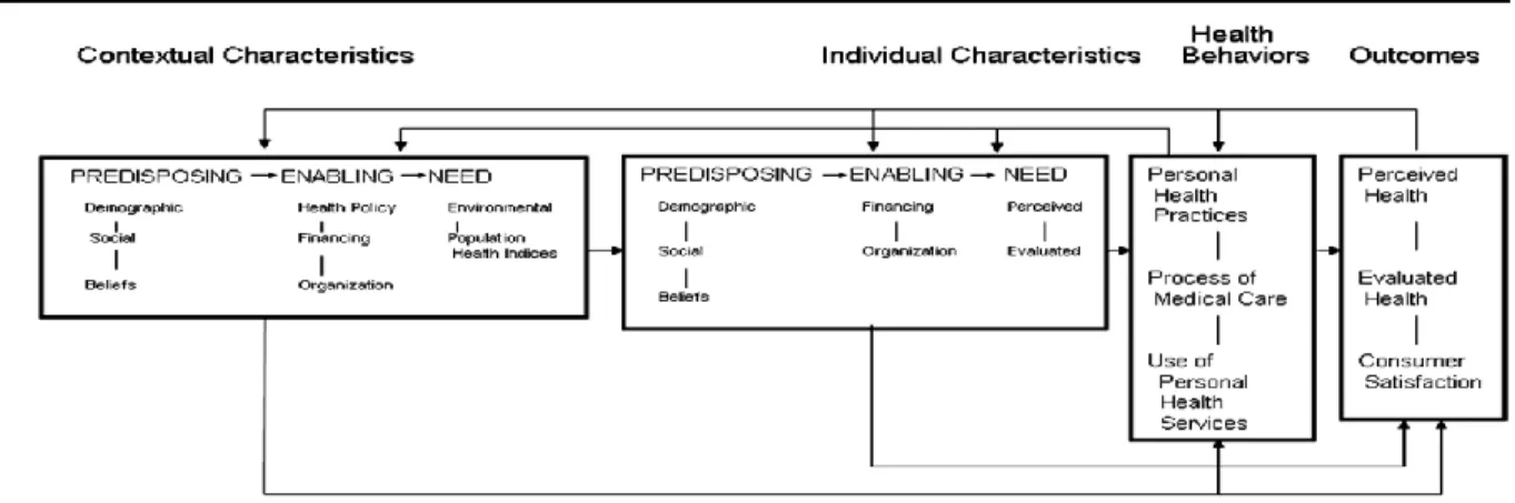
Figure 3. Andersen’s behavioral model of health services use
The most recent version of the model is depicted in Figure 3 and incorporates macro level factors influencing health behavior. Contextual characteristics represent the aggregate health system, organizational, community- and provider- level determinants of health services use.113 With this latest iteration of the model, a different set of variables are assigned to the predisposing, enabling, and need categories, differentiating contextual and individual characteristics. At the aggregate level, contextual characteristics include predisposing factors like community structure, enabling factors like number of medical facilities and need factors like community disability rates that impact individual health services use.
At the individual-level, predisposing characteristics refer to demographic (e.g., age, gender), social structure (e.g., race, education, occupation), and health belief related factors.7 Enabling characteristics at this level include financial and organizational factors such as whether an individual has a regular source of care, income, and whether an individual has health insurance. Need characteristics describe both perceived and evaluated indicators of an individual’s health that include factors such as number of illnesses and mental health status.
Predisposing, enabling, and need population characteristics subsequently determine health behavior, comprising of personal practices, use of health services, and processes of medical care.113 Personal practices include diet, tobacco use, exercise and other self-care activities that affect an individual’s health. Use of health services include doctor and emergency room visits and processes of medical care describe prescriptions, test ordering and other activities that define the interaction between providers and patients.113