TARGETING MUTANT KRAS IN PANCREATIC CANCER
Tikvah Katheryn Hayes
A dissertation submitted to the faculty of the University of North Carolina at Chapel Hill in partial fulfillment of the requirements for the degree of Doctor of Philosophy in the Curriculum in
Genetics and Molecular Biology.
Chapel Hill 2015
Approved by:
Albert S. Baldwin
© 2015
ABSTRACT
Tikvah Katheryn Hayes: Targeting mutant KRAS in pancreatic cancer (Under the direction of Channing Der)
The development of pharmacologic inhibitors of the KRAS oncoprotein, which is mutated
in ~30% of all human cancers, has been at the forefront of drug discovery for the last three
decades. Despite intensive efforts by the pharmaceutical industry, no effective anti-KRAS
strategies have reached the cancer patient. While many approaches to achieve this are being
pursued, arguably inhibition of mutant KRAS effector signaling is considered the most promising to block KRAS-driven cancer growth. The best-validated downstream effector of KRAS is a
three-tiered protein kinase cascade, the Raf-MEK-ERK protein kinase cascade, where KRAS activates
Raf, which then activates MEK, and MEK then activates ERK. Activated ERK then activates a
complex spectrum of signaling events that then drive cancer growth. Unfortunately, inhibitors of
the first two levels, targeting Raf or MEK, have proven ineffective in mutant KRAS cancers. The ineffectiveness of anti-Raf and –MEK therapies has been attributed to inhibitor-induced resistance
mechanisms, where the majority cause reactivation of ERK signaling to bypass the action of these
employed a drug sensitivity screen to identify novel inhibitor combinations that enhanced ERK
inhibitor sensitivity. We identified the PI3K-AKT-mTOR signaling cascade as a potent modulator
of ERK inhibitor sensitivity, which was consistent with our previous finding where concurrent PI3K
inhibition combination enhanced ERK inhibitor sensitivity. We unexpectedly found that long-term
treatment of sensitive cell lines caused cellular senescence, a type of irreversible growth arrest,
mediated in part by causing degradation of Myc and activation of the p16-RB tumor suppressor
pathway. Next, we performed a novel genetic gain-of-function screen to identify mechanisms of
acquired resistance to ERK inhibition. Interestingly, we identified, once again, the PI3K-AKT
signaling cascade, as modulator of ERK inhibitor sensitivity. We also found p38 to be an important
modulator or ERK inhibitor sensitivity. Finally, to investigate de novo resistance to ERK inhibition, we used a loss-of-function screen to identify kinases whose inhibition in combination with ERK
inhibitor treatment resulted in sensitivity. Future studies will be needed to elucidate the
mechanisms behind these modulators of pharmacological ERK inhibition. Collectively, our
findings not only revealed distinct consequences of inhibiting this kinase cascade at the level of
ACKNOWLEDGEMENTS
First, I would like to thank Channing J. Der and the Der Lab members, past and present,
for supporting my scientific development over the last 6 years. Specifically, I would like to
acknowledge Jeran S. Stratford, Timothy D. Martin, Tai-Young Kim, David J. Reiner, and Nicole
F. Neel for guidance and support, without which I would not be the scientist I am today.
Additionally, I would like to acknowledge Kirsten L. Bryant, Samuel D. George, and Swapnil
Kher for their enthusiasm and willingness to collaborate, which significantly improved the Der
Lab work environment during the later half of my graduate studies. I would also like to
recognize members of the Baldwin, Davis, Kim, and Yeh labs for letting me dig through their
freezers and supplies to in order to attain reagents. My extensive network of collaborators really
perpeled by Furthermore, I would like to thank my committee members Albert S. Baldwin,
Christopher M. Counter, Gary L. Johnson, and William Y. Kim for their time, interest, and
insightful discussion.
Next, I would like to acknowledge IMSD (Initiative for Maximizing Student Diversity) for
support me during my graduate studies. As a part of this organization I was fortunate enough to
I would like to acknowledge all the administrative support I received from IMSD, GMB
(Curriculum in Genetics and Molecular Biology), and Lineberger Comprehensive Cancer Center
(LCCC). Each of these organizations made it easier for me to focus solely on my science.
Chapel Hill is home to a vibrant rugby community. The last 5 years I have been
privileged to be part of the University of North Carolina Woman’s Rugby Football Club
(UNCWRFC). There really aren’t words that can accurately capture what the members and this
experience have meant to me, and my development as a person and a professional. I would
like to specifically recognize the members that have been here with me from the beginning:
Johnathan R. Atkeison, Ariel Esperancilla, Cameron Gunn, Brittany N. Lademann, D’Chante
McKenzie, and H. James Morrison. I would also like to acknowledge 2016 four year graduating
seniors: Lindsey J. Broadwell, Nicole R. Davis, Tanya K. Houston, Katherine D. Lutton, Lindsey
C. Oliver, and Naya E. Tapper. I would also like to acknowledge members of the the Chapel Hill
Warriors and Highlanders for filling my summers with touch rugby.
I would not have been successful without the support of my friends outside of UNC
Chapel Hill community. I would like to acknowledge my friends from California, Leanna R.
Corpus, Kristen Driskell, Victoria Gargiso, Nancy H. Godsk, Sarah Harstrom, Emily Ogata, and
Katielee S. Pallis for all their love and support throughout the years. I would also like to
recognize the Raleigh crew, Brie Butler, Kira Cervenka, Erin Gillikin, Kacy L. Hunt, Deanna
Tesch, and Katie Wrigglesworth for our many adventures. Their love and support helped me to
adjust to North Carolina living and made it a place I could call home. Anaiah L. Younger and
Amber B. Cardenas have been my best friends for the better part of two decades, their constant
love and unwavering support has been instrumental through this process.
Finally, I would like to thank my mom, Sarajane Hayes. She truly is the wind beneath
TABLE OF CONTENTS
LIST OF TABLES ... vii
LIST OF FIGURES ... xi
LIST OF ABRREVIATIONS AND SYMBOLS ... xiii
CHAPTER 1: INTRODUCTION ... 1
1.1 RAS genes and Ras proteins ... 1
1.2 Ras and cancer ... 2
1.3 RAS mutations are early events in cancer development and progression ... 3
1.4 RAS mutations in human cancers ... 5
1.5 Ras Effectors ... 6
1.6 The Raf-MEK-ERK kinase cascade ... 7
1.6.1 Raf serine/threonine kinases ... 7
1.6.2 MEK dual specificity kinases ... 9
1.6.3 ERK serine /threonine kinases ... 9
CANCER IS ASSOCIATED WITH MYC DEGRADATION AND SENESCENCE-
LIKE GROWTH SUSPRESSON1 ... 40
INTRODUCTION ... 40
RESULTS ... 41
MEK Inhibitor-Resistant PDAC Cell Lines Are Sensitive to ERK Inhibitor ... 41
ERK Inhibitor Sensitivity Is Not Associated with KRAS Dependency or with K-Ras-dependent Effector Signaling ... 42
Short-term Treatment with SCH772984 Enhances Apoptosis and Alters Cell Cycle Regulation ... 43
Mechanistically Diverse Inhibitor Combinations Synergistically Enhance the Growth Inhibitory Activity of ERK Inhibitor versus MEK Inhibitor ... 45
Long-term ERK Inhibition Causes a Senescence-like Phenotype ... 46
ERK inhibition-induced Degradation of MYC Is Necessary and Sufficient for Induction of Senescence ... 47
SCH772984 Treatment Reduces Tumor Xenograft Growth and MYC Protein Levels in vivo ... 49
Identification of Resistance Mechanisms to Short-term SCH772984 Treatment ... 50
DISCUSSION ... 52
MATERIALS AND METHODS ... 54
CHAPTER 3: DISCUSSION ... 97
3.1 Mutant RAS Dependency ... 98
3.2 ERK inhibitors in mutant Ras cancer ... 103
3.3 Ras effectors and ERK inhibition ... 107
3.5 Resistance mechanisms for ERK inhibition in mutant RAS cancer ... 109
3.5.1 Acquired Resistance Mechanisms ... 109
3.5.2 De Novo Resistance Mechanisms ... 111
3.6 Inhibitor Dosing ... 112
LIST OF TABLES
LIST OF FIGURES
Figure 1.1 Human Ras proteins ... 19
Figure 1.2. Regulation of the Ras GDP-GTP cycle ... 20
Figure 1.3. Ras proteins and membrane association ... 21
Figure 1.4. PDAC genetic progression model ... 22
Figure 1.5. Ras effector signaling ... 24
Figure 1.6. Components of the Raf-MEK-ERK protein kinase cascade ... 25
Figure 1.7. Regulation of Raf dimerization and activation ... 26
Figure 1.8. Receptor tyrosine kinase-mediated activation of wild type Ras ... 27
Figure 1.9. Approaches for the development of anti-Ras drugs ... 28
Figure 1.10. Mechanisms of RAS mutant cancer cell resistance to Raf or MEK inhibitors ... 29
Figure 1.11. Pharmacologic inhibitors of Raf-MEK-ERK under clinical evaluation ... 30
Figure 2.1. PDAC Cell Line Sensitivity to the ERK-Selective Inhibitor SCH772984 Is Not Associated with KRAS Dependency ... 64
Figure 2.2. Related to Figure 2.1 ... 66
Figure 2.8. Long-term SCH772984 Treatment Induces Markers of Senescence
and Ubiquitination in Sensitive but Not Resistant Cell Lines ... 77
Figure 2.9. Related to Figure 2.8 ... 79
Figure 2.10. Induction of Senescence Is Dependent on Proteasomal
Degradation of cMYC ... 81
Figure 2.11. Related to Figure 2.10 ... 84
Figure 2.12. SCH772984 Inhibition of Tumor Growth Is Associated with
Suppression of Myc and Aurora B Abundance ... 86
Figure 2.13. Related to Figure 2.12 ... 88
Figure 2.14. Related to Figure 2.15 ... 91
Figure 2.15. Identification of Protein Kinases That Regulate Resistance of
PDAC Cell Lines to SCH772984 ... 92
Figure 3.1. KRAS stable knockdown leads to changes in phospho-tyrosine,
threonine, and serine ... 115
Figure 3.2. ERK-inhibitor treatment in PDAC cell lines causes
widespread ubiquitination ... 116
Figure 3.4. MYC amplification in cancer ... 118
LIST OF ABRREVIATIONS AND SYMBOLS
A-Raf, A- rapidly accelerated fibrosarcoma
AKT (PKB), protein kinase b
AML, acute myeloid leukemia
ATP, adenosine triphosphate
AURKB, aurora kinase b
B-Raf, B- rapidly accelerated fibrosarcoma
C-terminal, carboxy-terminal
CDKN2A/INK4A (p16), cyclin dependent kinase inhibitor 2A
CFP, cyan fluorescent protein
CK2, casein kinase 2
COSMIC, catalogue of somatic mutations in cancer
COT (Tpl2 or MAP3K8), cancer osaka thyroid
CRD, cysteine-rich domain
CRISPR, clustered regularly interspaced short palindromic repeats
DMBA, 7,12-dimethylbenz[a]anthracene DNAseq, deoxyribonucleic acid sequencing
DSS, drug sensitivity screen
(OX)
FOS, FBJ murine osteosarcoma viral oncogene homolog
FTI, farnesyltransferase inhibitor
GEMM, genetically engineered mouse model
GFP, green fluorescent protein
GGTase, geranylgeranyltransferase-I enzyme
GI50, growth inhibition at 50%
GSK3β, glycogen synthase kinase 3 beta
GDP, guanine diphosphate
GTP, guanine triphosphate
GTPase, guanine triphosphate enzyme
HRAS, Harvey Rat sarcoma
HVR, hypervariable region
ICMT, isoprenylcysteine carboxyl methyltransferase
JAK, janus kinase
JNK, c-Jun N-terminal protein kinase
KPC, K-Ras LSLG12D/+ p53R172H/+ Pdx1:Cre
KRAS, Kirsten Rat sarcoma
K-Ras4A, KRAS splice variant
K-Ras4B, KRAS splice variant
KSR-1, kinase suppressor of Ras 1
MAPK, mitogen activated protein kinase
MAP2K, mitogen activated protein kinase kinase
MAP3K, mitogen activated protein kinase kinase kinase
MEK1/2, mitogen/extracellular signal-regulated kinase
MLK-3 (MAP3K11), mitogen activated protein kinase kinase kinase 11
MNK (ATP7A), menkes
MOS, moloney murine sarcoma
mRNA, messenger ribonucleic acid
M/S, mass spectrometry
MSK, mitogen and stress activated protein kinase
mTOR, mammalian target of rapamycin
MTT, 3-(4,5-dimethylthiazol-2-yl)-2,5-diphenyltetrazolium bromide
MYC, avian myelocytomatosis viral oncogene homolog
NFκB, nuclear factor kappa b
NRAS, Neuroblastoma Rat sarcoma
p21, protein 21
p27, protein 27
p38, protein 38
PAK, p21 associated kinase
PanINs, pancreatic intraepithelial neoplasia
PARP, poly ADP ribose polymerase
PDAC, pancreatic ductal adenocarcinoma
Rac1, Ras-related C3 botulium toxin substrate 1
Raf, rapidly accelerated fibrosarcoma
Raf-1 (c-Raf), rapidly accelerated fibrosarcoma 1
Ral, Ras-related protein
RalGDS, ral guanine nucleotide dissociation stimulator
RAS, Rat Sarcoma
RasGAP, Ras GTP activating protein
RasGEF, Ras guanine exchange factor
RB, retinoblastoma
RBD, Ras binding domain
Rce1, Ras converting CAAX endopeptidase 1
RFP, red fluorescent protein
RNAi, ribonucleic acid interference
RNAseq, ribonucleic acid sequencing
RPPA, reverse phase protein array
RSK, ribosomal S6 kinase
RT-PCR, real-time polymerase chain reaction
RTK, receptor tyrosine kinase
shRNA, short hairpin ribonucleic acid
siRNA, small interfering ribonucleic acid
SMAD4 (DPC4), deleted in pancreatic carcinoma, locus 4
SOS1, Son of Sevenless protein
SRC, rous sarcoma
Tiam1, T-cell lymphoma invasion and metastasis-inducing protein 1
TP53, tumor protein 53
YAP1, yes-associated protein 1
YFP, yellow fluorescent protein
β beta
µ, mu
CHAPTER 1: INTRODUCTION
1.1 RAS genes and Ras proteins
The three human RAS genes encode four highly related 188-189 amino acid 21 kDa small GTPases (Figure 1.1). K-Ras4A and K-Ras4B are splice variants encoded by alternative exon 4
usage and differ primarily at their C-terminal sequences. The RAS genes comprise one the most frequently mutated gene family in human cancer (Cox and Der, 2010). In cancer and various
developmental disorders (referred to as Rasolopaties), RAS genes harbor missense mutations that encode mutant proteins that are altered in their biochemical properties (Prior et al., 2012;
Rauen, 2013).
Ras proteins act as binary switches regulating a number of biological processes that
include cellular proliferation, survival and differentiation. Ras proteins share 90% sequence
identity in the G domain (1-164), which is important for guanine nucleotide binding and GTP
hydrolysis, and interaction with regulators and effectors. In contrast, the C-terminal 24/25 residues
exhibit significant sequence divergence (8% amino acid identity) and is therefore referred to as
the hypervariable region (HVR).
Ras activity is regulated by two distinct classes of regulatory proteins: Ras-selective
guanine exchange factors (RasGEFs) and GTPases activating proteins (RasGAPs) (Figure 1.2)
(Vigil et al., 2010). RasGEFs (e.g., Sos1) accelerate the slow intrinsic guanine nucleotide
exchange rate of Ras proteins. Since the intracellular concentration of GTP is 10 times more
abundant than GDP, this favors formation of the active Ras-GTP complex. Once bound to GTP,
effectors to regulate a diversity of cytoplasmic signaling networks. In order to terminate Ras
signal transduction, RasGAPs (e.g., neurofibromin, p120 RasGAP) bind to Ras and accelerate
its weak intrinsic GTP hydrolysis activity, returning Ras to the inactive GDP-bound form.
However, when Ras proteins are mutated, they display altered intrinsic and GAP-stimulated
GTPase activity, favoring a GTP-bound state and promotion of aberrant signal transduction.
The C-terminal sequence is crucial for Ras membrane association and subcellular
localization (Figure 1.3). The terminal four residues comprise the CAAX motif (C = cyteine, A =
aliphatic amino acid, X = terminal amino acid), which signals for three sequential posttranslational
modifications that increase hydrophobicity and promote membrane association (Ahearn et al.,
2012). The sequences immediately upstream of the CAAX motif contain a second membrane
targeting signal. For H-Ras, K-Ras4A and N-Ras, cysteine residues signal for covalent addition
of a palmitate fatty acid, whereas Ras4B has a polybasic stretch that serves a similar role.
K-Ras4B additionally contains a serine residue (S181) that is phosphorylated by protein kinase C.
This modification regulates a dynamic trafficking between the plasma and endomembranes. In
addition to association with the plasma membrane, Ras proteins are also found in other cellular
endomembrane compartments that include the Golgi, endoplasmic reticulum, mitochondria, and
endosomes.
1.2 Ras and cancer
PDAC is the fourth leading cancer related death in the United States (Siegel et al., 2014).
Among 45,220 individuals diagnosed with PDAC in 2014, approximately 38,000 died of the
disease (Siegel et al., 2014). While the mortality rate for most cancers is declining, PDAC is
projected to become the second most common cause of cancer-related death by 2020. Given
the vague clinical symptoms and the lack of effective screening methods, only 10-20% of PDAC
patients are candidates for curative resection at the time of diagnosis. Even with surgical resection
followed by adjuvant therapy, the median overall survival of those patients is around two years
(Conroy et al., 2011; Neoptolemos et al., 2010; Oettle et al., 2007). Among the rest of the PDAC
patients who are not candidates for resection, half of them have localized disease (borderline
resectable and locally-advanced disease) and half have metastatic disease at the time of
diagnosis. Patients with local disease are most often treated with chemotherapy with or without
radiation, and their median overall survivals are around 15 months (Huguet et al., 2007). In
patients with metastatic PDAC, the prognosis is extremely dismal. With the recent development
of chemotherapy regimens such as FOLFIRINOX (Conroy et al., 2011) and gemcitabine plus
nab-paclitaxel (Von Hoff et al., 2013), the survival of metastatic PDAC has moved beyond 6 months
(Burris et al., 1997), but remains less than one year. At present, targeted therapies have not
provided any meaningful clinical benefit for PDAC patients despite the approval of the EGFR
inhibitor erlotinib (Moore et al., 2007). The poor prognosis associated with PDAC reflects an
urgent need for novel drug development.
1.3 RAS mutations are early events in cancer development and progression
Ras mutations are generally early events in cancer development and progression (Figure
1.4). For pancreatic cancer, KRAS mutations are the initiating genetic event, followed by a progression of mutations in three tumor suppressor genes. The early onset of RAS mutations in cancer emphasizes their key role in promoting the initiation and progression of cancer. This role
mutational activation initiates the early stages of cancer development (Hingorani et al., 2003b).
However, when coupled with additional mutations in tumor suppressors, Kras-initiated tumor formation was accelerated and advanced to invasive and metastatic disease. For example, Kras (G12D) activation alone induced ductal lesions that recapitulated the full spectrum of human
pancreatic intraepithelial neoplasias (PanINs), the putative precursors to invasive pancreatic
cancer (Hingorani et al., 2003b). At low frequency, these lesions also progressed to invasive and
metastatic adenocarcinomas. However, when combined with a mutation in the Tp53 (R172H) tumor suppressor, rapid onset of invasive and widely metastatic carcinoma was seen (Hingorani
et al., 2005). Decreased latency and acceleration of metastatic PDAC was seen when mutational
activation of Kras (G12D) was coupled with the deletion of either of the other two key tumor suppressor lesions in this cancer (CDKN2A/INK4A and SMAD4) (Bardeesy et al., 2006a; Bardeesy et al., 2006b; Waddell et al., 2015).
Despite their clear role in tumor initiation and progression, continued expression of mutant
KRAS is still required for maintenance of the primary and advanced metastatic cancer. For pancreatic cancer, this was first demonstrated by RNA interference suppression of mutant KRAS (G12V) expression in the KRAS mutant Capan-1 human PDAC cell line, causing loss of loss of anchorage-independent growth in vitro and impaired tumorigenic growth in vivo (Brummelkamp
1.4 RAS mutations in human cancers
Data in COSMIC shows that the RAS genes are mutated in 33% of all cancers evaluated, making it the most frequently mutated oncogene family in cancer
(http://cancer.sanger.ac.uk/cancergenome/projects/cosmic/). There are three common sites for
Ras mutational activation, residues G12, G13, and Q61, which together account for >95% of
identified mutations (Cox and Der, 2010). Mutational activation at G12 and G13 interferes with
the ability of Ras to be stimulated by GAPs, as any other residue aside from glycine creates steric
hindrance (Scheffzek et al., 1997). However, mutational activation at Q61 disrupts the
coordination of the water molecule necessary for GTP-hydrolysis (Scheidig et al., 1999). Once
mutated at G12, G13, or Q61 Ras becomes constitutively active, leading to aberrant activation of
downstream effectors. Of the Ras isoforms, KRAS is the most frequently mutated accounting for ~85% of all RAS mutations found in cancer (Cox and Der, 2010).
The frequency of mutation of the three RAS genes varies significantly with KRAS is the most commonly mutated isoform. Mutation data available in COSMIC v68
(http://cancer.sanger.ac.uk/cancergenome/projects/cosmic/) shows that KRAS mutations were
found in 29,557 of 139, 474 unique samples analyzed (21.2%), followed by NRAS, found in 3,587 of 62,609 samples (5.7%), with HRAS mutations relatively rare, found in 1,127 of 35,366 samples (3.2%) (Table 1.1). The simple summation of these data is the basis for the ~30% RAS mutation frequency found in all human cancers. Mutation frequencies from other databases (e.g.,
cBioPortal; (http://www.cbioportal.org/public-portal/), representing a smaller dataset, although
with more accurate data restricted to deep sequencing studies, come up with a reduced
frequency. Since the cancers represented in each database are different, no one source provides
There is preferential mutation of a specific isoform in different cancers, with near exclusive
mutation of KRAS in PDAC. In contrast, NRAS is the predominant isoform mutated in melanoma and acute myelogenous leukemias, whereas HRAS is the predominant isoform mutated in bladder and head and neck squamous cell carcinomas. The majority of missense mutations found
in RAS occur at G12, G13 and Q61. This pattern contrasts with RAS mutations found in developmental disorders. There are also RAS isoform distinct frequencies in these mutations, with Q61 mutations rare in KRAS, yet the predominant mutation seen in NRAS. Finally, the mutation spectrum found in KRAS varies widely in different cancers. For example, the G12C is the predominant mutation found in lung cancer, it is rarely seen in PDAC.
1.5 Ras Effectors
There are at least 11 catalytically-distinct classes of Ras effectors (Vigil et al., 2010). Ras
effectors typically possess either a Ras binding (RBD) or Ras association (RA) domain that
facilitates preferential binding to activated GTP-bound Ras. Of these effector classes, six have
been validated roles in Ras-mediated oncogenesis: Raf serine/threonine kinases, class I
phosphatidylinositol-4,5-bisphosphate 3-kinases (PI3K), and GEFs for the Ral (RalGEFs) and
Rac1 (Tiam1) small GTPases (Figure 1.5) (Cox et al., 2014). Of these, Raf and PI3K are the best
validated, in part, because of their frequent mutational activation in human cancer (Davies et al.,
2002; Karakas et al., 2006). In addition to their mutational prevalence in cancer, both Raf and
1.6 The Raf-MEK-ERK kinase cascade
The Raf-MEK-ERK three-tiered protein kinase cascade is one of three major MAPK kinase
kinase (MAPKKK/MAP3K)- MAPK kinase (MAPKK/MAP2K)-MAPK modules involved in
cytoplasmic signaling downstream of active cell surface receptors. The other two MAPKs, p38
and JNK, have less significant roles in oncogenesis and are not known to be directly activated
downstream of Ras. Therefore, in this thesis, we focus on the Raf-MEK-ERK cascade in Ras
signaling.
1.6.1 Raf serine/threonine kinases
There are three distinct Raf (MAPKKK/MAP3K) isoforms: A-Raf, B-Raf, and Raf-1 (c-Raf).
Like Ras isoforms, Raf isoforms share similar sequence identity and conserved domain topology
(Figure 1.6). The N-terminal region contains an RBD followed by a cysteine-rich domain
(CRD/C1), while the C-terminal region contains the serine/threonine kinase domain. Generally,
N-Ras, H-Ras, and K-Ras activate Raf-1 and B-Raf similarly, whereas A-Raf is only weakly
activated by Ras (Rodriguez-Viciana et al., 2004). Ras-GTP binding to the RBD relieves the
N-terminal auto-inhibitory activity and additionally recruits Raf to the plasma membrane, where
additional phosphorylation events and interactions facilitate activation of Raf catalytic function
(Figure 1.7). How Ras causes activation of Raf is complex and still not completely understood,
with most studies focused on Raf-1, the isoform found originally transduced and activated in the
3611-MSV oncogenic retrovirus that caused rapidly accelerated fibrosarcomas (Matallanas et al., 2011)
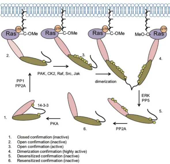
Inactive Raf-1 exists in a closed confirmation stabilized by the 14-3-3 complex interacting
RTKs recruit the Sos1 RasGEF to the plasma membrane where it catalyzes the formation of
Ras-GTP (Figure 1.8). PP1 and PP2A phosphatases dephosphorylate Raf-1 at residue S259
permitting Ras-GTP to bind the Raf-1 RBD effectively resulting in an open yet inactive
confirmation. Several known (PAK, CK2, Raf, Src, and Jak) and unknown kinases phosphorylate
the C-terminal region of Raf-1 at residues S338 and Y341. Once activated, Raf proteins either
homo- or heterodimerize, which is stabilized by either the 14-3-3 complex, KSR-1, or MLK-3, all
well validated scaffolds.
Once Raf-1 is activated, it can phosphorylate its only known physiological downstream
substrates, the closely related MEK1 and MEK2 dual specificity protein kinases. Raf kinases
phosphorylate MEK1 and MEK2 at two sites (S218 and S222), which are located in the activation
loop. Once active MEK1/2 phosphorylate ERK, which also has two distinct isoforms. As a
consequence, the field has relied heavily on changes to levels of phosphorylated ERK1 and ERK2
to determine Ras-Raf signaling activation.
B-Raf activation is similar to Raf-1 activation, though several observations have suggested
that B-Raf activation may require fewer components (Matallanas et al., 2011). Currently, the
model for B-Raf activation is thought to require Ras and 14-3-3 complex interactions for activation.
Unlike Raf-1, B-Raf contains a negatively charged N-terminal domain due to the presence of both
aspartate at the position corresponding to Raf-1 residue Y341 (D448/449) and constitutive
residues found in the N-terminal domain of A-Raf, most important being Y296, which if mutated
results in a constitutively active kinase. This residue, in particular, is thought to stabilize the
N-terminal domain interaction with the catalytic domain promoting a closed kinase confirmation. In
the C-terminal domain, residue S432, located between the ATP-binding motif and the activation
loop, is crucial for both MEK activation and A-Raf signaling. Unlike B-Raf, A-Raf contains a cluster
of phosphorylation sites between residues 248 and 267, which, once activated, contribute to
dissociation from the plasma membrane. Thus, A-Raf signaling has been found at several
subcellular compartments, including the Golgi apparatus, endosomes and mitochondria.
1.6.2 MEK dual specificity kinases
MEK1 and MEK2 (MAP/ERK kinase; MAPKK/MAP2K) are highly related dual specificity
kinases that catalyze the phosphorylation of both threonine and tyrosine residues in the TxY motif
their only known substrates, ERK1 and ERK2 (Roskoski, 2012b)(Figure 6). MEK1/2 structure can
be split into three distinct functional domains: N-terminal domain, protein kinase domain, and a
short C-terminal domain (Fischmann et al., 2009). The N-terminal region consists of an inhibitory
domain, a nuclear export domain, and a domain that aids in the ability to bind the ERK kinases.
The kinase domain comprises the majority of MEK1/2 structure and includes the activation
segment and the proline-rich segment. Raf activates MEK1/2 by dual phosphorylation at tandem
serine residues (Figure 1.6). Two other known activators of MEK1/2 are the COT/Tpl2 and Mos
serine/threonine kinases. Like Raf, Tpl2 and Mos were also identified originally as retroviral
oncogenes and act as MAPKKK/MAP3Ks (Moloney, 1966; Salmeron et al., 1996). The PAK1
serine/threonine kinase can phosphorylate and modulate MEK1 at S298, promoting Raf activation
of MEK (Coles and Shaw, 2002; Slack-Davis et al., 2003).
The only well-established MEK1 and MEK2 substrates are the highly related ERK1 and ERK2
serine/threonine kinases (Figure 1.6). Phosphorylation of the ERK kinases is the most common
readout for Ras activation of the Raf-MEK-ERK signaling cascade (Roskoski, 2012a). Like many
protein kinases ERK1 and ERK2 have short N-terminal and C-terminal domains, with the protein
kinase domain making up the largest region. All known cellular activators of ERK1 and ERK2
lead to phosphorylation and activation of both kinases in parallel (Lefloch et al., 2009). To become
active MEK1/2 phosphorylate ERK1 and ERK2 at residues T202 and Y204, starting with the
tyrosine residue.
Whether ERK1 and ERK2 have unique biological functions has been under evaluation for the
last decade. There is evidence that genetic ablation of Erk2 but not Erk1 causes embryonic lethality (Hatano et al., 2003; Pages et al., 1999; Saba-El-Leil et al., 2003; Yao et al., 2003).
Furthermore, ERK2 but not ERK1 was necessary for H-Ras-induced epithelial-to-mesenchymal
transformation in MCF-10A breast epithelial cells (Shin et al., 2010). RNAi silencing of either
ERK1 or ERK2 impaired the growth of BRAF mutant melanoma cells (Qin et al., 2012). Together these observations suggest that ERK1 and ERK2 have distinct biological functions. Future studies
will be needed to further characterize these distinct biological functions.
Unlike the highly restricted substrates of the Raf and MEK isoforms, the ERK1 and ERK2
(extracellular-signal regulated kinase) kinases are thought to share up to 200 substrates, in both
phosphorylate many nuclear transcription factors that include Ets family transcription factors (e.g.,
Elk-1), Fos and Myc. ERK phosphorylation of Myc at S62 stabilizes Myc and prevents FBW7 E3
ligase-mediated proteasomal degradation (Farrell and Sears, 2014).
1.7 Raf-MEK-ERK target validation in Ras mutant cancers
The Raf-MEK-ERK signaling cascade has been rigorously validated as a necessary effector for Ras transformation (Cuadrado et al., 1993; Khosravi-Far et al., 1995; Khosravi-Far et
al., 1996; White et al., 1995). In early focus formation and clonogenic growth assays, several
laboratories observed that dominant-negative mutants of Raf, MEK, or ERK effectively inhibited
Ras-driven transformation (Cowley et al., 1994; Kolch et al., 1991; Schaap et al., 1993).
Consistent with the importance of Raf-MEK-ERK signaling, it was also demonstrated that the Ras
effector domain mutant T35G, which preferentially impairs Ras-Raf interaction relative to PI3K
and RalGEF, inhibited H-Ras transforming capabilities in NIH 3T3 mouse fibroblasts (White et al.,
1995). Expression of activated Raf-1 could overcome growth inhibition associated with loss of
Ras or expression of Ras dominant negative mutant S17N (Feig and Cooper, 1988). Finally,
genetic loss of all three RAS isoforms causes growth cessation of mouse embryo fibroblasts and only activated Raf (and not PI3K and/or RalGEF) could rescue the growth defect of “Rasless”
cells (Drosten et al., 2010). Activated MEK or ERK could also partially restore growth. Together
these initial observations demonstrated that the Raf-MEK-ERK signal cascade was both
downstream of Ras in mammalian cells and necessary for Ras transformation.
Raf has been validated extensively in human cancer cell lines as a target for therapies
(Hingorani et al., 2003a; Hoeflich et al., 2006; Karasarides et al., 2004; Sharma et al., 2005;
Sumimoto et al., 2006). This is a direct consequence of its mutational activation, as well as,
mutual exclusivity from Ras mutations in cancers (Davies et al., 2002; Karasarides et al., 2004;
Rajagopalan et al., 2002; Sieben et al., 2004; Singer et al., 2003). The non-overlapping
suggests that Raf is likely the most significant downstream effector in these Ras mutant cancers.
This contrasts with activating mutations in PIK3CA (encodes p110α that can occur together with
RAS mutations. Consistent with this observation, several studies have validated the role of Raf downstream of mutant Ras in colorectal, pancreatic and lung tumor cell lines (Campbell et al.,
2007; Li et al., 2013; Subramanian and Yamakawa, 2012). Together these data suggest that
therapies for Ras mutant cancer treatments should be, at least partially, focused on targeting the
Raf-MEK-ERK signaling cascade.
While RNAi use in cell culture is a strong tool for validating the importance of a cancer
target, there are several caveats associated with genetic manipulation and its translation into
actual cancer therapies. First, in vivo RNAi targeting is still under investigation and far from use as an effective therapy. As such, our most effective tools for cancer treatment are still targeted
small molecule inhibitors. Inhibitors, generally, block catalytic function or prevent protein-protein
activation, however RNAi targets the entire protein for depletion, which is vastly distinct from
catalytic or allosteric inhibition. Though RNAi is far from ideal, it still remains a powerful tool for
targeted drug discovery.
Raf has also been sufficiently validated as an in vivo target for mutant Ras-driven cancer initiation and progression. The two-stage chemical carcinogenesis model, where a single
studies observed that Craf but not Braf deficiency impaired Kras G12D-induced lung tumor formation (Blasco et al., 2011; Karreth et al., 2011). However, in contrast to these studies, Craf was found to be dispensable for Kras G12D-induced pancreatic cancer formation (Eser et al., 2013). While a role for Braf was not addressed in this study, the finding by McMahon and colleagues that activated BrafV600E alone could phenocopy activated Kras G12D and induce pancreatic cancer formation suggests that different RAF isoforms may drive KRAS-driven cancer development in different tissues (Collisson et al., 2012).
The studies above provide validation that Raf is necessary for tumor initiation and
progression. However, whether Raf plays a role in mutant Ras tumor maintenance remains
partially answered. Counter and colleagues observed that ERK plays a role in tumor maintenance
of Ras transformed cells, as an inducible dominant-negative MEK prevented continued tumor
growth in a xenograft mouse model (Lim and Counter, 2005). However, their key finding was that
a membrane-targeted, activated, variant of p110α, not c-Raf, was sufficient to maintain
tumorigenic growth of KRAS mutant human colon and pancreatic cancer cell lines when KRAS expression was ablated. This result suggests that PI3K rather than Raf inhibition will be required
to block the KRAS mutant tumor growth.
Use of genetic knockout mouse models where effector function is ablated concurrently
with RAS activation addresses the role of that effector in tumor initiation and progression but not maintenance. Additionally, genetic ablation of an effector, resulting in loss of protein expression,
is not an accurate modeling of the consequences of pharmacologic inhibition of the catalytic
function of the effector. With the development of potent and selective pharmacologic inhibitors of
the Raf-MEK-ERK cascade, the limitations in these studies can be overcome. However, they still
face the limitations of our current mouse models of cancer (Colvin and Scarlett, 2014). Orthotopic
tumors induced by implantation of human tumor cells into immunocompromised mice provide
another model. However, with the obvious importance of the immune system in host response to
response. Genetically engineered mouse models overcome these limitations. However, since
tumor develop is initiated by one or two genetic alterations, they are genetically less complex than
bona fide human cancers.
1.8 Targeting RAS for cancer Treatment
As indicated above, the current therapeutic options for PDAC are limited and ineffective.
With the high frequency of KRAS mutations in this cancer and strong preclinical evidence that disruption of KRAS function will impair cancer growth, the development of effective anti-KRAS
inhibitors has been actively pursued. However, despite more than three decades of intensive
effort by the pharmaceutical industry and academia, to date, no effective therapeutic strategies
have reached the clinic (Bryant et al., 2014; Stephen et al., 2014). In this section, we provide a
summary of past and ongoing efforts to develop anti-Ras therapeutic strategies (Figure 1.9).
As described above, the Ras C-terminal CAAX motif signals for posttranslational
modifications that promote Ras membrane association. That mutation of the cysteine residue to
serine (SAAX) to prevent the addition of the farnesyl isoprenoid lipid or truncation of the AAX
residues results in completely inactive Ras proteins supported the rationale to target
farnesyltransferase as a therapeutic strategy (Berndt et al., 2011). Numerous companies
successfully developed potent and selective farnesyltransferase inhibitors (FTIs), with two
(tipifarnib and lonafarnib) advancing to Phase III clinical evaluation (Basso et al., 2006). Despite
Currently, alternative strategies to disrupt Ras membrane association are being
considered. These include targeting the other two CAAX motification enzymes, ICMT and Rce1
(Figure 1.3). Another approach are farnesyl lipid mimics, salirasib, that act apparently by
competing for Ras membrane association (Bustinza-Linares et al., 2010). More recently,
inhibitors of a chaperone protein, the prenyl-binding protein phosphodiesterase 6 delta that
modulates Ras trafficking to the plasma membrane have been described (Chandra et al., 2012;
Zimmermann et al., 2013).
The most aggressively pursued anti-Ras strategy involves inhibition of Ras downstream
effector signaling. However, these efforts are complicated by the fact that Ras uses multiple
effectors to promote cancer growth (Mitin et al., 2005). Of these effector pathways, the Raf
(A-Raf, B-Raf and C-Raf) and class I phosphatidylinositol-3 kinases (PI3K; p110α, γ and δ) effector
pathways have attracted the greatest interest (Fritsch et al., 2013; Nissan et al., 2013), with
multiple inhibitors of components of each pathway currently under clinical evaluation. That these
effector pathways have driver functions in KRAS-dependent cancer growth is supported by their frequent mutational activation in cancer: BRAF (20%) and PIK3CA (encodes p110α; 12%)
(COSMIC). However, when applied as monotherapies, these inhibitors have shown limited to no
clinical activity in RAS mutant cancers. There are numerous ongoing clinical trials evaluating whether concurrent inhibition of Raf and PI3K effector signaling will be more effective
(http://clinicaltrials.gov/).
An approach once thought impossible involves direct inhibition of mutant Ras. Initial
efforts to disrupt GTP binding were not successful, due to the picomolar affinity of GTP binding to
Ras. This contrasts with the low micromolar binding affinity of ATP to protein kinases, where
effective ATP-competitive protein kinase inhibitors have been developed successfully. Similarly,
efforts to identify small molecules that can act as a GAP for mutant Ras proteins did not succeed.
binding have been described (Maurer et al., 2012; Ostrem et al., 2013; Sun et al., 2012). Whether
these early stage Ras binders can be advanced to more potent and selective Ras binding
molecules and whether they can effectively block the critical functions of Ras to have a clinical
consequence remains to be determined.
Other directions considered for anti-Ras drug discovery include targeting the metabolic
changes in glucose and glutamine metabolism found in RAS mutant cancers (Ahearn et al., 2012; Sun et al., 2012). RNAi targeting of RAS gene expression is also being pursued. Here, whether
these can be effectively delivered to the cancer, and whether sufficient suppression of RAS gene expression can be achieved, are the current uncertainties in these directions. Finally, unbiased
RNA interference screening has been applied to search for synthetic lethal interactors of mutant
RAS. However, these studies have been hampered by the lack of reproducibility in the findings (Luo et al., 2012; Weiwer et al., 2012).
1.8.1 Pharmacologic Inhibition of Raf-MEK-ERK signaling in mutant RAS cancers
Pharmacologic inhibitors of Raf have not been effective against RAS mutant cancers. Their ineffectiveness is due to the paradoxical activation rather than inactivation of ERK signaling
(Figure 1.10A). Studies in cell culture and mouse models determined that Raf inhibitor treatment
caused the formation of B-Raf/C-Raf heterodimers that are dependent on activated Ras (Heidorn
et al., 2010). Ras activation promotes Raf dimerization, primarily B-Raf/C-Raf heterodimers. In
Although developed originally as a C-Raf inhibitor, sorafenib is a multi-kinase inhibitor that also
inhibitors RTKs involved in tumor angiogenesis. Therefore, its clinical activity is not attributed to
its Raf inhibitory activity. A number of additional Raf inhibitors are currently under clinical
evaluation (Figure 1.11). Currently, efforts to develop Raf inhibitors that do not promote Raf
dimerization or have more pan-Raf inhibitory activities, or inhibitors of Raf dimerization, are being
pursued to overcome the limitation of first generation Raf inhibitors.
MEK inhibitors have also shown limited to no anti-tumor efficacy in RAS mutant cancers. For example, Rosen and colleagues found that MEK inhibitor treatment was effective against
BRAF but not RAS mutant human cancer cell lines (Daouti et al., 2010; Solit et al., 2006). MEK inhibition alone was not effective in a mouse model of Kras-driven lung cancer formation (Engelman et al., 2008). The ineffectiveness of MEK inhibition is attributed to the loss of ERK
activation induced feedback inhibitory mechanisms (Figure 1.10B). Flux through the
Raf-MEK-ERK cascade requires critical regulation, with high levels of activated Raf-MEK-ERK causing growth
suppression; ERK activation induces feedback inhibition mechanisms that dampen upstream
activators of the pathway (Figure 6). These mechanisms include ERK phosphorylation of Raf to
dampen Ras activation of Raf (Dougherty et al., 2005). Other feedback mechanisms include ERK
phosphorylation of Sos1 or the EGFR, or transcription factor-mediated induction of gene
expression of negative regulators such as DUSP protein phosphatases (Little et al., 2011; Pratilas
et al., 2009; Wagle et al., 2014) or Sprouty (Roskoski, 2010). In a recent unbiased approach to
define mechanisms that drive resistance to MEK inhibition, Johnson and colleagues showed that
MEK inhibition of KRAS mutant breast cancer cell lines resulted in the activation of multiple RTKs (Duncan et al., 2012). They further showed that concurrent inhibition of RTK activation then
at least 16 additional MEK1/2 inhibitors under clinical evaluation (Figure 8), many also allosteric
non-ATP competitive inhibitors. One unique inhibitor, RO5126766, inhibits MEK and additionally
Raf, making it less susceptible to the feedback activation of Raf caused by ERK inhibition.
With the ineffectiveness of anti-Raf and –MEK therapies in RAS mutant cancers due largely to kinome reprogramming mechanisms that caused reactivation of ERK, it prompted
studies to address whether inhibition of ERK directly may overcome these limitations. Recently,
it was shown that an ERK1/2-selective inhibitor, SCH772984, was active in Raf- and
MEK-resistant BRAF mutant melanoma in preclinical models (Morris et al., 2013). SCH772984 binds ERK, preventing MEK phosphorylation and activation of ERK and additionally preventing ATP
binding and ERK phosphorylation of its substrates. Additionally, another group described another
ERK inhibitor capable of overcoming resistance to MEK inhibitors (Hatzivassiliou et al., 2012).
Furthermore, Genetech also produced an ERK inhibitor that is currently being evaluated in
pre-clinical models.
ERK inhibition represents a new approach to blocking an old pathway; however, the
question remains whether ERK inhibition as a therapy will be successful in combating RAS mutant cancers, or succumb to some of the limitations associated with Raf and MEK inhibition. Currently,
three ERK inhibitors are under clinical evaluation (MK-8353/SCH 900353 is an orally available
Figure 1.1
Figure 1.2.
Figure 1.2. Regulation of the Ras GDP-GTP cycle. Ras proteins act as molecular switches alternating between GTP- (active state) and GDP- (inactive state) bound states, where Ras-GTP binds preferentially to downstream effectors (E). There are two classes of regulatory proteins that regulate this cycling process: RasGEFs (guanine exchange factors) and RasGAPs (GTPase activating proteins). In resting cells, normal Ras is predominantly GDP-bound (~95%). Upon growth factor stimulation and activation of RasGEF, rapid and transient GDP-GTP exchange is stimulated. RasGAP stimulation of the intrinsic GTPase activity and GTP hydrolysis restores the inactive Ras-GDP state. Mutant Ras proteins are impaired in their intrinsic and GAP-stimulated GTP hydrolysis activities, resulting in stimulus-independent, persistent Ras-GTP formation (~80%).
JKL
JML
L
7#$%JGN
#$%J,L
GOO15<>9%
#$%
#$%
JKL JML
Figure 1.3.
Figure 1.3. Ras proteins and membrane association. Ras proteins are synthesized initially as cytosolic and inactive proteins. Within minutes, they undergo a series of posttranslational modifications signaled by the C-terminal CAAX motif. First, cytosolic farnesyltransferase (FTase) catalyzes covalent, irreversible addition of a C15 isoprenoid lipid to the cysteine residue of the C-terminal CAAX motif. This then allows Rce1-catalyzed proteolytic removal of the AAX residues and Icmt-catalyzed, reversible carboxylmethylation (-OMe) of the now terminal farnesylated cysteine. H-Ras is the only Ras isoform that is solely modified by FTase. Although normally also FTase substrates, when FTase activity is blocked by FTase inhibitor (FTI) treatment, K-Ras and N-Ras, can now be modified by geranylgeranyltransferase-I (GGTase-I)-catalyzed addition of a related C20 geranylgeranyl isoprenoid, resulting in membrane association.
#$%
NK$%1
#51&
F5@<
"R,,S
#$% "R,,S
"R"TU1
#$% "R,,S
#$%
NKF
/0R&H0O$941%V;07%>W914>76
*"#$% "R"TU1
JJK$%1"F
#51&
F5@<
Table 1.1.
Table 1.1. Frequency of RAS mutations in human cancers
K$[;10&d&00N9123145V0>O0"#$@3<$<7>4%0740Y3@$405$4519%$
Figure 1.5.
Figure 1.5. Ras effector signaling. Ras-GTP binds preferentially to 11 catalytically-distinct classes of effectors. Cell culture and/or mouse model studies have been implicated six classes in Ras-mediated tumor initiation, progression and/or maintenance. This includes the Raf, p110 catalytic subunits of class I PI3Ks, GEFs for the Ral small GTPases (RalGEFs; RalGDS, Rgl, Rgl2 and Rgl3) and Rac small GTPase (Tiam1) and PLCe whose functions are necessary for tumor growth. In contrast, RASSF1A family members are negative regulators and their expression is lost in cancer.
#$%
#$O
LFI* K7$@& #$;JGN
LM*& ,*K #$5 L,* UG* G#* #$; K.*& R1;;0%39878$; R1;;0:9>X<Y K9$4%597W<7>4 RV<>%Z1;1<>4 R1;;0@7:9$<7>4 R1;;0:9>X<Y R1;;05V5;10 K9$4%597W<7>4 G46>5V<>%7% U1<$[>;7%@ L\R! M,J L*R
R$D]%7:4$;74:
Figure 1.6.
Figure 1.6. Components of the Raf-MEK-ERK protein kinase cascade. Shown are the human proteins, with domain structure determined in SMART. The percentages of overall protein and kinase domain amino acid identities are indicated (%) and were determined by ClustalW multiple sequence alignment analyses. The phosphorylation sites that regulate Raf kinase activity are complex and include both positive (green) and negative (red) phosphorylation events. We have not included all known phosphorylation sites and have included only select key phosphorylation sites. The negative regulatory sites are conserved in all Raf isoforms and serve as recognition sites for 14-3-3 dimer binding and inhibition of Raf. Phosphorylation of S432(A-Raf)/S579(B-Raf)/S471(C-Raf) is important for Raf interaction with MEK. Raf phosphorylation sites in MEK1/2 and MEK1/2 phosphorylation sites in ERK1/2 are indicated. The V600E amino acid substitution comprises ~80% of cancer-associated activating mutations in B-Raf. Abbreviations are: RBD, Ras-binding domain, CRD, cysteine-rich domain; S/T, serine/threonine; S/T/Y, serine/threonine/tyrosine. Figure 1.7.
R"#$O
C+'."#$O
^CC E_K0*74$%1UG*&
I(IUG*D
G#*&
I^(G#*D
ICA KD AD K& 'H ED &' #.M,"#$O
& CAC#.M E_K_`0*74$%1 & & L L & E_K0*74$%1 +AA ED DD E_K0*74$%1 L L & E_K_`0*74$%1 L L L L & & E_K0*74$%1 'A_'^B 'C_''B H'_^^B HH_'&B R#M
R#M L L
Figure 1.8.
Figure 1.8. Receptor tyrosine kinase-mediated activation of wild type Ras. Wild-type Ras activation occurs when ligands (e.g., epidermal growth factor; EGF) stimulate activation of receptor tyrosine kinases (RTK; e.g., EGF receptor). Once stimulated, RTKs autophosphorylate tyrosine residues in the cytoplasmic domain, creating docking sites for Src homology 2 (SH2) domain-containing proteins (e.g., Grb2). The tandem SH2 domains of Grb2 interact with proline-rich sequences in the Sos1 RasGEF, promoting Sos1 translocation to the plasma membrane, leading to activation of membrane-associated Ras. Sos1-mediated formation of Ras-GTP then promotes Ras association with Raf, leading to activation of the ERK MAPK cascade. Shown here is KSR association with Raf, MEK and ERK. KSR is but one of a number of scaffolding proteins that associate with one or more components of the three-tiered protein kinase cascade. Scaffolds modulate the composition of the pathway and additional influence temporal and spatial activity of ERK signaling. Other ERK scaffolds include IQGAP1, MP1, Sef and b-arrestin
.
#$%
JKL"R"TU1
#$%
JML"R"TU1
J9[D
E>%&
L
L L
L L
L L
L
435;1$90<$9:1<%
5V<>%>;750<$9:1<%
#$O
UG*
G#*
*E#
L
L L
Figure 1.9.
Figure 1.10.
Figure 1.10. Mechanisms of RAS mutant cancer cell resistance to Raf or MEK inhibitors. (A). RAS mutant cancer cells exhibit paradoxical activation rather than inactivation of ERK signaling. (B) RAS mutant cancer cells exhibit multiple mechanisms of resistance to MEK inhibition. The mechanisms that relate to RTK and Sos activation of Ras are not expected to be relevant for mutant (*) Ras activity, since it is already persistently GTP-bound due to GAP insensitivity. However, since there is evidence that wild type (WT) Ras proteins support mutant Ras in cancer growth, these feedback mechanisms are then still important even in RAS mutant cancer cells. #$% JKL "R"TU1 ."#$O L a1@39$O147[ M$[9$O147[ , R"#$O L UG* L G#* L #$% JKL "R"TU1 #$O UG* L
L 1 3@1 747[
Figure 1.11.
Figure 1.11. Pharmacologic inhibitors of Raf-MEK-ERK under clinical evaluation. Compiled from ClinicalTrials.gov. Past and/or ongoing approaches for targeting Ras include direct Ras binders and inhibitors of Ras function and inhibition of Ras membrane association. Functional si/shRNA library screens have been applied to identify genes (x), that when silenced, impair the growth of RAS mutant but not wild type tumor cell lines (aka synthetic lethal interactors of mutant Ras). *FDA-approved for the treatment of renal cell, hepatocellular and thyroid carcinoma; +
FDA-approved for the treatment of BRAF-mutant melanoma.
#$O UG* G#* M7915<0#$%074Y7[7<>9% U1@[9$410$%%>57$<7>4 EV4<Y1<75074<19$5<>9 ,##`"IAA ,E^AI('' ,jM'IIA_,##`"+D+^A+ R>[7@1<747[_JMR"A(^I_S\H&'_#J^+D& GCDA& JMR"ACDI_#J^+DA UG*&CD_,##`"+I'&CD LM"AIDH(A& L7@$%19<7[_,E^AIADC_UER&(ICIC(. #1O$@1<747[_.,`'C"(^CC_#MG,&&( #T+('^CHH_R!+('^CHH E1;3@1<747[_,jMCD++_,##`"&+D''C K,*"^II K9$@1<747[_JE*&&DAD&D] kS"HH+ ,#l0^IC .J."D'I .UE"(A'CCD_S\D'& M$[9$O147[_JE*D&&'+IC] \JS'&' \`IAA(&DA U\)D+'A #,NDCH_R!F#"DCH #1:>9$O147[ E>9$O147[_.,`0+I"(AAC -a1@39$O147[_L\S+AID] #TH&DC^CC_R!H&DC^CC000 .aM"HDI JMRmA((+ U*"'IHI_ER!0(AAIHI000000
#$%JKL"R"TU1
S
L
L
REFERENCES
Ahearn, I.M., Haigis, K., Bar-Sagi, D., and Philips, M.R. (2012). Regulating the regulator: post-translational modification of RAS. Nat Rev Mol Cell Biol 13, 39-51.
Bai, Y., Edamatsu, H., Maeda, S., Saito, H., Suzuki, N., Satoh, T., and Kataoka, T. (2004). Crucial role of phospholipase Cepsilon in chemical carcinogen-induced skin tumor development. Cancer research 64, 8808-8810.
Bardeesy, N., Aguirre, A.J., Chu, G.C., Cheng, K.H., Lopez, L.V., Hezel, A.F., Feng, B., Brennan, C., Weissleder, R., Mahmood, U., et al. (2006a). Both p16(Ink4a) and the p19(Arf)-p53 pathway constrain progression of pancreatic adenocarcinoma in the mouse. Proc Natl Acad Sci U S A 103, 5947-5952.
Bardeesy, N., Cheng, K.H., Berger, J.H., Chu, G.C., Pahler, J., Olson, P., Hezel, A.F., Horner, J., Lauwers, G.Y., Hanahan, D., et al. (2006b). Smad4 is dispensable for normal pancreas development yet critical in progression and tumor biology of pancreas cancer. Genes Dev 20, 3130-3146.
Bass, A.J., Lawrence, M.S., Brace, L.E., Ramos, A.H., Drier, Y., Cibulskis, K., Sougnez, C., Voet, D., Saksena, G., Sivachenko, A., et al. (2011). Genomic sequencing of colorectal adenocarcinomas identifies a recurrent VTI1A-TCF7L2 fusion. Nat Genet 43, 964-968. Basso, A.D., Kirschmeier, P., and Bishop, W.R. (2006). Lipid posttranslational modifications. Farnesyl transferase inhibitors. J Lipid Res 47, 15-31.
Berndt, N., Hamilton, A.D., and Sebti, S.M. (2011). Targeting protein prenylation for cancer therapy. Nature reviews Cancer 11, 775-791.
Biankin, A.V., Waddell, N., Kassahn, K.S., Gingras, M.C., Muthuswamy, L.B., Johns, A.L., Miller, D.K., Wilson, P.J., Patch, A.M., Wu, J., et al. (2012). Pancreatic cancer genomes reveal aberrations in axon guidance pathway genes. Nature 491, 399-405.
Blasco, R.B., Francoz, S., Santamaria, D., Canamero, M., Dubus, P., Charron, J., Baccarini, M., and Barbacid, M. (2011). c-Raf, but not B-Raf, is essential for development of K-Ras oncogene-driven non-small cell lung carcinoma. Cancer cell 19, 652-663.
Brummelkamp, T.R., Bernards, R., and Agami, R. (2002). Stable suppression of tumorigenicity by virus-mediated RNA interference. Cancer Cell 2, 243-247.
Bryant, K.L., Mancias, J.D., Kimmelman, A.C., and Der, C.J. (2014). KRAS: feeding pancreatic cancer proliferation. Trends Biochem Sci 39, 91-100.
Burris, H.A., 3rd, Moore, M.J., Andersen, J., Green, M.R., Rothenberg, M.L., Modiano, M.R., Cripps, M.C., Portenoy, R.K., Storniolo, A.M., Tarassoff, P., et al. (1997). Improvements in survival and clinical benefit with gemcitabine as first-line therapy for patients with advanced pancreas cancer: a randomized trial. J Clin Oncol 15, 2403-2413.
Campbell, P.M., Groehler, A.L., Lee, K.M., Ouellette, M.M., Khazak, V., and Der, C.J. (2007). K-Ras promotes growth transformation and invasion of immortalized human pancreatic cells by Raf and phosphatidylinositol 3-kinase signaling. Cancer research 67, 2098-2106.
Chandra, A., Grecco, H.E., Pisupati, V., Perera, D., Cassidy, L., Skoulidis, F., Ismail, S.A., Hedberg, C., Hanzal-Bayer, M., Venkitaraman, A.R., et al. (2012). The GDI-like solubilizing factor PDEdelta sustains the spatial organization and signalling of Ras family proteins. Nat Cell Biol 14, 148-158.
Coles, L.C., and Shaw, P.E. (2002). PAK1 primes MEK1 for phosphorylation by Raf-1 kinase during cross-cascade activation of the ERK pathway. Oncogene 21, 2236-2244.
Collins, M.A., Bednar, F., Zhang, Y., Brisset, J.C., Galban, S., Galban, C.J., Rakshit, S., Flannagan, K.S., Adsay, N.V., and Pasca di Magliano, M. (2012a). Oncogenic Kras is required for both the initiation and maintenance of pancreatic cancer in mice. J Clin Invest 122, 639-653. Collins, M.A., Brisset, J.C., Zhang, Y., Bednar, F., Pierre, J., Heist, K.A., Galban, C.J., Galban, S., and di Magliano, M.P. (2012b). Metastatic pancreatic cancer is dependent on oncogenic Kras in mice. PLoS One 7, e49707.
Collisson, E.A., Trejo, C.L., Silva, J.M., Gu, S., Korkola, J.E., Heiser, L.M., Charles, R.P., Rabinovich, B.A., Hann, B., Dankort, D., et al. (2012). A central role for RAF-->MEK-->ERK signaling in the genesis of pancreatic ductal adenocarcinoma. Cancer Discov 2, 685-693. Colvin, E.K., and Scarlett, C.J. (2014). A Historical Perspective of Pancreatic Cancer Mouse Models. Semin Cell Dev Biol.
Conroy, T., Desseigne, F., Ychou, M., Bouche, O., Guimbaud, R., Becouarn, Y., Adenis, A., Raoul, J.L., Gourgou-Bourgade, S., de la Fouchardiere, C., et al. (2011). FOLFIRINOX versus gemcitabine for metastatic pancreatic cancer. N Engl J Med 364, 1817-1825.
Cowley, S., Paterson, H., Kemp, P., and Marshall, C.J. (1994). Activation of MAP kinase kinase is necessary and sufficient for PC12 differentiation and for transformation of NIH 3T3 cells. Cell 77, 841-852.
Davies, H., Bignell, G.R., Cox, C., Stephens, P., Edkins, S., Clegg, S., Teague, J., Woffendin, H., Garnett, M.J., Bottomley, W., et al. (2002). Mutations of the BRAF gene in human cancer. Nature 417, 949-954.
Dougherty, M.K., Muller, J., Ritt, D.A., Zhou, M., Zhou, X.Z., Copeland, T.D., Conrads, T.P., Veenstra, T.D., Lu, K.P., and Morrison, D.K. (2005). Regulation of Raf-1 by direct feedback phosphorylation. Molecular cell 17, 215-224.
Drosten, M., Dhawahir, A., Sum, E.Y., Urosevic, J., Lechuga, C.G., Esteban, L.M., Castellano, E., Guerra, C., Santos, E., and Barbacid, M. (2010). Genetic analysis of Ras signalling
pathways in cell proliferation, migration and survival. The EMBO journal 29, 1091-1104. Duncan, J.S., Whittle, M.C., Nakamura, K., Abell, A.N., Midland, A.A., Zawistowski, J.S.,
Johnson, N.L., Granger, D.A., Jordan, N.V., Darr, D.B., et al. (2012). Dynamic reprogramming of the kinome in response to targeted MEK inhibition in triple-negative breast cancer. Cell 149, 307-321.
Ehrenreiter, K., Kern, F., Velamoor, V., Meissl, K., Galabova-Kovacs, G., Sibilia, M., and Baccarini, M. (2009). Raf-1 addiction in Ras-induced skin carcinogenesis. Cancer cell 16, 149-160.
Engelman, J.A., Chen, L., Tan, X., Crosby, K., Guimaraes, A.R., Upadhyay, R., Maira, M., McNamara, K., Perera, S.A., Song, Y., et al. (2008). Effective use of PI3K and MEK inhibitors to treat mutant Kras G12D and PIK3CA H1047R murine lung cancers. Nat Med 14, 1351-1356. Eser, S., Reiff, N., Messer, M., Seidler, B., Gottschalk, K., Dobler, M., Hieber, M., Arbeiter, A., Klein, S., Kong, B., et al. (2013). Selective requirement of PI3K/PDK1 signaling for Kras oncogene-driven pancreatic cell plasticity and cancer. Cancer cell 23, 406-420.
Farrell, A.S., and Sears, R.C. (2014). MYC Degradation. Cold Spring Harb Perspect Med 4. Feig, L.A., and Cooper, G.M. (1988). Inhibition of NIH 3T3 cell proliferation by a mutant ras protein with preferential affinity for GDP. Mol Cell Biol 8, 3235-3243.
Fischmann, T.O., Smith, C.K., Mayhood, T.W., Myers, J.E., Reichert, P., Mannarino, A., Carr, D., Zhu, H., Wong, J., Yang, R.S., et al. (2009). Crystal structures of MEK1 binary and ternary complexes with nucleotides and inhibitors. Biochemistry 48, 2661-2674.
Flaherty, K.T., Puzanov, I., Kim, K.B., Ribas, A., McArthur, G.A., Sosman, J.A., O'Dwyer, P.J., Lee, R.J., Grippo, J.F., Nolop, K., et al. (2010). Inhibition of mutated, activated BRAF in metastatic melanoma. N Engl J Med 363, 809-819.
Fritsch, R., de Krijger, I., Fritsch, K., George, R., Reason, B., Kumar, M.S., Diefenbacher, M., Stamp, G., and Downward, J. (2013). RAS and RHO families of GTPases directly regulate distinct phosphoinositide 3-kinase isoforms. Cell 153, 1050-1063.
Hatano, N., Mori, Y., Oh-hora, M., Kosugi, A., Fujikawa, T., Nakai, N., Niwa, H., Miyazaki, J., Hamaoka, T., and Ogata, M. (2003). Essential role for ERK2 mitogen-activated protein kinase in placental development. Genes Cells 8, 847-856.
Hatzivassiliou, G., Liu, B., O'Brien, C., Spoerke, J.M., Hoeflich, K.P., Haverty, P.M., Soriano, R., Forrest, W.F., Heldens, S., Chen, H., et al. (2012). ERK inhibition overcomes acquired
resistance to MEK inhibitors. Mol Cancer Ther 11, 1143-1154.
Hauge, C., and Frodin, M. (2006). RSK and MSK in MAP kinase signalling. J Cell Sci 119, 3021-3023.
Heidorn, S.J., Milagre, C., Whittaker, S., Nourry, A., Niculescu-Duvas, I., Dhomen, N., Hussain, J., Reis-Filho, J.S., Springer, C.J., Pritchard, C., et al. (2010). Kinase-dead BRAF and
oncogenic RAS cooperate to drive tumor progression through CRAF. Cell 140, 209-221. Hingorani, S.R., Jacobetz, M.A., Robertson, G.P., Herlyn, M., and Tuveson, D.A. (2003a). Suppression of BRAF(V599E) in human melanoma abrogates transformation. Cancer Res 63, 5198-5202.
Hingorani, S.R., Petricoin, E.F., Maitra, A., Rajapakse, V., King, C., Jacobetz, M.A., Ross, S., Conrads, T.P., Veenstra, T.D., Hitt, B.A., et al. (2003b). Preinvasive and invasive ductal pancreatic cancer and its early detection in the mouse. Cancer Cell 4, 437-450.
Hingorani, S.R., Wang, L., Multani, A.S., Combs, C., Deramaudt, T.B., Hruban, R.H., Rustgi, A.K., Chang, S., and Tuveson, D.A. (2005). Trp53R172H and KrasG12D cooperate to promote chromosomal instability and widely metastatic pancreatic ductal adenocarcinoma in mice. Cancer Cell 7, 469-483.
Hoeflich, K.P., Gray, D.C., Eby, M.T., Tien, J.Y., Wong, L., Bower, J., Gogineni, A., Zha, J., Cole, M.J., Stern, H.M., et al. (2006). Oncogenic BRAF is required for tumor growth and maintenance in melanoma models. Cancer research 66, 999-1006.
Hou, J., Lam, F., Proud, C., and Wang, S. (2012). Targeting Mnks for cancer therapy. Oncotarget 3, 118-131.
Karreth, F.A., Frese, K.K., DeNicola, G.M., Baccarini, M., and Tuveson, D.A. (2011). C-Raf is required for the initiation of lung cancer by K-Ras(G12D). Cancer Discov 1, 128-136.
Khosravi-Far, R., Solski, P.A., Clark, G.J., Kinch, M.S., and Der, C.J. (1995). Activation of Rac1, RhoA, and mitogen-activated protein kinases is required for Ras transformation. Mol Cell Biol 15, 6443-6453.
Khosravi-Far, R., White, M.A., Westwick, J.K., Solski, P.A., Chrzanowska-Wodnicka, M., Van Aelst, L., Wigler, M.H., and Der, C.J. (1996). Oncogenic Ras activation of Raf/mitogen-activated protein kinase-independent pathways is sufficient to cause tumorigenic transformation. Mol Cell Biol 16, 3923-3933.
Kolch, W., Heidecker, G., Lloyd, P., and Rapp, U.R. (1991). Raf-1 protein kinase is required for growth of induced NIH/3T3 cells. Nature 349, 426-428.
Lefloch, R., Pouyssegur, J., and Lenormand, P. (2009). Total ERK1/2 activity regulates cell proliferation. Cell Cycle 8, 705-711.
Li, Y., Takahashi, M., and Stork, P.J. (2013). Ras-mutant cancer cells display B-Raf binding to Ras that activates extracellular signal-regulated kinase and is inhibited by protein kinase A phosphorylation. The Journal of biological chemistry 288, 27646-27657.
Lim, K.H., and Counter, C.M. (2005). Reduction in the requirement of oncogenic Ras signaling to activation of PI3K/AKT pathway during tumor maintenance. Cancer Cell 8, 381-392.
Little, A.S., Balmanno, K., Sale, M.J., Newman, S., Dry, J.R., Hampson, M., Edwards, P.A., Smith, P.D., and Cook, S.J. (2011). Amplification of the driving oncogene, KRAS or BRAF, underpins acquired resistance to MEK1/2 inhibitors in colorectal cancer cells. Sci Signal 4, ra17. Luo, T., Masson, K., Jaffe, J.D., Silkworth, W., Ross, N.T., Scherer, C.A., Scholl, C., Frohling, S., Carr, S.A., Stern, A.M., et al. (2012). STK33 kinase inhibitor BRD-8899 has no effect on KRAS-dependent cancer cell viability. Proceedings of the National Academy of Sciences of the United States of America 109, 2860-2865.
Malliri, A., van der Kammen, R.A., Clark, K., van der Valk, M., Michiels, F., and Collard, J.G. (2002). Mice deficient in the Rac activator Tiam1 are resistant to Ras-induced skin tumours. Nature 417, 867-871.
Matallanas, D., Birtwistle, M., Romano, D., Zebisch, A., Rauch, J., von Kriegsheim, A., and Kolch, W. (2011). Raf family kinases: old dogs have learned new tricks. Genes Cancer 2, 232-260.
Maurer, T., Garrenton, L.S., Oh, A., Pitts, K., Anderson, D.J., Skelton, N.J., Fauber, B.P., Pan, B., Malek, S., Stokoe, D., et al. (2012). Small-molecule ligands bind to a distinct pocket in Ras and inhibit SOS-mediated nucleotide exchange activity. Proceedings of the National Academy of Sciences of the United States of America.
![cis {trans 1,2 Bis[2 (diphenylphosphino)benzamido]cyclohexane κ2P,P′}dichloroplatinum(II) chloroform trisolvate](data:image/gif;base64,R0lGODlhAQABAIAAAP///wAAACH5BAEAAAAALAAAAAABAAEAAAICRAEAOw==)