
Cover Page
The handle http://hdl.handle.net/1887/37294 holds various files of this Leiden University dissertation
Author: Melo Bernardo, Ana de
Primordial Germ Cells and Amnion
Development in the Avian Embryo
Primordial Germ Cells and Amnion Development in the Avian Embryo © Ana de Melo Bernardo, 2015, Leiden, The Netherlands. All rights reserved.
No parts of this thesis may be reproduced, distributed, stored in retrieval system or transmitted in any form or by any mean, without permission of the author.
ISBN: 978-94-6295409-0
Published by: Uitgeverij BOXPress, ‘s-Hertogenbosch
Cover and chapter photography by: Van Vincent
Primordial Germ Cells and Amnion
Development in the Avian Embryo
Proefschrift
ter verkrijging van de graad van Doctor aan de Universiteit Leiden, op gezag van Rector Magnificus prof.mr. C.J.J.M. Stolker,
volgens besluit van het College voor Promoties te verdedigen op dinsdag 26 januari 2016
klokke 16:15 uur
door
Ana de Melo Bernardo geboren te Covilhã, Portugal
Promotor Prof. Dr. C. L. Mummery
Co-promotor Dr. S. M. Chuva de Sousa Lopes
Overige leden Prof. Dr. M.J. Goumans
Prof. Dr. M. de Ruiter
Principles for the Development of a Complete Mind: Study the science of art. Study the art of science. Develop your senses - especially learn how to see. Realize that everything connects to everything else.
CONTENTS
Chapter 1
General Introduction and Outline of this Thesis
Chapter 2
Chicken Primordial Germ Cells Use the Anterior Vitelline Veins to Enter the Embryonic Circulation
Chapter 3
Meiotic Wave Adds Extra Asymmetry to the Development of Female
Chicken Gonads
Chapter 4
Avian as a Model for Human Ovarian Cancer: Advantages Reviewed
Chapter 5
The Involvement of the Proamnion in the Development of the Anterior Amnion Fold in Chicken
Chapter 6
General Discussion and Future Perspectives
Appendix
Summary
Samenvatting List of Peer-reviewed Publications About the Author
Acknowledgements
127
10
26
46
72
94
112
1
THE “IMMORTAL” CYCLE OF THE GERMLINE
The question of how humans and other organisms originate, has fascinated scientists and society throughout history. In 1651, the English physician William Harvey claimed “ex ovo Omnia” - all life originates from an egg – suggesting the importance of the egg in the origin of all living animals, and refuting the idea of spontaneous generation [1]. A few years later, Antoni van Leeuwenhoek was able to build a microscope that allowed him to observe spermatozoa [2]. Van Leeuwenhoek would become the first scientist to suggest that the spermatozoid penetrates the egg in a process that we now call fertilization. His ideas contradicted Harvey’s and the then common belief that fertilization occurred due to “vapors” originating in the seminal fluid. Nevertheless, Van Leeuwenhoek was never able to observe the spermatozoid penetrating the egg under his microscopes [3]. Only towards the end of the next century, in 1891, did Oscar Hertwig observe the process of fertilization in the sea urchin [4]. Even now, after almost 400 years of research, we continue to unravel the most fascinating mechanisms underlying the gametes and their progenitors: the primordial germ cells (PGCs).
Different animal models have been used to study different aspects of germline biology, ranging from flies, frogs and fish to birds and mammals. This dissertation focuses on PGC migration as they move to the gonads (Chapter 2) and meiosis (Chapter 3) in the chicken embryo.
ON THE ORIGIN OF PGCs: EPIGENESIS AND PREFORMATION Due to the importance of PGCs in development and evolution, the mechanisms regulating germline specification are strictly regulated during development. PGCs are specified early in development, a processed defined by the segregation of the germline from the somatic line [8]. Although the germline represents a major evolutionary step that enabled sexual reproduction in the metazoan species, PGC specification mechanisms are not conserved. Two main mechanisms are responsible for the development of PGCs in vertebrates: epigenesis and preformation (reviewed in [13]).
Preformation.In the late nineteenth century August Weissman introduced the concepts of germline and soma. He suggested that the oocyte contained germ plasm from the mother and that this plasm was responsible for the specification of germ cells after fertilization [14]. Robert Hegner was the first to identify germ granules, in which the germ plasm is transported, in the germ cells of beetles. They introduced the first concepts of “preformation”, the process by which germ cell precursors are defined by maternal factors contained in the egg [8]. This was later shown to be responsible for the development of germ cells in C. elegans, D. melanogaster, D. renio, X. laevis and G. gallus [15].
the basal part of the first embryonic cleavage [22].
In chicken embryos, two systems are used to define developmental stages: Eyal-Giladi & Kochav´s staging is commonly used from the first cleavage (stage I) until pre-primitive streak stage (stage X) [23]; and Hamburger and Hamilton staging that considers chicken development from early primitive streak (stage HH2) until just before hatching (stage HH45) [24]. Both systems consider morphological landmarks of the chicken embryo. The first embryonic cleavage is therefore defined by Eyal-Giladi & Kochav system as stage I [23]. Following the expression of CVH, after the first embryonic cleavages, it was shown that CVH remained in 6-8 cells located in the center of the blastocyst at stage V. Moreover, in vitro experiments have shown that inducing vasa overexpression in chicken stem cells (cSC) upregulates the expression of other germline markers, such as TUDOR, SDF1, CXCR4 and DAZZL [25]. Interestingly, these same cells, induced in vitro, were able to migrate to the embryonic gonad [25]. Although this seems to be evidence for the origin of PGCs by preformation in the chicken, functional studies are still necessary to prove this. However, this is presently not possible to do due to the lack of molecular and genetic tools for the chicken.
signaling cues necessary to produce eggs from mouse embryonic stem cells (mESC) and mouse-induced pluripotent stem cells (mIPSC) [36, 37]. Although there are differences between mouse and human PGCs, studies in mice have been fundamental in bringing us closer to producing human gametes in vitro [38].
ON THE ROAD: THE MIGRATORY ROUTE OF CHICKEN PGCs
After specification, PGCs are maintained outside the developing embryo, most likely to avoid signals that could compromise their strictly-controlled developmental program. When the basic pattern of the organism is formed, PGCs migrate from their place of origin to meet the somatic cells of the gonads [39]. In the gonads, PGCs continue their development into oogonia or spermatogonia in the female or male gonads respectively [40]. PGC migration mechanisms are also not conserved between organisms.
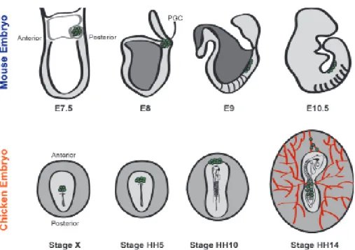
In the mouse, at E7.25 PGCs are localized in the base of the allantois from where they start to migrate to where the genital ridges will form [41]. From the base of the allantois, PGCs migrate to the adjacent endoderm, which will develop into the hindgut [42]. At E8.5 PGCs are found migrating along the midline of the embryo, from the hindgut through the dorsal mesentery. At around E10.5, PGCs reach the genital ridges [42] (Figure 2). The interaction of Sdf-1 and its receptor Cxcl12 is fundamental to chemotaxis of PGCs toward the genital ridges. In mice it was shown that the use of Sdf-1-coated beads in slice cultures of mouse embryos caused defective movements of PGCs and decreased survival [43, 44]. Moreover, homozygous knockout mice lacking Cdx2 show a dramatic decrease in the number of germ cells colonizing the genital ridges. This interaction seems to be important for the behavior of germ cells once they leave the hindgut [44].
In contrast to what happens in the mouse, in the chicken embryo the migration of PGCs occurs through the vascular system and starts from the anterior part of the embryo. Tsunekawa et al. [22]. From the germinal crescent, PGCs start to accumulate in the extraembryonic mesoderm between stages HH4-8. At stage HH10 blood islands start to form in the splanchnopleure, and since PGCs are localized in the mesoderm, from there they ingress into the vascular system. At around stage HH12, PGCs begin to appear in the extra-embryonic blood, and at stage HH14 they start to colonize the gonads (reviewed in [45]) (Figure 2).
between mice and chicken. In the chicken a role for SDF-1/CXCL2 has been shown in directing the migration of chicken PGCs toward the genital ridges, at stages HH11-16, when the cells are already migrating through the vasculature [47]. In Chapter 2 we describe the migration of chicken PGCs from the extraembryonic circulation into the embryo using CVH as a PGC marker. We show for the first time the role of the anterior vitelline veins in this process.
THE CURIOUS CASE OF CHICKEN GONADOGENESIS: ASYMMETRY, MEIOSIS AND CANCER DEVELOPMENT
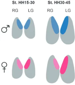
The genital ridges, precursors of the gonads, arise at stage HH20 in the ventromedial surface of the mesonephros, and constitute two macrosymmetrical structures, right and left, before sexual differentiation [48] (Figure 3). Undifferentiated female and male gonads are composed of the inner cortex and outer medulla [49]. The migration of PGCs toward the genital ridges occurs before sexual differentiation in an asymmetric way, since the right gonad generally presents more cells than the left gonad, in males and females, in early stages of development stages HH15-30 [50]. Although there are differences in terms of germ cell colonization, before the sexual differentiation there is almost no detectable morphological asymmetry between right and left. The development of the cortex, which is thicker in the left gonad in both sexes, is an exception to this [51] .
Only during sexual differentiation, which starts at around stages HH29-30, do differences in morphology and size between right and left gonads start to be more pronounced in females compared to males (Figure. 3) [49]. As a result, in males both gonads develop into a functional testis while in females only the left gonad develops into a functional ovary [49, 51].
Differential genetic expression in males and females leads to differences in the sexual morphology of the gonads. On the one hand, embryological testes have a greater medullary development, and the testicular cords containing germ cells will develop in that layer. On the other hand, the ovary presents a pronounced development of the cortex that will host the female germ cells [52]. DMRT1 [53] and SOX9 [54] seem to contribute to the male phenotype, while HINTW [55], FET1 [56] and FOXL2 [57] contribute to the female phenotype.
and phosphorylated histone H2A (H2AFX) - in both, right and left gonads, in both, male and female. Our results are presented in Chapter 4. We suggest that the localization of germ cells with respect to the left versus right gonad, cortex versus medulla of the left gonad, and central part versus the extremities versus the left cortex, influences meiotic maturation of the germ cells.
The adult hen is a model for epithelial ovarian cancer (EOC) in humans [64]. EOC is the most lethal gynaecological cancer, and the fifth cause of death in cancer-related mortality in women [65]. This is due to lack of treatments that specifically target EOC, difficulty in recognizing the symptoms and the wide spread of the disease in the peritoneal cavity [66]. Various models have been used to understand the mechanisms underlying the disease, including fruit flies, mice, in vitro models and the hen. In Chapter 5 we discuss the advantages of the avian model compared with other models in EOC. Moreover, we suggest that studying genes that are differentially expressed in chickens during asymmetric gonadogenesis and expressed in human EOC, such as PITX2, could offer a model to study the molecular basis of EOC in humans.
THE DEVELOPMENT OF EXTRAEMBRYONIC MEMBRANES IN AMNIOTES
As discussed previously, in the mouse embryo the PGCs are localized in the base of the allantois before they start to migrate, while avian PGCs use the vessels of the yolk sac to migrate into the embryo. Allantois and yolk sac are part of the extraembryonic membranes of amniotes and there is a tight relationship between their development and the initial developmental dynamics of PGCs. How does extraembryonic development occur? In amniotes, the embryo develops in a blastodisc on top of the yolk mass, and during gastrulation, the borders between embryo and extraembryonic membranes are not clearly defined [64]. Only after gastrulation is complete, when the embryo is already taking shape, do the endoderm and mesoderm (splanchnopleure) form the yolk sac and allantois, while the ectoderm and mesoderm (somatopleure) start to form the amnion and the chorion. In between the somatopleure and splanchnopleure, a cavity is formed – the extraembryonic coelom [67].
The splanchnopleure of the yolk sac contributes to the development of a vascular network, that is part of the first functional organ system in the embryo – the cardiovascular system. The activation of FGF-receptor in the splanchnopleure activates the differentiation of blood islands: angioblasts in the area pellucida and angioblasts and hematopoietic cells in the area opaca and paraaortic clusters [68]. The endothelial cells are then responsible for connecting the blood islands and remodeling blood vessels into a branched network that will cover the entire yolk sac. The process of vasculogenesis, the formation of the blood vessels, is followed by angiogenesis – capillary sprouting, splitting and remodeling – that leads to a reorganization of the vascular network. Moreover, after the heart starts beating, the resulting hemodynamics will have an important role in remodeling its branching and growth [68]. The development of the vascular network is a complex process, which starts at stages HH8-13 and is only defined at stage HH18 [69]. At the same time, the allantois, also derived from the splanchnopleure, connects to the extraembryonic coelom, and stores toxic by-products [70].
the chicken are described in detail in Chapter 5. Our results revisit an old model [71] for amnion formation, where the proamnion plays an important role.
AIM AND OUTLINE OF THIS THESIS
The mechanisms underlying PGC biology have been extensively studied in different organisms over the last several centuries. Nonetheless, many underlying mechanisms governing PGC behaviour are still unclear and deserve a more detailed analysis. The aim of this thesis is to study the migration of PGCs and meiosis in the chicken embryo. We also discuss the advantages and applicability of the avian model for ovarian cancer research in humans. Furthermore, we provide experimental evidence for the role of the proamnion in amnion development in the chicken, a structure that is often neglected in the literature.
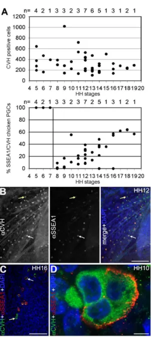
In Chapter 2 the migration route of germ cells from the extraembryonic circulation into the chicken embryo is described in detail. We show that SSEA1 is not a good marker for chicken PGCs at this time of development, since not all CVH-positive cells stain with an anti-SSEA1 antibody. Focusing on CVH as a marker for PGCs, we analyze the position of PGCs in the chicken embryo between stages HH10-19. We redefine the migration route of PGCs, providing evidence that the anterior vitelline veins are the main vehicles of transportation of germ cells from the anterior region of the extraembryonic vasculature into the genital ridges.
In Chapter 3 we show a detailed analysis of the expression of two different markers SYCP3 and H2AFX. We conclude that there is no evidence for apoptosis of germ cells localized in the right ovary as had been suggested by other authors. Moreover we demonstrate that differences in the expression of meiotic markers reveal three different aspects influencing the meiotic maturation of germ cells localized in female chicken gonads: their localization in the left or right gonad, cortex or medulla in the right gonad, and their position in the left cortex.
Chapter 4 provides a review of the use of different models in the study of epithelial ovarian cancer. We focus on the advantages of using the avian model, such as the similarities with the disease in humans, in improving the outcome of clinical research.
the first time, we show, through functional assays, the importance of sinking of the head in the proamnion for the development of the anterior amnion fold.
Finally, Chapter 6 provides a general discussion of the results obtained in the previous chapters. Moreover, we discuss the study of germ cell development in chickens and also its importance in pluripotency studies in a non-mammalian model.
REFERENCES
1. Surani MA. Genetics: immaculate misconception. Nature. 2002;416(6880):491-3.
2. Ruestow EG. Images and ideas: Leeuwenhoek’s perception of the spermatozoa. Journal of the history of biology. 1983;16(2):185-224.
3. Karamanou M, Poulakou-Rebelakou E, Tzetis M, Androutsos G. Anton van Leeuwenhoek (1632-1723): father of micromorphology and discoverer of spermatozoa. Rev Argent Microbiol. 2010;42(4):311-4.
4. Briggs E, Wessel GM. In the beginning...animal fertilization and sea urchin development. Dev Biol. 2006;300(1):15-26.
5. Richardson BE, Lehmann R. Mechanisms guiding primordial germ cell migration: strategies from different organisms. Nature reviews Molecular cell biology. 2010;11(1):37-49. 6. Moore H, Aflatoonian B. From stem cells to spermatozoa and back. Soc Reprod Fertil
Suppl. 2007;65:19-32.
7. Surani MA. Germ cells: the eternal link between generations. Comptes rendus biologies. 2007;330(6-7):474-8.
8. Lesch BJ, Page DC. Genetics of germ cell development. Nature reviews Genetics. 2012;13(11):781-94.
9. Ward JO, Reinholdt LG, Hartford SA, Wilson LA, Munroe RJ, Schimenti KJ, et al. Toward the genetics of mammalian reproduction: induction and mapping of gametogenesis mutants in mice. Biology of reproduction. 2003;69(5):1615-25.
10. Clift D, Schuh M. Restarting life: fertilization and the transition from meiosis to mitosis. Nat Rev Mol Cell Biol. 2013;14(9):549-62.
11. Stevens LC. Germ cell origin of testicular and ovarian teratomas. Transplant Proc. 1984;16(2):502-4.
12. Leitch HG, Blair K, Mansfield W, Ayetey H, Humphreys P, Nichols J, et al. Embryonic germ cells from mice and rats exhibit properties consistent with a generic pluripotent ground state. Development. 2010;137(14):2279-87.
13. Strome S, Updike D. Specifying and protecting germ cell fate. Nature reviews Molecular cell biology. 2015;16(7):406-16.
14. Crother BI, White ME, Johnson AD. Inferring developmental constraint and constraint release: primordial germ cell determination mechanisms as examples. J Theor Biol. 2007;248(2):322-30.
16. Sinsimer KS, Jain RA, Chatterjee S, Gavis ER. A late phase of germ plasm accumulation during Drosophila oogenesis requires lost and rumpelstiltskin. Development. 2011;138(16):3431-40.
17. Gavis ER, Lehmann R. Localization of nanos RNA controls embryonic polarity. Cell. 1992;71(2):301-13.
18. Edgar BA, Schubiger G. Parameters controlling transcriptional activation during early
Drosophila development. Cell. 1986;44(6):871-7.
19. Okada M. Germline cell formation in Drosophila embryogenesis. Genes & genetic systems. 1998;73(1):1-8.
20. Rongo C, Broihier HT, Moore L, Van Doren M, Forbes A, Lehmann R. Germ plasm assembly and germ cell migration in Drosophila. Cold Spring Harbor symposia on quantitative biology. 1997;62:1-11. P
21. Williamson A, Lehmann R. Germ cell development in Drosophila. Annu Rev Cell Dev Biol. 1996;12:365-9
22. Tsunekawa N, Naito M, Sakai Y, Nishida T, Noce T. Isolation of chicken vasa homolog gene and tracing the origin of primordial germ cells. Development. 2000;127(12):2741-5 23. Eyal-Giladi H, Kochav S. From cleavage to primitive streak formation: a complementary
normal table and a new look at the first stages of the development of the chick. I. General morphology. Dev Biol. 1976;49(2):321-3
24. Hamburger V, Hamilton HL. A series of normal stages in the development of the chick embryo. J Morphol. 1951;88(1):49-92.
25. Lavial F, Acloque H, Bachelard E, Nieto MA, Samarut J, Pain B. Ectopic expression of Cvh (Chicken Vasa homologue) mediates the reprogramming of chicken embryonic stem cells to a germ cell fate. Dev Biol. 2009;330(1):73-82.
26. Nieuwkoop P. SL. Primordial Germ Cells in the Chordates: Embryogenesis and phylogenesis 1979.
27. Ohinata Y, Payer B, O’Carroll D, Ancelin K, Ono Y, Sano M, et al. Blimp1 is a critical determinant of the germ cell lineage in mice. Nature. 2005;436(7048):207-13.
28. Magnusdottir E, Dietmann S, Murakami K, Gunesdogan U, Tang F, Bao S, et al. A tripartite transcription factor network regulates primordial germ cell specification in mice. Nat Cell Biol. 2013;15(8):905-15.
29. Lawson KA, Dunn NR, Roelen BA, Zeinstra LM, Davis AM, Wright CV, et al. Bmp4 is required for the generation of primordial germ cells in the mouse embryo. Genes Dev. 1999;13(4):424-36.
30. Ying Y, Zhao GQ. Cooperation of endoderm-derived BMP2 and extraembryonic ectoderm-derived BMP4 in primordial germ cell generation in the mouse. Dev Biol. 2001;232(2):484-92.
31. Ying Y, Liu XM, Marble A, Lawson KA, Zhao GQ. Requirement of Bmp8b for the generation of primordial germ cells in the mouse. Mol Endocrinol. 2000;14(7):1053-63. 32. Hayashi K, Kobayashi T, Umino T, Goitsuka R, Matsui Y, Kitamura D. SMAD1 signaling
is critical for initial commitment of germ cell lineage from mouse epiblast. Mechanisms of development. 2002;118(1-2):99-109.
33. Tremblay KD, Dunn NR, Robertson EJ. Mouse embryos lacking Smad1 signals display defects in extra-embryonic tissues and germ cell formation. Development. 2001;128(18):3609-21.
35. de Sousa Lopes SM, Roelen BA, Monteiro RM, Emmens R, Lin HY, Li E, et al. BMP signaling mediated by ALK2 in the visceral endoderm is necessary for the generation of primordial germ cells in the mouse embryo. Genes & development. 2004;18(15):1838-36. Hayashi K, Ohta H, Kurimoto K, Aramaki S, Saitou M. Reconstitution of the mouse germ
cell specification pathway in culture by pluripotent stem cells. Cell. 2011;146(4):519-32. 37. Hayashi K, Saitou M. Generation of eggs from mouse embryonic stem cells and induced
pluripotent stem cells. Nature protocols. 2013;8(8):1513-24.
38. Irie N, Weinberger L, Tang WW, Kobayashi T, Viukov S, Manor YS, et al. SOX17 is a critical specifier of human primordial germ cell fate. Cell. 2015;160(1-2):253-68.
39. Starz-Gaiano M, Lehmann R. Moving towards the next generation. Mech Dev. 2001;105(1-2):5-18.
40. Tanaka SS, Nishinakamura R. Regulation of male sex determination: genital ridge formation and Sry activation in mice. Cell Mol Life Sci. 2014;71(24):4781-802.
41. Saitou M, Yamaji M. Primordial germ cells in mice. Cold Spring Harb Perspect Biol. 2012;4(11).
42. Molyneaux KA, Stallock J, Schaible K, Wylie C. Time-lapse analysis of living mouse germ cell migration. Dev Biol. 2001;240(2):488-98.
43. Doitsidou M, Reichman-Fried M, Stebler J, Koprunner M, Dorries J, Meyer D, et al. Guidance of primordial
44. Molyneaux KA, Zinszner H, Kunwar PS, Schaible K, Stebler J, Sunshine MJ, et al. The chemokine SDF1/CXCL12 and its receptor CXCR4 regulate mouse germ cell migration and survival. Development. 2003;130(18):4279-86.
45. Nakamura Y, Kagami H, Tagami T. Development, differentiation and manipulation of chicken germ cells. Development, growth & differentiation. 2013;55(1):20-40.
46. Fujimoto T, Ukeshima A, Kiyofuji R. The origin, migration and morphology of the primordial germ cells in the chick embryo. Anat Rec. 1976;185(2):139-45.
47. Stebler J, Spieler D, Slanchev K, Molyneaux KA, Richter U, Cojocaru V, et al. Primordial germ cell migration in the chick and mouse embryo: the role of the chemokine SDF-1/ CXCL12. Dev Biol. 2004;272(2):351-61.
48. Carlon N, Stahl A. Origin of the somatic components in chick embryonic gonads. Arch Anat Microsc Morphol Exp. 1985;74(1):52-9.
49. Smith CA, Sinclair AH. Sex determination: insights from the chicken. Bioessays. 2004;26(2):120-32.
50. Nakamura Y, Yamamoto Y, Usui F, Mushika T, Ono T, Setioko AR, et al. Migration and proliferation of primordial germ cells in the early chicken embryo. Poult Sci. 2007;86(10):2182-93.
51. Zaccanti F, Vallisneri M, Quaglia A. Early aspects of sex differentiation in the gonads of chick embryos. Differentiation. 1990;43(2):71-80.
52. Smith CA, Roeszler KN, Hudson QJ, Sinclair AH. Avian sex determination: what, when and where? Cytogenet Genome Res. 2007;117(1-4):165-73.
53. Smith CA, Roeszler KN, Ohnesorg T, Cummins DM, Farlie PG, Doran TJ, et al. The avian Z-linked gene DMRT1 is required for male sex determination in the chicken. Nature. 2009;461(7261):267-71.
54. Kent J, Wheatley SC, Andrews JE, Sinclair AH, Koopman P. A male-specific role for SOX9 in vertebrate sex determination. Development. 1996;122(9):2813-22.
Biol. 2009;53(1):59-67.
56. Reed KJ, Sinclair AH. FET-1: a novel W-linked, female specific gene up-regulated in the embryonic chicken ovary. Mech Dev. 2002;119 Suppl 1:S87-90.
57. Hudson QJ, Smith CA, Sinclair AH. Aromatase inhibition reduces expression of FOXL2 in the embryonic chicken ovary. Dev Dyn. 2005;233(3):1052-5.
58. Guioli S, Lovell-Badge R. PITX2 controls asymmetric gonadal development in both sexes of the chick and can rescue the degeneration of the right ovary. Development. 2007;134(23):4199-208.
59. Hoshino A, Koide M, Ono T, Yasugi S. Sex-specific and left-right asymmetric expression pattern of Bmp7 in the gonad of normal and sex-reversed chicken embryos. Dev Growth Differ. 2005;47(2):65-74.
60. Mattsson A, Olsson JA, Brunstrom B. Activation of estrogen receptor alpha disrupts differentiation of the reproductive organs in chicken embryos. General and comparative endocrinology. 2011;172(2):251-9.
61. Levin M. Left-right asymmetry in embryonic development: a comprehensive review. Mech Dev. 2005;122(1):3-25.
62. Nakabayashi O, Kikuchi H, Kikuchi T, Mizuno S. Differential expression of genes for aromatase and estrogen receptor during the gonadal development in chicken embryos. J Mol Endocrinol. 1998;20(2):193-202.
63. Ukeshima A. Germ cell death in the degenerating right ovary of the chick embryo. Zoolog Sci. 1996;13(4):559-63.
64. Johnson PA, Giles JR. The hen as a model of ovarian cancer. Nat Rev Cancer. 2013;13(6):432-6.
65. Lowe KA, Chia VM, Taylor A, O’Malley C, Kelsh M, Mohamed M, et al. An international assessment of ovarian cancer incidence and mortality. Gynecol Oncol. 2013;130(1):107-14. 66. Vaughan S, Coward JI, Bast RC, Jr., Berchuck A, Berek JS, Brenton JD, et al.
Rethinking ovarian cancer: recommendations for improving outcomes. Nat Rev Cancer. 2011;11(10):719-25.
67. A. GSaR. Embryology, Constructing the Organism: Sinauer Associates, Inc.; 1997. 68. le Noble F, Fleury V, Pries A, Corvol P, Eichmann A, Reneman RS. Control of arterial
branching morphogenesis in embryogenesis: go with the flow. Cardiovasc Res. 2005;65(3):619-28.
69. Styp-Rekowska B, Hlushchuk R, Pries AR, Djonov V. Intussusceptive angiogenesis: pillars against the blood flow. Acta Physiol (Oxf). 2011;202(3):213-23.
70. Adamstone FB. Experiments on the development of the amnion in the chick. J Morphol. 1948;83(3):359-71.
2
Chicken primordial germ cells use the
anterior vitelline veins to enter the
embryonic circulation
Ana de Melo Bernardo1, Kaylee Sprenkels1, Gabriela Rodrigues2, Toshiaki Noce3 and Susana M. Chuva De Sousa Lopes1
1Department of Anatomy and Embryology, Leiden
University Medical Center, Leiden, The Netherlands; 2Centro de Biologia Ambiental e Departamento de Biologia Animal,
Faculdade de Ciências, Universidade de Lisboa, Lisboa, Portugal; 3Department of Physiology, Keio University
School of Medicine, Tokyo, Japan
ABSTRACT
During gastrulation, chicken primordial germ cells (PGCs) are present in an extraembryonic region of the embryo from where they migrate towards the genital ridges. This is also observed in mammals, but in chicken the vehicle used by the migratory PGCs is the vascular system. We have analysed the migratory pathway of chicken PGCs, focusing on the period of transition from the extraembryonic region to the intraembryonic vascular system.
Our findings show that at Hamburger and Hamilton developmental stage HH12–HH14 the majority of PGCs concentrate axially in the sinus terminalis and favour transport axially via the anterior vitelline veins into the embryonic circulation. Moreover, directly blocking the blood flow through the anterior vitelline veins resulted in an accumulation of PGCs in the anterior region and a decreased number of PGCs in the genital ridges. We further confirmed the key role for the anterior vitelline veins in the correct migration of PGCs using an ex ovo culture method that resulted in defective morphogenetic development of the anterior vitelline veins.
INTRODUCTION
Early during the development of amniotes, the germline is segregated from the somatic cell lineages. This is an important event because the primordial germ cells (PGCs), the precursors of the oocytes and sperm, carry the genetic information throughout generations and are therefore the engine of evolution, contributing to genetic variability in sexually reproducing animals [1]. Even though PGCs can be formed by two distinct mechanisms, epigenesis and preformation, they show some common characteristics, including early segregation, similar morphology cross-species [2] and a distinct migratory period from a peripheral or extraembryonic location to the place where the somatic gonad compartments are formed. Understanding the details surrounding the migration of PGCs is important because an aberrant migration can cause cancer and infertility (reviewed by [3]). Interestingly, in Gallus gallus the PGCs migrate from an anterior location towards the genital ridge compartment, whereas in Mus musculus the PGCs migrate from a posterior/caudal location towards the genital ridges [4].
In chicken, the staining method classically used to distinguish PGCs from the somatic cells was the periodic acid-Schiff (PAS) staining [5]. There are also immunological markers against cell-surface glycoproteins present in PGCs, like SSEA1, which is commonly used to identify mammalian and chicken PGCs. However, SSEA1 is not restricted to chicken or mammalian PGCs, but is found in several types of undifferentiated multipotent mouse and chicken cells [6, 7]. More recently, Tsunekawa and colleagues identified the chicken vasa homolog (CVH) gene and have shown its germline-specific expression [8]. The function of VASA is not well understood, but it has been shown that vasa is indispensable for germ cell development and it is present in the germline of many animal species, suggesting a conserved role throughout evolution (reviewed by [3]). Immunohistochemical analyses, using specific antibodies against CVH protein, demonstrated that CVH-expressing cells were detectable during early embryogenesis of chicken embryos, starting from the first cleavage of fertilized eggs,[8], suggesting that a preformation mode of germline specification was adopted in chicken.
The PGCs move from the hypoblast layer to accumulate in the extraembryonic mesoderm localized between the ectoderm and hypoblast. Subsequently, the PGCs become lodged in the vascular system as the blood islands are formed in the yolk sac around HH10 and by HH12 use those extraembryonic blood vessels as a vehicle to reach the embryo [5, 12, 13]. By HH15, the PGCs start leaving the vascular system close to the genital ridges, just caudally from the vitelline arteries and by HH17 the majority of the PGCs have settled in the genital ridges [5, 13-16]. The mechanism by which the PGCs enter the vascular system is less well understood than the mechanism by which the PGCs exit the vascular system (SDF1–CXCR4) to colonize the gonads [17] that has clear similarities with the process of homing of lymphocytes during inflammation and tumor metastasis [18, 19].
MATERIAL AND METHODS Embryo collection and manipulation
Fertilized White Leghorn chicken (Gallus gallus) eggs were incubated in a humidified atmosphere at 37.0°C until the desired HH stage [11]. Embryos were washed and manipulated on 2% agar-coated petri dishes containing phosphate buffer solution (PBS). The vitelline membrane was removed, the embryos were isolated with intact area opaca and pellucida and fixed overnight (o/n) at 4°C either in 4% paraformaldehyde (PFA) for whole mount immunofluorescence or in Bouin’s solution (Sigma) for immunohistochemistry and stored in PBS at 4°C until further use.
Whole mount immunofluorescence
Fixed embryos were permeabilized with 0.5% Triton (Sigma) in PBS (PBT) o/n at 4°C with rotation. Thereafter, they were washed in PBS and incubated 24 hours at 4°C with the first antibodies diluted in 1% bovine serum albumin (BSA, Fraction V) (Gibco) in PBS. The first antibodies used were rabbit anti-CVH IgG at 1:500 and mouse anti-SSEA1 IgM (TG1) at 1:10. Next, the embryos were washed in PBS for 1 hour and incubated with the respective secondary antibodies diluted in 1% BSA/PBS for 24 hours at 4°C. The secondary antibodies used were Alexa Fluor 488 donkey anti-rabbit IgG (Molecular Probes) and Alexa Fluor 568 goat anti-mouse IgM (Molecular Probes), both used at 1:1000. The embryos had a final rinse in PBS and were enclosed with Vectashield with Dapi (Vector), covered with a cover glass and sealed with nail polish. For this analysis, we considered only embryos that showed normal morphology, including the presence of PGCs. The total number of PGCs per embryo was counted and plotted.
Immunohistochemistry
sections were blocked for 1 hour at RT in fresh 1% BSA/PBS. The slides were incubated with rabbit anti-CVH IgG at 1:500 diluted in blocking o/n at 4°C, washed in PBS and incubated with BrightVision Poly-HRP anti-rabbit (Immunologic) for 30 minutes at RT. Thereafter, the slides were washed first with PBS, than with 0.05M Tris-maleate buffer (pH 7.6), revealed with a solution of 0.4 mg/ml 3,3-diaminobenzidine (DAB) and finally counterstained with Mayer’s Hematoxylin. The sections were washed in water, dehydrated in an increasing series of ethanol and finally xylene. Thereafter, the samples were mounted in Entellan (Merck). For this analysis, we considered only embryos that showed normal morphology, including the presence of PGCs. The total number of PGCs per embryo was counted and plotted.
In ovo clamp experiments
At HH14, an opening was made in the shell of eggs and part of the vitelline membrane was removed to expose the embryo and some drops of PBS were added to avoid embryo drought. To block blood flow though the anterior vitelline veins a knot was tied using a small semicircular multipass needle attached to a prolene monofilament (Ethicon). The opening made in the eggs was closed and the eggs incubated for 6 hours (until HH15). The control embryos were treated similarly, but the vitelline veins were not clamped. After the incubation time, the embryos were isolated, fixed in 4% PFA o/n and processed for whole mount immunofluorescence. The total number of PGCs per embryo was counted and statistical analysis to compare the distribution of PGCs in the two groups of embryos was performed using the non-parametric Mann–Whitney test.
Ex ovo culture of chicken embryos
After the incubation time, the embryos were isolated, fixed in 4% PFA o/n and processed for whole mount immunofluorescence.
Image acquisition and analysis
RESULTS
The number of chicken PGCs remained constant, but increasing numbers of PGCs expressed SSEA1 between HH8–HH19
We analysed the number of PGCs in White Leghorn chicken embryos between HH5– HH19 (n = 42) by whole mount double immunofluorescence for CVH and SSEA1, counting the total number of PGCs present in the embryo, area pellucida and area opaca. We observed a high variation in the total number of PGCs between embryos of the same developmental stage; however, the average number of PGCs present in the germinal crescent at HH5 was similar to the average number of PGCs present in the genital ridges between HH16–HH19. The majority of the embryos exhibited between 200 and 450 PGCs (Fig. 1A). Between HH5–HH7, all the CVH-positive cells were positive for SSEA1 (Fig. 1B), but SSEA1 was observed in many other cells and tissues in the embryo, making SSEA1 an inadequate marker of the germline at those stages. At HH8, SSEA1 was drastically downregulated in the CVH-positive PGCs and was then slowly upregulated in a fraction of the germ cells (Fig. 1B,C) until it stabilized at 60% of the cells by the time the PGCs colonized the genital ridges at HH16–HH19 (Fig. 1B,D). Interestingly, in the typically 3–4 cell clusters of germ cells, already described in 1914 by Swift [13], we often observed both SSEA1-positive and SSEA1-negative cells (Fig. 1E). In agreement, Swift noticed pronounced differences in the yolk content and yolk coloration among PGCs [13] and this may be directly linked to the heterogeneity observed in SSEA1 staining.
At HH13, the PGCs localized to the sinus terminalis and anterior vitelline veins
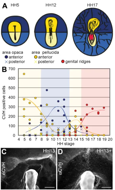
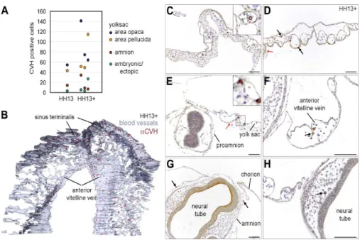
At HH13, the PGCs start to transit between the anterior region of area opaca, area pellucida and the genital ridges and this coincides with the period of initiation of the (vitelline) blood circulation. Therefore, we zoomed in at HH13 and defined two developmental sub-stages, HH13 and HH13circ. At HH13, the PGCs were relatively dispersed in the anterior central part of the area opaca and anterior central area pellucida (Fig. 2C). However, in some HH13 embryos, the PGCs concentrated in a narrower continuous axial region between the area opaca and the area pellucida (Fig. 2D). Both HH13 and HH13circ embryos contained about 17–19 somites and approximately the same number of PGCs (with similar medians) in the yolk sac in the area opaca and in the area pellucida, the amnion and ectopically, in particular in the head vasculature (Fig. 3A).
The histological analysis and 3D reconstruction of the vasculature and the position of the PGCs confirmed that at HH13circ the majority of PGCs are concentrated in specific blood vessels in the yolk sac, namely in the sinus terminalis (in the area opaca) and continuous to the developing anterior vitelline veins (in the area pelucida) (Fig. 3B). Our observations contrast with the current model where the PGCs were thought to be scattered broadly throughout the yolk sac vasculature and enter the embryo through the omphalomesenteric veins. Also of note was the fact that, at HH13circ the PGCs were clearly present both inside (in the lumen of the vessel) and outside the blood vessels in the yolk sac (Fig. 3C–F), suggesting that they are not simply engulfed by the blood vessels as they form. In addition, PGCs were also observed frequently in the amnion (somatopleura) (Fig. 3G) and ectopically in the vasculature of the embryo head (Fig. 3H). The PGCs in the amnion probably mislocated when the somatopleura (amnion/ chorion) and splanchnopleura (yolk sac) separated.
Finally, we also report that the head (at the level of the prosencephalon) at stage HH13circ, while extending anteriorly, becomes transiently enveloped in the proamnion, a bilaminar tissue consisting of hypoblast and epiblast (and no mesoderm); and, as a result, both the somatopleura (amnion/chorion) and splanchnopleura (yolk sac) are transiently localized above the developing head (Fig. 3E).
The PGCs migrated towards the embryo primarily using the anterior vitelline veins
After 6 hours of culture, in control embryos (n = 7) the majority of PGCs found their way to the embryo and colonized the genital ridges (Fig. 4B–D). However, in experimental embryos (n = 6), the PGCs remained clustered in the region of the clamped anterior vitelline veins in the axial anterior area pellucida (Fig. 4B,E) and showed a reduced number of PGCs transiting through the embryo and in the genital ridges when compared to the controls (Fig. 4B,F). Our results indicate that blocking the blood flow from the anterior vitelline veins at HH14 has a significant effect in the correct migration of PGCs towards the genital ridges. We concluded that the anterior vitelline veins are the main vehicle used by the PGCs during their migration from the extraembryonic vasculature into the intraembryonic vasculature.
To further confirm the role of the anterior vitelline veins in the migration of PGCs from the extraembryonic to the intraembryonic circulation, we analyzed embryos cultured ex ovo using a modified Cornish pasty method that results in primary defects in the morphogenesis of the anterior extraembryonic structures [20]. Using this method, the somatopleura (amnion/chorion) and the splanchnopleura (yolk sac) separate, but the amniotic folds from the head, lateral and tail do not form leaving the embryo exposed. Moreover, the anterior axial conversion of the sinus terminalis and the anterior vitteline veins does not occur and therefore we investigated whether the PGCs were able to find their way into the genital ridges from the germinal crescent. For the modified Cornish pasty method, embryos at HH5 were removed from the egg, folded in two by their anterior–posterior axis and the edges of the semi-circle were pressed together to create a “mini yolk sac-like” (Fig. 5A) and cultured in suspension.
After 30 hours ex ovo, embryos at stage HH13 were collected and immunostained for CVH (Fig. 5B,C). In general, the head showed a normal development, the embryos had 17–19 somites (n = 10), the heart was beating and the extraembryonic circulation well-established in the posterior region of the embryo. As expected, the headfold of the amnion did not form leaving the head exposed and tilting upwards. We observed many PGCs (± 200) dispersed in the splanchnopleura, anterior to the head at the border between the area opaca and pellucida and some PGCs mislocated in the somatopleura (Fig. 5C).
several HH17 ex ovo embryos (n = 5) and the majority of the PGCs (± 150) were present in the region of the splanchnopleura between the left and right vitelline veins and anterior to the heart. In those embryos, only a very low number of PGCs (< 10) was observed in the region of the genital ridges. We concluded that due to the developmental defects in the position of the sinus terminalis and the anterior vitelline veins in the HH13 and HH17 ex ovo embryos, the PGCs fail to find and ingress these blood vessels and therefore remained ectopically in the “germinal crescent” region. We propose a novel model for the migration of PGCs in the chicken with a key role for the sinus terminalis and the anterior vitelline veins (Fig. 5E) as part of a defined or preferred vascular pathway used by the PGCs to travel from their position in the germinal crescent into the heart via the omphalomesenteric veins.
DISCUSSION
SSEA1 has been used as an appropriate marker to identify and isolate PGCs from chicken embryos [22-27]. However, by performing double immunofluorescence analyses, using antibodies against CVH and SSEA1, we have now demonstrated that although SSEA1 marked PGCs, it is only expressed by a fraction, albeit increasing, of CVH-positive PGCs between HH8–HH19, but not in the entire population of PGCs. This perhaps explains why the fraction of circulating PGCs at HH13–HH15 isolated by fluorescence-activat-ed cell sorting (FACS) on the basis of SSEA1 expression by Mozdziak and colleagues was smaller than the fraction of circulating PGCs isolated using a Nycodenz density gradient [28] or found in chicken blood by PAS staining [29]. Interesting, pluripotent mouse embryonic stem cells, which are closely related to PGCs, also show pronounced heterogeneity for SSEA1 staining [30].
It will be important to identify additional lineage-specific markers, like NANOG
and DEAD END [31-33] and in particular novel cell surface markers to study chicken PGCs. This will be important to investigate both embryonic pluripotency and PGC development at very early developmental stages, where the CVH antibody is the only available to identify PGCs.
We and others [29] have observed a large individual variation in the number of PGCs between HH5 and HH19. This results from genetic variation as demonstrated by Tajima and colleagues that showed that specific hens layed eggs with embryos containing consistently high or low number or circulating PGCs [29]. Maybe due to this high variability in PGC numbers, we were unable to observe an increasing number of PGCs between HH5 and HH19 as reported [12]. In our hands, the number of PGCs remained approximately constant between HH5 and HH19. In agreement, Fujimoto and colleagues described 312 PGCs at HH10 [5], Motono and colleagues referred to about 300 PGCs at HH13–HH16 [24] and Nakamura described embryos containing on average 300 PGCs from HH14 to HH20 [34]. Together, we concluded that from HH5, when the PGCs are present in the germinal crescent, until HH19, when the PGCs have reached the genital ridges, the number of PGCs remained constant and range from 200–400.
Two studies have mapped the position of PGCs during their migration from the germinal crescent (HH4) to the genital ridges (HH17) by analysis of whole amount embryos [5, 12]. None of them mentioned the vitelline veins, even though there is a clear concentration of PGCs visible at the junction between the sinus terminalis and the vitelline veins at HH10–11. Curiously, neither Fujimoto nor Nakamura analysed embryos at stage HH13 and this could be the reason why the concentration of PGCs in the anterior vitelline veins has remained unnoticed.
Different groups have collected circulating PGCs at HH13–HH14 using blood from the sinus terminalis, the vitelline vessels, the heart and the dorsal aorta [12, 15, 25, 26, 28, 29, 35, 36]. Although none referred to a specific vascular route used by the PGCs, their methods to collect circulating PGCs support the idea that the majority of the PGCs indeed concentrate and use the sinus terminalis and anterior vitelline veins as an effective way to reach the embryo via the omphalomesenteric veins that enter the heart. Circumstantial evidence was also provided by Nakamura and colleagues by injecting quail PGCs in chick vitelline vessels at HH15 and later observing those quail PGCs in the recipient chicks’ genital ridges [37]. We now provide functional data indicating a key role for the anterior vitelline veins transporting PGCs towards the embryo.
before it turns caudally, via the first aortic arch, into the dorsal aorta. It is therefore not surprising that we and others [34, 38] observed that HH13–HH15 PGCs had the tendency to become trapped in the cephalic capillary network when being pumped out of the heart, instead of performing the U-turn towards the dorsal aorta. In embryos in which the posterior part, including the gonads, have been excised, the PGCs still accumulate in the head capillaries [34]. Interestingly, stromal cell-derived factor-1 (SDF1/CXCL12), a chemokine involved in the extravasation of the PGCs from the vascular system to the mesenchyme of the genital ridges [17], is expressed at HH12–HH15 specifically both in the area of the genital ridges and the head region [39] and could promote migration of PGCs into both areas.
The signaling pathway involved in attracting the PGCs into the vascular system is less understood. Having a better understanding of the vascular route taken by the PGCs and the markers that can be used to follow the population of PGCs will greatly facilitate the investigation of the mechanisms used by PGCs to enter the vascular system. In turn this may prove an important model to understand how metastatic cells behave on their way to form secondary tumors and how leukocytes behave during processes like infection and inflammation.
ACKNOWLEDGMENTS
We thank the following: P. Beverly for mouse anti-SSEA1; S. Maas, C. van Munsteren and L. van Iperen for help with the egg-logistics; B. Wisse for help with the Amira software; and R. Poelman for discussions. This research was supported by the European Commission’s Livelong Learning Programme (Leonardo da Vinci 2011-1-PT1-LEO02-08199) to A.M.B. and by the Netherlands Organization for Scientific Research (NWO Aspasia 015.007.037) to S.M.C.S.L.
REFERENCES
1. Johnson AD, Richardson E, Bachvarova RF, Crother BI. Evolution of the germline-soma relationship in vertebrate embryos. Reproduction. 2011;141(3):291-300.
2. de Sousa Lopes S, Roelen BA. An overview on the diversity of cellular organelles during the germ cell cycle. Histol Histopathol. 2010;25(2):267-76.
3. Chuva de Sousa Lopes SM, Roelen BA. On the formation of germ cells: The good, the bad and the ugly. Differentiation. 2010;79(3):131-40.
5. Fujimoto T, Ukeshima A, Kiyofuji R. The origin, migration and morphology of the primordial germ cells in the chick embryo. Anat Rec. 1976;185(2):139-45.
6. Pain B, Clark ME, Shen M, Nakazawa H, Sakurai M, Samarut J, et al. Long-term in vitro
culture and characterisation of avian embryonic stem cells with multiple morphogenetic potentialities. Development. 1996;122(8):2339-48.
7. Solter D, Knowles BB. Monoclonal antibody defining a stage-specific mouse embryonic antigen (SSEA-1). Proc Natl Acad Sci U S A. 1978;75(11):5565-9.
8. Tsunekawa N, Naito M, Sakai Y, Nishida T, Noce T. Isolation of chicken vasa homolog gene and tracing the origin of primordial germ cells. Development. 2000;127(12):2741-50. 9. Eyal-Giladi H, Kochav S. From cleavage to primitive streak formation: a complementary
normal table and a new look at the first stages of the development of the chick. I. General morphology. Dev Biol. 1976;49(2):321-37.
10. Ginsburg M. Primordial germ cell formation in birds. Ciba Found Symp. 1994;182:52-61; discussion -7.
11. Hamburger V, Hamilton HL. A series of normal stages in the development of the chick embryo. 1951. Dev Dyn. 1992;195(4):231-72.
12. Nakamura Y, Yamamoto Y, Usui F, Mushika T, Ono T, Setioko AR, et al. Migration and proliferation of primordial germ cells in the early chicken embryo. Poult Sci. 2007;86(10):2182-93.
13. C. Swift. Origin and early history of the primordial germ cells in the chick. Am J Anat. 1914;15:483–516.
14. Meyer DB. The Migration of Primordial Germ Cells in the Chick Embryo. Dev Biol. 1964;10:154-90.
15. Yamamoto Y, Usui F, Nakamura Y, Ito Y, Tagami T, Nirasawa K, et al. A novel method to isolate primordial germ cells and its use for the generation of germline chimeras in chicken. Biol Reprod. 2007;77(1):115-9.
16. Ukeshima AK, M. Fujimoto, T. Relationship between genital ridge formation and settlement site of primordial germ cells in chick embryos. . Anat Rec. 1987;219:311–4
17. Stebler J, Spieler D, Slanchev K, Molyneaux KA, Richter U, Cojocaru V, et al. Primordial germ cell migration in the chick and mouse embryo: the role of the chemokine SDF-1/ CXCL12. Dev Biol. 2004;272(2):351-61.
18. Alsayed Y, Ngo H, Runnels J, Leleu X, Singha UK, Pitsillides CM, et al. Mechanisms of regulation of CXCR4/SDF-1 (CXCL12)-dependent migration and homing in multiple myeloma. Blood. 2007;109(7):2708-17.
19. Ueda Y, Yang K, Foster SJ, Kondo M, Kelsoe G. Inflammation controls B lymphopoiesis by regulating chemokine CXCL12 expression. J Exp Med. 2004;199(1):47-58.
20. Nagai H, Lin MC, Sheng G. A modified cornish pasty method for ex ovo culture of the chick embryo. Genesis. 2011;49(1):46-52.
21. Streit A, Stern CD. Operations on primitive streak stage avian embryos. Methods Cell Biol. 2008;87:3-17.
22. Lee SI, Lee BR, Hwang YS, Lee HC, Rengaraj D, Song G, et al. MicroRNA-mediated posttranscriptional regulation is required for maintaining undifferentiated properties of blastoderm and primordial germ cells in chickens. Proc Natl Acad Sci U S A. 2011;108(26):10426-31.
24. Motono M, Ohashi T, Nishijima K, Iijima S. Analysis of chicken primordial germ cells. Cytotechnology. 2008;57(2):199-205.
25. Motono M, Yamada Y, Hattori Y, Nakagawa R, Nishijima K, Iijima S. Production of transgenic chickens from purified primordial germ cells infected with a lentiviral vector. J Biosci Bioeng. 2010;109(4):315-21.
26. Mozdziak PE, Angerman-Stewart J, Rushton B, Pardue SL, Petitte JN. Isolation of chicken primordial germ cells using fluorescence-activated cell sorting. Poult Sci. 2005;84(4):594-600.
27. Petitte JN, Karagenc L, Ginsburg M. The origin of the avian germline and transgenesis in birds. Poult Sci. 1997;76(8):1084-92.
28. Zhao DF, Kuwana T. Purification of avian circulating primordial germ cells by nycodenz density gradient centrifugation. Br Poult Sci. 2003;44(1):30-5.
29. Tajima A, Hayashi H, Kamizumi A, Ogura J, Kuwana T, Chikamune T. Study on the concentration of circulating primordial germ cells (cPGCs) in early chick embryos. J Exp Zool. 1999;284(7):759-64.
30. Hayashi K, Lopes SM, Tang F, Surani MA. Dynamic equilibrium and heterogeneity of mouse pluripotent stem cells with distinct functional and epigenetic states. Cell Stem Cell. 2008;3(4):391-401.
31. Aramaki S, Sato F, Kato T, Soh T, Kato Y, Hattori MA. Molecular cloning and expression of dead end homologue in chicken primordial germ cells. Cell Tissue Res. 2007;330(1):45-52. 32. Canon S, Herranz C, Manzanares M. Germ cell restricted expression of chick Nanog. Dev
Dyn. 2006;235(10):2889-94.
33. Fernandez-Tresguerres B, Canon S, Rayon T, Pernaute B, Crespo M, Torroja C, et al. Evolution of the mammalian embryonic pluripotency gene regulatory network. Proc Natl Acad Sci U S A. 2010;107(46):19955-60.
34. Nakamura M, Kuwana T, Miyayama Y, Yoshinaga K, Fujimoto T. Ectopic colonization of primordial germ cells in the chick embryo lacking the gonads. Anat Rec. 1991;229(1):109-15.
35. Nakamura Y, Usui F, Ono T, Takeda K, Nirasawa K, Kagami H, et al. Germline replacement by transfer of primordial germ cells into partially sterilized embryos in the chicken. Biol Reprod. 2010;83(1):130-7.
36. Yasuda Y, Tajima A, Fujimoto T, Kuwana T. A method to obtain avian germ-line chimaeras using isolated primordial germ cells. J Reprod Fertil. 1992;96(2):521-8.
37. Nakamura M, Yoshinaga K, Fujimoto T. Histochemical identification and behavior of quail primordial germ cells injected into chick embryos by the intravascular route. J Exp Zool. 1992;261(4):479-83.
38. Nakamura M, Kuwana T, Miyayama Y, Fujimoto T. Extragonadal distribution of primordial germ cells in the early chick embryo. Anat Rec. 1988;222(1):90-4.
3
Meiotic Wave adds Extra Asymmetry to the
Development of Female Chicken Gonads
Ana de Melo Bernardo§1, A. Marijne Heeren§1,2, Liesbeth van Iperen1, Maria
Fernandes1, Nannah He1, Stanford Anjie1, Toshiaki Noce3, Ester S. Ramos4
and Susana M. Chuva de Sousa Lopes1,5
1Dept. of Anatomy and Embryology, Leiden University Medical Center, Leiden, The Netherlands; 2Current address: Dept. of Obstetrics and Gynaecology, VU University Medical Center,
Amsterdam, The Netherlands; 3Dept. of Physiology, Keio University School of Medicine, Tokyo,
Japan 4Department of Genetics, Ribeirao Preto Medical School, University of Sao Paulo, Sao
Paulo, Brazil; 5Dept. for Reproductive Medicine, Ghent University Hospital, Ghent, Belgium; § Equal first authorship contribution
In Molecular Reproduction and Development. 2015;9999:1-13
3
Meiotic wave adds extra asymmetry to the
development of female chicken gonads
Ana de Melo Bernardo§1, A. Marijne Heeren§1,2, Liesbeth van Iperen1,
Maria Fernandes1, Nannan He1, Stanford Anjie1, Toshiaki Noce3,
Ester S. Ramos4 and Susana M. Chuva de Sousa Lopes1,5
1Dept. of Anatomy and Embryology, Leiden University Medical Center, Leiden,
The Netherlands; 2Current address: Dept. of Obstetrics and Gynaecology, VU
University Medical Center, Amsterdam, The Netherlands; 3Dept. of Physiology,
Keio University School of Medicine, Tokyo, Japan 4Department of Genetics,
Ribeirao Preto Medical School, University of Sao Paulo, Sao Paulo, Brazil;
5Dept. for Reproductive Medicine, Ghent University Hospital, Ghent, Belgium; § Equal first authorship contribution
ABSTRACT
Development of female gonads in chicken is asymmetric. This asymmetry affects gene expression, morphology and germ cell development. As a result only the left ovary develops into a functional organ, whereas the right ovary remains vestigial. In males, both gonads develop into functional testes.
Here, we revisited the development of asymmetric traits in female (and male) chicken gonads between Hamburger Hamilton stage 16 (HH16) and hatching. At HH16, primordial germ cells (PGCs) migrated preferentially to the left gonad independently of gender, accumulating in the left coelomic hinge between the gut mesentery and developing gonad. Using the meiotic markers SYCP3 and (phosphorylated) H2AFX, we were able to identify a pronounced asymmetry regarding meiotic progression in the germ cells located in the central, lateral and extremity part of the cortex of the left female gonad from HH38 until hatching. This has not been previously described. Moreover, in contrast to the current view that medullary germ cells enter apoptosis during development in the right female gonad, we show that medullary germ cells are not apoptotic, but arrested in pre-leptotene until hatching.
INTRODUCTION
In chicken, primordial germ cells (PGCs), the progenitors of the gametes, are of extraembryonic origin and, at Hamburger and Hamilton stage (HH)10-12 [1], they are found in the developing blood islands in a region of the yolk sac anterior to the head. From there, they migrate axially through the bloodstream, concentrating in the sinus terminalis, and enter the embryo mainly through the anterior vitelline veins [2]. The PGCs then travel through the embryonic vasculature to reach the gonadal ridges. After the PGCs colonize both left and right gonadal ridges, those undergo sex differentiation, to become morphologically different according to their gender developing as ovaries or testes. In males, both gonads develop into functional testes but in females, as in most birds, it is only the left gonad that develops into a functional ovary, while the right gonad remains rudimentary [3,4]. Before any signs of sex differentiation, the number of germ cells present in the gonadal ridges shows a sex-independent asymmetrical distribution with a preference for the left side in both males and females at HH15-HH17 [5] and at HH22-26 [6]. This sex-independent left-right asymmetry in the number of germ cells is still present at HH35 [7]. In addition to the asymmetry in the number of germ cells, the thickness of surface epithelium of the gonadal ridges also has pronounced sex-independent left-right asymmetry, being consistently thicker in the left gonad until HH36 [8,9].
MATERIAL AND METHODS Embryo collection and sexing
Fertilized White Leghorn chicken (Gallus gallus) eggs were incubated in a humidified atmosphere at 37°C. Eggs were windowed and embryos staged [1]. The sex of HH35 embryos until prior to hatching was determined by eye, whereas the sex of HH16-30 embryos was determined by genomic polymerase chain reaction (PCR) as described [37]. Embryos were isolated and used whole from HH16-HH30, whereas from HH35 until prior to hatching, the paired gonads were further dissected out of the embryo.
Immunofluorescence on whole mount and paraffin section
Embryos and gonads were fixed in 4% paraformaldehyde (PFA) (MERCK, Germany)
in phosphate-buffered saline (PBS) overnight (o/n) at 4˚C. Immunofluorescence on
HH16-HH19 embryos (n=7) was performed as whole amount and on HH25-HH42 embryos (n=14) on paraffin sections was performed essentially as described [2,38]. Primary antibodies used were rabbit anti-DDX4 (1:500, gift from T. Noce), mouse anti-PCNA (1:500, sc-56, Santa Cruz Biotechnology, USA), rabbit anti-cleaved CASP3 (Asp 175) (1:300, 9661S, Cell Signaling Technology, USA), mouse anti-H2AFX (Ser139) (1:500, 05-636, Millipore, USA) and rabbit anti-FASLG (1:100, sc-6237, Santa Cruz Biotechnology, USA). Secondary antibodies used were Alexa Fluor 568 goat anti-mouse (1:500, A-21124, Life Technologies, UK) and Alexa Fluor 488 donkey anti-rabbit (1:500, A11008, Life Technologies, UK). DNA fragmentation was determined by TUNEL assay with TMR red In Situ Cell Death Detection Kit (Roche Applied Science, Germany) for 1 hour at 37°C following manufacturer’s instructions. Slides were counterstained with 1:1000 dilution of 4’,6-diamidino-2-phenylindole dihydrochloride (DAPI) (Life Technologies, UK) in PBS for 1 minute and mounted with ProLongGold antifade reagent (Life Technologies, UK).
Germ cell counting and FACS-analysis
Germ cells were counted in the right and left gonad in whole mount HH16-HH19 embryos (n=3 females; n=4 males) and sequential paraffin sections of HH25-HH30 embryos (n=3 females; n=3 males) immunostained for DDX4.
and then incubated in TRIPLE 5x (Life Technologies, UK) at 37°C for 30 minutes, with pipetting from time to time. The single cell suspension was washed in DPBS0 and cells were fixed in 4% PFA for 10 minutes on ice. Thereafter, cells were permeabilized using 0.1% Triton/DPBS0 for 30 minutes on ice, incubated with 1:1000 rabbit anti-DDX4 in blocking solution [1% bovine serum albumin (BSA, Life Technologies, UK) in 0.05% Tween/DPBS0] for 1 hour on ice, washed with DPBS0 and incubated with 1:500 Alexa fluor goat anti-rabbit 488 in blocking solution for 1 hour on ice. After a final wash in DPBS0 cells were resuspended in FACS buffer (1%BSA, 10mM EDTA in PBS) and FACS analysis was performed using BD FACSAriaTM III (BD BioSciences, the Netherlands). Results were processed using the software BD FACSDivaTM version 6.0 (BD BioScience, the Netherlands).
The percentage of PGCs in the left and right gonad was calculated in relation to the total number of germ cells counted per embryo. The Student’s T-test (two-tailed distribution, two-sample unequal variance) was used to compare the percentage of DDX4 positive germ cells between the right and left gonads. *, P<0.05; **, P<0.01; ***, P<0.001.
Quantitative reverse transcription-PCR (qPCR)
The left and right gonads were harvested individually from HH35 females (n=3), HH35 males (n=3), HH42 females (n=5) and HH42 males (n=5). RNA was isolated using RNeasy Micro Kit (Qiagen, Netherlands) and cDNA made using the iScript™ cDNA Synthesis Kit (Biorad, USA). QPCR was performed using iQ™ SYBR® Green Supermix (Biorad, USA) on a CFX96TM Real-time system, C1000TM Thermal Cycler (Biorad, USA) with the condition 1x (95°C, 3 minutes), 40x (95°C, 15 seconds; 60°C, 30 seconds; 72°C, 45 seconds) and 1x (95°C, 10 seconds; 65°C, 5 seconds; 95°C, 50 seconds). The primers used here for DDX4, DAZL and the housekeeping genes RPS17 and ACTB were described elsewhere [39,40]. All reactions were performed in triplicate. Data was normalized using the DDCt method. Data from HH35 and HH42 is relative to the right male gonads. Immunofluorescence on cryosections
Paired gonads from HH35 to prior to hatching (n=19) were fixed in 4% PFA/PBS for 10 minutes at room temperature (RT), washed three times in PBS, cryoprotected in 30%
sucrose in PBS o/n at 4˚C, included in optimal cutting temperature (OCT) compound (Tissue-Tek, The Netherlands) and frozen at -80˚C. The gonads were sectioned
above for paraffin sections. Primary antibodies used were rabbit anti-SYCP3 (1:500, NB300-232, Novus Biologicals, USA), rabbit anti-FASLG (1:100, sc-6237, Santa Cruz Biotechnology, USA) and mouse anti-H2AFX (Ser139) (1:500, 05-636, Millipore, USA) and secondary antibodies used were as above.
Imaging
RESULTS
Preferential asymmetric distribution and localization of germ cells between HH16-42
It has been reported that until HH35 both male and female chicken embryos contained a larger number of germ cells in their left gonads [5,7]. This led us to investigate the precise distribution of the germ cells in the developing gonadal ridges from the time of their arrival in the gonads at HH16 until HH42. Using immunostaining for the germ cell-marker DDX4 (also known as CVH), we either counted or FACS-analysed (DDX4)-positive germ cells in embryos from HH16 to HH42 and confirmed a consistently higher number in the left gonad independent of the gender until HH42, even though this differences were less pronounced in males (Figure 1A, 1B). At HH42, the left female gonad contains about 80% of the gonadal germ cells, whereas the male left gonad contains about 60%. This asymmetry was also observed by quantitative reverse transcriptase-PCR for DDX4 (Figure 1C) and similarly for DAZL, another germ cell marker using dissected whole gonads (Supplementary Figure S1). As expected [2,5,7], the number of germ cells in each gonad shows some variability. Between HH35-42, left-right differences in germ cell numbers persisted in the female gonads, but got less pronounced in the male gonads. In terms of distribution, at HH25, germ cells strikingly accumulated underneath the coelomic epithelium at the hinge between the gut mesentery and the developing left gonad in both males and females (red arrows Figure 1D); this preferential localization just underneath the thick epithelial layer of the left gonad remained at HH28-30 (Figure 1D). Interestingly, outside the gonads, ectopic tight clusters of germ cells were observed both in females and males (white arrows in Figure 1D), where many remained localized in the gut mesentery. At HH35-42, the localization of germ cells to the “cortex” developing in the left female gonad became very pronounced, even though many germ cells were scattered in the “medulla” of the left and right female gonad (Figure 2A, 2B). Interestingly, between HH45 and just before hatching (BH), the germ cells in the medulla of both the right and left female gonad remained numerous (Figure 2C-2F). In the right medulla, germ cells also localized to the surface, however those did not become enclosed in germ cells cords as in the cortex of the left gonad (Figure 2C-2F).
Germ cells undergo a meiotic wave in the left cortex between HH38 and hatching
the timing of meiosis entry and progression until hatching. For this, we analysed the expression pattern of the meiotic-specific marker SYCP3 together with that of (phosphorylated) H2AFX, a marker of DNA double-strand breaks not only associated with DNA damage in apoptosis during mitosis [13], but also those DNA double-strand breaks that occur early during meiotic recombination [14,15].
At HH38, germ cells along the whole length of the left cortex as well as in the medulla of both the left and right female gonads expressed punctated H2AFX, but showed overall no/low levels of (nuclear) SYCP3 (Figure 3A, 3B; Supplementary Figure S2). However, in the cortex center of the left female gonad between HH38 and HH42, H2AFX is downregulated and at HH42, in the cortex center germ cells showed either only (punctated nuclear) SYCP3 or both SYCP3 and H2AFX (Figure 3C, 3Ci, 3Cii). In the extremities of the left cortex and in the medulla of both the left and right female gonads, germ cells maintained the characteristic pattern of H2AFX (Figure 3C, 3Ciii, 3Civ, 3D, 3Di) as observed at HH38. Combining our localization data using the intact gonads with previous data from chromosome spreads from dispersed germ cells [16-18], we conclude that the expression pattern of H2AFX and SYCP3 at HH38 corresponded to that of germ cells in pre-leptotene and, at HH42, germ cells located in the central and lateral part of the left cortex either in early leptotene (punctated SYCP3 and low/no H2AFX) or are in late leptotene/early zygotene (punctated SYCP3 and H2AFX), and those at the extremities of the left cortex and all medullary germ cells are still in pre-leptotene (no SYCP3 and high H2AFX). At HH45, the expression pattern of H2AFX and SYCP3 along the left cortex showed that germ cells in late leptotene/early zygotene (punctated SYCP3 and H2AFX) not only occupied the central part of the cortex, but now also occupied most of the lateral parts of the cortex, whereas germ cells in early leptotene (punctated SYCP3 and low/no H2AFX) became fewer and restricted to a narrow band, adjacent to the pre-leptotene germ cells (no SYCP3 and high H2AFX) in the cortex extremities (Figure 4A, 4Ai-4Aiii; representative higher magnifications in Supplementary Figure S3). This was less pronounced in the long axes of the cortex of the left gonad, but we could still find pockets of H2AFX-positive SYCP3-negative germ cells at the cortex extremities (Supplementary Figure S4).





![Effect of Ambient UV B on Stomatal Density, Conductance and Isotope Discrimination in Four Field Grown Soybean [Glycine max (L ) Merr ] Isolines](data:image/gif;base64,R0lGODlhAQABAIAAAP///wAAACH5BAEAAAAALAAAAAABAAEAAAICRAEAOw==)