THE MECHANISM OF MODULATION OF THE ESCHERICHIA COLI DNA HELICASE II (UVRD) UNWINDING ACTIVITY, A STUDY OF THE 2B SUBDOMAIN
Matthew J. Meiners
A dissertation submitted to the faculty of the University of North Carolina at Chapel Hill in partial fulfillment of the requirements for the degree of Doctor of Philosophy in the
Department of Biology.
Chapel Hill 2014
iii ABSTRACT
Matthew J. Meiners: The mechanism of modulation of the Escherichia coli DNA Helicase II (UvrD) unwinding activity, a Study of the 2B subdomain
(Under the direction of Steve Matson)
Helicases are ubiquitous motor proteins which catalyze the separation of double stranded DNA into single stranded DNA for the purposes of replication, recombination and repair. Escherichia coli DNA helicase II, also known as UvrD, is a Superfamily 1 helicase involved in post-replicative mismatch repair, nucleotide excision repair and conjugative recombination. These roles that UvrD fills in the cell require dramatically different unwinding activities to be performed accurately. Nucleotide excision repair only requires a 12-13 bp section of DNA to be unwound for efficient repair, while methyl-directed mismatch repair and recombination can require upwards of 1 kilobase of DNA to be traversed for the processes to be faithfully completed. These contrasting activity requirements are even more confusing when one examines the in vitro processivity of UvrD, which has been measured to be on the order of 40 of bp unwound in a single event. This begs the question how is the activity of UvrD regulated to perform these roles which require converse activity levels in the cell?
iv
subdomain is the interaction site between UvrD and other proteins which have been shown to have a stimulatory effect on the unwinding activity of UvrD. However, deletion of this subdomain in its entirety does not seem to stimulate the helicase activity of UvrD as it has been shown to do in other Superfamily 1 helicases.
v
vi
ACKNOWLEDGEMENTS
I would like to thank my advisor, Steve Matson, for all the effort and work he has dedicated to my graduate career. Without doubt, I could not have asked for a better advisor, collaborator, and friend. I fully realize how fortunate I have been to benefit from his years of experience and excellence in the field. Likewise, I want to offer my deepest feelings of appreciation to my committee for their tireless efforts. Your valuable input has shaped me into a better scientist capable of more than I ever thought possible.
I also want to thank my family, specifically my parents and sisters who always believed in and never doubted my capabilities. This work represents the fruition of my formal education process to this date and I want to thank you all for your help and guidance these last 25 years of my scholastic endeavors.
vii
TABLE OF CONTENTS
LIST OF TABLES ... ix
LIST OF FIGURES ... x
LIST OF ABBREVIATIONS...xii
CHAPTER 1: DNA HELICASES ... 1
Methyl-Directed Mismatch Repair and UvrD ... 6
MutL ... 9
Nucleotide Excision Repair and UvrD ... 10
The Structure of SF1 Helicases and the 2B subdomain ... 11
References ... 17
CHAPTER 2: THE UVRD303 HYPER-HELICASE EXHIBITS INCREASED PROCESSIVITY ... 27
Abstract ... 27
Introduction ... 28
Materials and Methods ... 30
Results ... 38
Discussion ... 47
References ... 55
CHAPTER 3: THE MISMATCH REPAIR PROTEIN MUTL IS A PROCESSIVITY FACTOR FOR THE UVRD HELICASE... 59
Abstract ... 59
Introduction ... 61
Materials and Methods ... 65
Results ... 69
Discussion ... 83
viii
Abstract ... 94
Introduction ... 95
Materials and Methods ... 100
Results ... 106
Discussion ... 116
References ... 123
CHAPTER 5: CONCLUDING REMARKS ... 126
Mutations within the 2B subdomain stimulate the helicase activity of UvrD ... 128
MutL stimulates the helicase activity of UvrD by increasing the processivity ... 130
The 2B subdomain of UvrD is required for effective DNA repair, but not for helicase activity ... 132
ix
LIST OF TABLES
Table 1. Oligonucleotides used in the UvrD303 study………... 34
Table 2. DNA-stimulated ATPase activity of UvrD and UvrD303………..…..….49
Table 3. The 303 mutation confers an increased processivity and step size compared to wild-type………...……….. 49
Table 4. Oligonucleotides used in the MutL stimulation of UvrD study….….…………... 67
Table 5. Strains examined in the UvrD2B study………. 114
x
LIST OF FIGURES
Figure 1. Model of the Escherichia coli methyl-directed mismatch repair pathway……...9 Figure 2. Strucutres of the UvrD Crystal……….14 Figure 3. The presence of a 2B subdomain in SF1 helicases………...16 Figure 4. Models of E. coli UvrD and the mutant UvrD303 helicase in open and closed conformations……….………....…...31 Figure 5. UvrD303 exhibits hyper-helicase activity compared to wild-type UvrD………...42 Figure 6. UvrD303 exhibits similar DNA binding properties to that of wild-type UvrD…. ..43 Figure 7. UvrD303 exhibits a stimulated helicase activity in single turnover rapid quench reactions……….………...45 Scheme 1. The sequential n-step kinetic model………..……...…....…....………....50 Figure 8. Modeled kinetics of UvrD303 helicase activity………...51 Figure 9. Representation of two models of MutL stimulated UvrD-catalyzed unwinding of
xi
Figure 18. Models of E. coli UvrD helicase in the open conformation and the mutant UvrD2B………..….102 Figure 19. Expression and purification of UvrD2B………....109 Figure 20. UvrD2B exhibits similar helicase activity compared to wild-type UvrD……..110 Figure 21. UvrD2B exhibits a decreased helicase activity in single turnover rapid quench reactions……….113 Figure 22. UvrD2B does not exhibit helicase activity as a monomer………..…...115
xii
LIST OF ABBREVIATIONS
AMP-PNP Adenosine-5ʹ-(-imido) triphosphate ATP Adenosine-5ʹ- triphosphate
ATP--S Adenosine-5ʹ-O-(3-thiotriphosphate) BER Base excision repair
BSA Bovine serum albumin
bp base pair
CBD Chitin binding domain
D403A 403rd amino acid, aspartic acid, changed to alanine D404A 404th amino acid, aspartic acid, changed to alanine
DNA Deoxyribonucleic acid
DSBR Double strand break repair
dsDNA Double-stranded deoxyribonucleic acid
DTT Dithiothreitol
E. coli Escherichia coli
EDTA Ethylenediamine tertaacetic acid EGTA Ethylene glycol tetraacetic acis
FRET Fluorescence resonance energy transfer
HEPES N-[2-hydroxyethel] piperazine -Nʹ-[2-ethanesculfonic acid]
IDL Insertion/deletion loops
IPTG Isopropyl--D-thiogalactopyranoside
LB Luria-Bertani broth
MMR Methyl-directed mismatch repair
NaPi Sodium Phosphate
xiii NTP Nucleotide triphosphate
OD Optical density
PCR Polymerase chain reaction PMSF Phenylmethanesulfonyl fluoride
PNK Polynucleotide kinase
rATP Riboadenosine triphosphate
SDS Sodium dodecyl sulfate
SDS-PAGE Sodium dodecyl sulfate polyacrylamide gel electrophoresis
SF Superfamily
ssDNA Single-stranded deoxyribonucleic acid TBE Tris base, boric acid, and EDTA
TE Tris-HCl (pH8.0) EDTA
Tris Tris [hydroxymethyl] aminomethane
ZY 1% N-Z-amine, 0.5% yeast extract
ZYM5052 1% N-Z-amine, 0.5% yeast extract, 50mM phosphate, 0.5% glycerol, 0.05% glucose, 0.2% lactose
-[32
P]ATP Adenosine-5ʹ- triphosphate 32P labeled on the phosphate -[32
P]dCTP 2ʹDeoxycytidine-5ʹ-triphosphate 32P labeled on the phosphate -ME 2-mercaptomethanol
-[32
CHAPTER 1: DNA HELICASES
Helicases are a class of motor proteins defined by their ability to catalyze the separation of complementary strands of a duplex nucleic acid. The “unwinding” of these substrates is powered by the energy derived from the hydrolysis of nucleoside 5ʹ-triphosphates (NTPs) (1-4). Helicases are ubiquitous and found in nearly every organism, from prokarya to ekuarya and in viruses. Helicases have been demonstrated to be required for a wide array of cellular events, from DNA replication and repair to RNA processing (5-7). Regardless of the specific task the helicase performs, this class of enzymes shares a common similarity, the NTP hydrolysis-driven disruption of hydrogen bonds between complementary base pairs of a duplex nucleic acid.
The first helicase discovered was purified from Escherichia coli and was shown to catalyze the separation of double stranded DNA (dsDNA) (8). DNA helicase I, as this enzyme was named, was subsequently found to be involved in the transfer of the F plasmid between E. coli cells (9). Since the discovery of the first helicase, hundreds of new helicases have been identified in nearly all forms of life. The universal presence of helicases reflects their importance in many fundamental nucleic acid metabolic processes, such as replication, recombination, DNA repair, and transcription. One common element these processes share is the need for double stranded nucleic acids to be separated into their respective single strands to provide the template or intermediate required for these cellular events.
2
coli DnaB can processively unwind thousands of base pairs in a single binding event (10). Others, such as UvrD, the subject of this dissertation, possess a processivity on the order of 40 base pairs unwound in a single event (11). In addition, the functional form of the enzyme can vary widely from one helicase to another. Some helicases, such as the T7 bacteriophage gene 4 protein, have been shown to function as hexamers of identical subunits (12). Others such as E. coli Rep helicase unwind DNA as a dimer or higher order oligomer (2, 13). Still others, such as E. coli RecQ, appear to unwind DNA as a monomer (14).
These differences, as well as similarities in amino acid sequence, have formed the basis of the classification of these enzymes into groups called superfamilies. Initially the delineation segmented helicases into three distinct groups based predominantly on amino acid sequence similarity (15-18). This initial classification proved to be insufficient to differentiate between bona fide helicases, which physically separate the duplex into its component strands and “putative” helicases, which couple the hydrolysis of NTPs to movement along nucleic acids but do not facilitate duplex separation. More recent investigations have expanded the number of categories to six: Superfamilies 1 (SF1), 2 (SF2), 3 (SF3), 4 (SF4), 5 (SF5), and 6 (SF6) (15, 19).
3
In SF1 helicases, studies of the seven motifs have clarified many of the roles played by these highly conserved regions of the proteins. Motifs I and II, which are the most highly conserved across all six superfamilies, are described as the Walker A and B motifs present in many NTP binding proteins (20). In helicases, these two motifs were shown to be crucial for the binding and hydrolysis of NTPs in order to unwind duplex nucleic acids. The mutation of the invariant lysine in motif I in the SF1 helicase UvrD resulted in a helicase with a large decrease in the rate of ATP hydrolysis and an inability to catalyze duplex DNA unwinding. (21). Additional mutations within UvrD motifs I and II did not impact ATP or DNA substrate binding, but directly affected the ability of the enzyme to hydrolyze ATP (22, 23). Mutations within motif III resulted in helicases that were unable to bind ATP or DNA ligands (24). Additionally, one point mutation in motif III of UvrD resulted in the loss of the ability of the helicase to initiate unwinding from a nicked DNA duplex, the biological substrate for UvrD (25). Studies in the SF1 helicases Rep and UvrD suggest that motif IV greatly decreases the helicases ability to bind ATP (26, 27). Motif V in Rep helicase was shown to interact with the DNA backbone of the substrate (28) and motif V of UL5, a SF1 helicase from herpes virus, was shown to have a role in ssDNA binding (29). Lastly, even though motif VI in the Rep helicase crystal structure was not observed to make contacts with either the nucleotide or ssDNA substrate, it was seen to make contacts with motifs IV and III. These data may indicate that motif VI is involved in the coupling of the energy derived from ATP hydrolysis to the unwinding of the duplex substrate.
4
be required for proper in vivo function of these helicases (30-33). There has been some evidence to suggest that even though these motifs are conserved between SF1 and SF2, there are some differences in their functions. Motifs I and II were seen to be functionally similar to their counterparts in SF1 helicases, while the other motifs were quite different. Motifs III and IV seem to share little, if any, sequence similarities between SF1 and SF2 helicases. Additionally, motifs III and IV from SF2 NS3 helicase from the hepatitis C virus and SF1 helicase such as Rep and UvrD appear to be located in different relative positions (34) and perform different tasks (28, 35). Motif V exhibits limited sequence similarity between SF2 and SF1 proteins, but mutations within this motif exhibit similar defects to those previously mentioned in SF1. Mutations within the NS3 helicase motif V have demonstrated DNA binding defects (36, 37). Motif VI is the third most conserved motif between SF2 and SF1 helicases. Defects within this motif in SF2 helicases resulted in nucleic acid binding defects (38). Studies (35, 39) suggested that motif VI in NS3 helicase is part of the energy transduction mechanism, coupling NTP hydrolysis to DNA unwinding, as described above for motif VI in SF1 helicases. These studies indicate that most, if not all, of the seven conserved motifs are associated with the NTPase activity of helicases. It would appear that the conserved motifs act as a functional unit and have been thought of as the components that form the NTP hydrolysis-driven motor of the helicase.
5
(1 and 2), each further divided into two subdomains (A and B) (28, 40-42). These helicases have been shown to play roles in DNA repair and genome maintenance, and will be discussed in more detail below. Eukaryotes contain SF1 helicases as well, but less in number than are found in bacteria. Saccharomyces cerevisiae, contains the well-studied Srs2 helicase, which is thought to have a role in regulating homologous recombination via Rad51 filament disruption (43). Humans contain Fbh1, which appears to be the functional analogue of Srs2 and is also involved in regulating homologous recombination (44, 45) .
One subset of SF2 is the RecQ protein family, consisting of RECQL1, BLM, WRN, RECQL4 and RECQL5 in most mammals. This group of helicases has been referred to as the “guardians of the genome” (5, 46) and defects within these helicase-encoding genes has been linked to premature aging disorders such as Werner’s Syndrome and Bloom’s Syndrome. These data suggest that RecQ helicases are vital for proper development, and additionally it has been shown that these helicases are active in many forms of DNA repair, including base excision repair (BER) (47-49), double strand break repair (DSBR) (50-53), and DNA replication restart (54, 55).
6
contains the mini chromosome maintenance helicase, the main replicative helicase in eukaryotes, and is essential for replication in humans (63).
This disertation will examine the activity of one member of SF1, E. coli DNA helicase II, also known as UvrD. This 82 kDa helicase exhibits a well characterized 3ʹ to 5ʹ directionality as both a translocase and a helicase (1, 64-66). It has been suggested that UvrD is involved in regulating recombination by hindering RecA filament formation, as well as having well documented roles in both methyl-directed mismatch repair and nucleotide excision repair (67-72)
Methyl-Directed Mismatch Repair and UvrD
7
Successful MMR requires two different types of detection within the cell. The machinery must be able to find the mispaired bases in a very large population of base pairs and it must be able to distinguish which of the two strands of DNA contains the wrong information. The strand discrimination signal in E. coli is a hemi-methylation of a newly synthesized DNA duplex in which only the parental DNA has been marked via deoxyadenine methyl-transferase (74). This signal is conveyed by the addition of a methyl group to the N6 of the Ade in a d(GATC) sequence on the template DNA, while the daughter DNA remains transiently unmethylated (75). The asymmetric methylation of the duplex DNA targets repair to the newly synthesized, error-containing daughter strand. The second mechanism of detection, the identification of mismatched bases, is performed by the repair protein MutS (76). The mismatched base pairs can generate distortions in the DNA backbone due to the poor stacking of the incorrectly incorporated bases (77). These distortions can be recognized by the MutS homodimer which will bind these mismatches and initiate the repair process. Structural and biochemical studies including atomic force microscopy and total internal reflection microscopy have shown the ability of MutS to interact with mismatched DNA specifically at the site of the base-base mispair (78-80).
8
daughter strand of DNA is cleaved by MutH, MutL facilitates the loading of UvrD onto the DNA in order to catalyze the separation of the duplex (81). The length of DNA that must be unwound by UvrD for repair is variable depending on the location of the d(GATC) site relative to the mispair but can be in excess of 1 kilobase from the nick induced by MutH. An inverse correlation has been observed between increasing distance of the mismatch from the site of unwinding initiation and the efficiency of repair (87). After the error containing nascent strand of DNA has been displaced by UvrD, it will be degraded by appropriate exonucleases. Finally, DNA polymerase III, assisted by ssDNA binding protein, will fill in the resulting gap and DNA ligase seals the nicked strand completing the repair event (Fig. 1) (88).
It has been suggested that defects in the MMR pathway are directly linked with the emergence of antibiotic resistant strains of bacteria (Reviewed in 89). Strains of bacteria have been isolated from clinical settings which seem to possess a 1000-fold increase in mutation frequency (90, 91). “Mutator” strains found in pathogenic E. coli and Salmonella typhimurium have been categorized as having defects in mutS, mutL, and uvrD genes (91-93), and even isolates of vancomycin resistant Staphylococcus aureus have been shown to contain frameshifts within the mutS gene (94). Any research into the effectiveness of the MMR pathway could elucidate new techniques for countering the increase in emergence of antibiotic resistant strains of bacteria.
9
stimulation of UvrD-catalyzed unwinding is accomplished (81, 86). The work in this dissertation seeks to clarify the relationship between UvrD and MutL to arrive a better understanding of the mechanism by which MutL stimulates the unwinding activity of UvrD.
Figure 1. Model of the Escherichia coli methyl-directed mismatch repair pathway.
MutL
10
as LN40 (MutL N-terminal 40 kDa) and LC20 (MutL C-terminal 20 kDa), respectively. Both the N –terminal and the C-terminal region of MutL are essential for dimerization of the protein (95). Using size exclusion chromatography in the presence of adenine nucleotides MutL exhibits a more compact structure, suggesting that the N-terminal regions dimerize in response to nucleotide occupancy (96). The MutL-mediated stimulation of UvrD and MutH is dependent on nucleotide binding but not hydrolysis (81, 103, 104). It has been suggested that the two subunits of a MutL dimer come together to form a ring containing a large central channel capable of encompassing duplex DNA (102). This, in turn, may suggest the ability of MutL to increase the processivity of a UvrD-DNA complex, by constraining the helicase on the DNA. If this were the case, MutL could be envisioned to act as a clamping processivity factor for UvrD similar to the beta clamp that increases the processivity of DNA polymerase III. It will be one of the aims of this thesis to examine if MutL is capable of increasing the processivity of UvrD and, if so, to determine how this increase is being conveyed.
Nucleotide Excision Repair and UvrD
11
The damage induced by ultraviolet radiation causes a distortion in the DNA backbone which can be recognized and targeted for repair. In order to facilitate efficient repair, the UvrA protein must recognize the DNA damage. Upon binding to the DNA, the UvrAdimer recruits UvrB (108). Recent atomic force microscopy studies suggest that as the UvrAB complex is searching for damage, the complex contains two molecules of both UvrA and UvrB (109). After the UvrAB complex has identified DNA damage, the UvrA dimer exits the complex allowing UvrC to be recruited (108, 110). UvrC is a dual nuclease and, incises the damaged DNA strand 3ʹ to the lesion using it N-terminal nuclease domain and 5ʹ to the lesion using its C-terminal nuclease domain (111, 112). UvrD then enters the process in a step that is not well understood, possibly recruited by UvrB (113), and displaces the 12-13-mer containing the DNA damage as stated above. It has been reported that UvrD exhibits a lower unwinding activity when unwinding from a nick, than from its preferred substrate of duplex DNA with a 3ʹ ssDNA tail. However, in vitro work has presented evidence for the UvrAB complex having a stimulatory effect on the helicase activity of UvrD (104, 114). As the unwinding processivity of UvrD has been well documented to be approximately 40 bases unwound in a single event (11), UvrD seems ideally suited to perform of performing the role it plays in NER.
The Structure of SF1 Helicases and the 2B subdomain
12
task. Yet studies have shown that the MMR system is capable of increasing the fidelity of the replication process by as much as 1000-fold, indicating that UvrD is proficient in this repair process (91). How then might UvrD be regulated to exhibit both controlled short length unwinding activity and long processive unwinding?
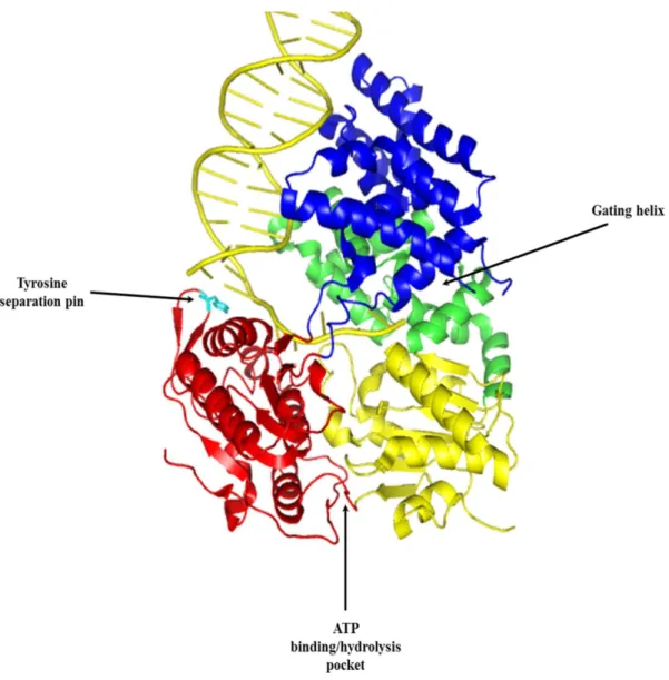
SF1 helicases have been shown to contain 2 domains, each with 2 subdomains: 1A, 1B, 2A, and 2B. Subdomains 1A and 2A contain part, if not all, of the seven conserved amino acid motifs described above to form a binding pocket for the hydrolysis of ATP. The energy derived from ATP hydrolysis forces the tyrosine separation pin in the 2A subdomain between the bases of duplex DNA, facilitating their separation. The 1B subdomain has been thought of as a site for ssDNA binding and contains a conserved phenylalanine residue which lies outside of the conserved motifs (42). In UvrD, as the duplex is unwound, one strand of the DNA is extruded though a channel between the 1A and 1B subdomains called the gating helix. The 1B subdomain has been shown to make contacts with the ssDNA as it passes through the gating helix (Fig. 2) (42).
13
much as 80% (117). However, a direct examination of the role of the 2B subdomain in UvrD, as was performed in Rep, was not possible as the deletion of the 2B subdomain appeared to be lethal to the cell upon expression (116). Ultimately, the role of the 2B subdomain in SF1 helicases is still not well understood.
As discussed above, there are many similarities between SF1 and SF2 helicases in their sequence, and there are similarities in their structures, as well. The crystal structures of SF1 helicases UvrD, Rep, and PcrA and SF2 helicase NS3 have been reported. (39, 41, 42, 116, 118). A common feature of these structures is the nucleotide binding pocket formed between domains 1A and 2A in SF1 helicases and between domains 1 and 2 in the SF2 helicase. In the UvrD crystal structure all seven conserved motifs are present in the 1A and 2A subdomains and make contacts with the ATP binding and hydrolysis pocket. The crystal structure of NS3 shows a nucleotide binding pocket nearly identical to that of the SF1 helicases. In fact, domains 1 and 2 from NS3 are structurally homologous to the 1A and 2A subdomains of UvrD and five (motifs I, Ia, II, V, and VI) of the seven conserved amino acid motifs appear to occupy the same relative position and function in both SF1 and SF2 helicases.
14
15
unwinding (115, 116). When the 2B subdomain was deleted from Rep helicase, the helicase activity of the monomeric form of Rep was activated. These studies suggest that the 2B subdomain in Rep helicase exhibits an autoregulatory control over the unwinding activity of the helicase. The crystal structures of the structurally similar UvrD and PcrA helicase monomer bound to a dsDNA/ssDNA junction show the 2B subdomain in contact with the duplex DNA. Although this 2B subdomain/duplex DNA interaction has been suggested to facilitate DNA unwinding (41, 42), the data from the Rep helicase studies clearly show that the 2B subdomain is not required for unwinding activity. An alternate interpretation is that the 2B subdomain might inhibit the DNA unwinding activity of the monomeric form of the helicase. Self-assembly into dimers or higher order oligomers might relieve this inhibition through the movement of the 2B subdomain (4). These hypotheses may present a new, but poorly understood mechanism by which SF1 helicases regulate their unwinding activity.
16
discuss the impact the 2B subdomain has on the activity of UvrD and propose experiments and future research directions.
17
REFERENCES
1. Matson SW, George JW. DNA helicase II of Escherichia coli. characterization of the single-stranded DNA-dependent NTPase and helicase activities. J Biol Chem 1987 Feb 15;262(5):2066-76.
2. Lohman TM. Helicase-catalyzed DNA unwinding. J Biol Chem 1993 Feb 5;268(4):2269-72.
3. Hall MC, Matson SW. Helicase motifs: The engine that powers DNA unwinding. Mol Microbiol 1999 Dec;34(5):867-77.
4. Lohman TM, Tomko EJ, Wu CG. Non-hexameric DNA helicases and translocases: Mechanisms and regulation. Nat Rev Mol Cell Biol 2008 May;9(5):391-401.
5. Larsen NB, Hickson ID. RecQ helicases: Conserved guardians of genomic integrity. Adv Exp Med Biol 2013;767:161-84.
6. Chu WK, Hickson ID. RecQ helicases: Multifunctional genome caretakers. Nat Rev Cancer 2009 Sep;9(9):644-54.
7. Bachrati CZ, Hickson ID. RecQ helicases: Guardian angels of the DNA replication fork. Chromosoma 2008 Jun;117(3):219-33.
8. Abdel-Monem M, Durwald H, Hoffmann-Berling H. Enzymic unwinding of DNA. 2. chain separation by an ATP-dependent DNA unwinding enzyme. Eur J Biochem 1976 Jun 1;65(2):441-9.
9. Frost LS, Ippen-Ihler K, Skurray RA. Analysis of the sequence and gene products of the transfer region of the F sex factor. Microbiol Rev 1994 Jun;58(2):162-210.
10. Mok M, Marians KJ. The escherichia coli preprimosome and DNA B helicase can form replication forks that move at the same rate. J Biol Chem 1987 Dec 5;262(34):16644-54. 11. Ali JA, Lohman TM. Kinetic measurement of the step size of DNA unwinding by
escherichia coli UvrD helicase. Science 1997 Jan 17;275(5298):377-80.
12. Singleton MR, Dillingham MS, Wigley DB. Structure and mechanism of helicases and nucleic acid translocases. Annu Rev Biochem 2007;76:23-50.
18
14. Zhang XD, Dou SX, Xie P, Hu JS, Wang PY, Xi XG. Escherichia coli RecQ is a rapid, efficient, and monomeric helicase. J Biol Chem 2006 May 5;281(18):12655-63. 15. Singleton MR, Dillingham MS, Wigley DB. Structure and mechanism of helicases and
nucleic acid translocases. Annu Rev Biochem 2007;76:23-50.
16. Hodgman TC. A new superfamily of replicative proteins. Nature 1988 May 5;333(6168):22-3.
17. Gorbalenya AE, Koonin EV, Donchenko AP, Blinov VM. A novel superfamily of nucleoside triphosphate-binding motif containing proteins which are probably involved in duplex unwinding in DNA and RNA replication and recombination. FEBS Lett 1988 Aug 1;235(1-2):16-24.
18. Gorbalenya AE, Koonin EV. Helicases: Amino acid sequence comparisons and structure-function relationships. Curr Opin Struct Biol 1993;3(3):419 <last_page> 429.
19. Fairman-Williams ME, Guenther UP, Jankowsky E. SF1 and SF2 helicases: Family matters. Curr Opin Struct Biol 2010 Jun;20(3):313-24.
20. Walker JE, Saraste M, Runswick MJ, Gay NJ. Distantly related sequences in the alpha- and beta-subunits of ATP synthase, myosin, kinases and other ATP-requiring enzymes and a common nucleotide binding fold. EMBO J 1982;1(8):945-51.
21. George JW, Brosh RM,Jr, Matson SW. A dominant negative allele of the escherichia coli uvrD gene encoding DNA helicase II. A biochemical and genetic characterization. J Mol Biol 1994 Jan 14;235(2):424-35.
22. Washburn BK, Kushner SR. Construction and analysis of deletions in the structural gene (uvrD) for DNA helicase II of escherichia coli. J Bacteriol 1991 Apr;173(8):2569-75. 23. Brosh RM,Jr, Matson SW. Mutations in motif II of escherichia coli DNA helicase II
render the enzyme nonfunctional in both mismatch repair and excision repair with differential effects on the unwinding reaction. J Bacteriol 1995 Oct;177(19):5612-21. 24. Brosh RM,Jr, Matson SW. A partially functional DNA helicase II mutant defective in
forming stable binary complexes with ATP and DNA. A role for helicase motif III. J Biol Chem 1996 Oct 11;271(41):25360-8.
25. Brosh RM,Jr, Matson SW. A point mutation in escherichia coli DNA helicase II renders the enzyme nonfunctional in two DNA repair pathways. evidence for initiation of unwinding from a nick in vivo. J Biol Chem 1997 Jan 3;272(1):572-9.
26. Hall MC, Matson SW. Mutation of a highly conserved arginine in motif IV of escherichia coli DNA helicase II results in an ATP-binding defect. J Biol Chem 1997 Jul
19
27. Velankar SS, Soultanas P, Dillingham MS, Subramanya HS, Wigley DB. Crystal structures of complexes of PcrA DNA helicase with a DNA substrate indicate an inchworm mechanism. Cell 1999 Apr 2;97(1):75-84.
28. Korolev S, Hsieh J, Gauss GH, Lohman TM, Waksman G. Major domain swiveling revealed by the crystal structures of complexes of E. coli rep helicase bound to single-stranded DNA and ADP. Cell 1997 Aug 22;90(4):635-47.
29. Graves-Woodward KL, Weller SK. Replacement of gly815 in helicase motif V alters the single-stranded DNA-dependent ATPase activity of the herpes simplex virus type 1 helicase-primase. J Biol Chem 1996 Jun 7;271(23):13629-35.
30. Naumovski L, Friedberg EC. Analysis of the essential and excision repair functions of the RAD3 gene of saccharomyces cerevisiae by mutagenesis. Mol Cell Biol 1986 Apr;6(4):1218-27.
31. Martinez R, Shao L, Weller SK. The conserved helicase motifs of the herpes simplex virus type 1 origin-binding protein UL9 are important for function. J Virol 1992 Nov;66(11):6735-46.
32. Ma L, Westbroek A, Jochemsen AG, Weeda G, Bosch A, Bootsma D, Hoeijmakers JH, van der Eb AJ. Mutational analysis of ERCC3, which is involved in DNA repair and transcription initiation: Identification of domains essential for the DNA repair function. Mol Cell Biol 1994 Jun;14(6):4126-34.
33. Richmond E, Peterson CL. Functional analysis of the DNA-stimulated ATPase domain of yeast SWI2/SNF2. Nucleic Acids Res 1996 Oct 1;24(19):3685-92.
34. Korolev S, Yao N, Lohman TM, Weber PC, Waksman G. Comparisons between the structures of HCV and rep helicases reveal structural similarities between SF1 and SF2 super-families of helicases. Protein Sci 1998 Mar;7(3):605-10.
35. Kim JL, Morgenstern KA, Griffith JP, Dwyer MD, Thomson JA, Murcko MA, Lin C, Caron PR. Hepatitis C virus NS3 RNA helicase domain with a bound oligonucleotide: The crystal structure provides insights into the mode of unwinding. Structure 1998 Jan 15;6(1):89-100.
36. Moolenaar GF, Visse R, Ortiz-Buysse M, Goosen N, van de Putte P. Helicase motifs V and VI of the escherichia coli UvrB protein of the UvrABC endonuclease are essential for the formation of the preincision complex. J Mol Biol 1994 Jul 22;240(4):294-307. 37. Hsu DS, Kim ST, Sun Q, Sancar A. Structure and function of the UvrB protein. J Biol
Chem 1995 Apr 7;270(14):8319-27.
20
39. Yao N, Hesson T, Cable M, Hong Z, Kwong AD, Le HV, Weber PC. Structure of the hepatitis C virus RNA helicase domain. Nat Struct Biol 1997 Jun;4(6):463-7.
40. Subramanya HS, Bird LE, Brannigan JA, Wigley DB. Crystal structure of a DExx box DNA helicase. Nature 1996 Nov 28;384(6607):379-83.
41. Velankar SS, Soultanas P, Dillingham MS, Subramanya HS, Wigley DB. Crystal structures of complexes of PcrA DNA helicase with a DNA substrate indicate an inchworm mechanism. Cell 1999 Apr 2;97(1):75-84.
42. Lee JY, Yang W. UvrD helicase unwinds DNA one base pair at a time by a two-part power stroke. Cell 2006 Dec 29;127(7):1349-60.
43. Macris MA, Sung P. Multifaceted role of the saccharomyces cerevisiae Srs2 helicase in homologous recombination regulation. Biochem Soc Trans 2005 Dec;33(Pt 6):1447-50. 44. Skarstad K, Katayama T. Regulating DNA replication in bacteria. Cold Spring Harb
Perspect Biol 2013 Apr 1;5(4):a012922.
45. Lorenz A, Osman F, Folkyte V, Sofueva S, Whitby MC. Fbh1 limits Rad51-dependent recombination at blocked replication forks. Mol Cell Biol 2009 Sep;29(17):4742-56. 46. Bohr VA. Rising from the RecQ-age: The role of human RecQ helicases in genome
maintenance. Trends Biochem Sci 2008 Dec;33(12):609-20.
47. Das A, Boldogh I, Lee JW, Harrigan JA, Hegde ML, Piotrowski J, de Souza Pinto N, Ramos W, Greenberg MM, Hazra TK, et al. The human werner syndrome protein stimulates repair of oxidative DNA base damage by the DNA glycosylase NEIL1. J Biol Chem 2007 Sep 7;282(36):26591-602.
48. Ahn B, Harrigan JA, Indig FE, Wilson DM,3rd, Bohr VA. Regulation of WRN helicase activity in human base excision repair. J Biol Chem 2004 Dec 17;279(51):53465-74. 49. Schurman SH, Hedayati M, Wang Z, Singh DK, Speina E, Zhang Y, Becker K, Macris
M, Sung P, Wilson DM,3rd, et al. Direct and indirect roles of RECQL4 in modulating base excision repair capacity. Hum Mol Genet 2009 Sep 15;18(18):3470-83.
50. Cooper MP, Machwe A, Orren DK, Brosh RM, Ramsden D, Bohr VA. Ku complex interacts with and stimulates the werner protein. Genes Dev 2000 Apr 15;14(8):907-12. 51. Parvathaneni S, Stortchevoi A, Sommers JA, Brosh RM,Jr, Sharma S. Human RECQ1
interacts with Ku70/80 and modulates DNA end-joining of double-strand breaks. PLoS One 2013 May 1;8(5):e62481.
21
two DNA end resection machineries for human DNA break repair. Genes Dev 2011 Feb 15;25(4):350-62.
53. Adams MD, McVey M, Sekelsky JJ. Drosophila BLM in double-strand break repair by synthesis-dependent strand annealing. Science 2003 Jan 10;299(5604):265-7.
54. Machwe A, Xiao L, Lloyd RG, Bolt E, Orren DK. Replication fork regression in vitro by the werner syndrome protein (WRN): Holliday junction formation, the effect of leading arm structure and a potential role for WRN exonuclease activity. Nucleic Acids Res 2007;35(17):5729-47.
55. Machwe A, Xiao L, Groden J, Orren DK. The werner and bloom syndrome proteins catalyze regression of a model replication fork. Biochemistry 2006 Nov
28;45(47):13939-46.
56. Davey MJ, O'Donnell M. Replicative helicase loaders: Ring breakers and ring makers. Curr Biol 2003 Aug 5;13(15):R594-6.
57. Hickman AB, Dyda F. Binding and unwinding: SF3 viral helicases. Curr Opin Struct Biol 2005 Feb;15(1):77-85.
58. Gorbalenya AE, Koonin EV, Wolf YI. A new superfamily of putative NTP-binding domains encoded by genomes of small DNA and RNA viruses. FEBS Lett 1990 Mar 12;262(1):145-8.
59. Singleton MR, Sawaya MR, Ellenberger T, Wigley DB. Crystal structure of T7 gene 4 ring helicase indicates a mechanism for sequential hydrolysis of nucleotides. Cell 2000 Jun 9;101(6):589-600.
60. Ilyina TV, Gorbalenya AE, Koonin EV. Organization and evolution of bacterial and bacteriophage primase-helicase systems. J Mol Evol 1992 Apr;34(4):351-7.
61. Gogol EP, Seifried SE, von Hippel PH. Structure and assembly of the escherichia coli transcription termination factor rho and its interaction with RNA. I. cryoelectron microscopic studies. J Mol Biol 1991 Oct 20;221(4):1127-38.
62. Skordalakes E, Berger JM. Structure of the rho transcription terminator: Mechanism of mRNA recognition and helicase loading. Cell 2003 Jul 11;114(1):135-46.
63. Labib K, Tercero JA, Diffley JF. Uninterrupted MCM2-7 function required for DNA replication fork progression. Science 2000 Jun 2;288(5471):1643-7.
64. Matson SW. Escherichia coli helicase II (urvD gene product) translocates
22
65. Maluf NK, Fischer CJ, Lohman TM. A dimer of escherichia coli UvrD is the active form of the helicase in vitro. J Mol Biol 2003 Jan 31;325(5):913-35.
66. Maluf NK, Ali JA, Lohman TM. Kinetic mechanism for formation of the active, dimeric UvrD helicase-DNA complex. J Biol Chem 2003 Aug 22;278(34):31930-40.
67. Hickson ID, Arthur HM, Bramhill D, Emmerson PT. The E. coli uvrD gene product is DNA helicase II. Mol Gen Genet 1983;190(2):265-70.
68. Centore RC, Leeson MC, Sandler SJ. UvrD303, a hyperhelicase mutant that antagonizes RecA-dependent SOS expression by a mechanism that depends on its C terminus. J Bacteriol 2009 Mar;191(5):1429-38.
69. Centore RC, Sandler SJ. UvrD limits the number and intensities of RecA-green fluorescent protein structures in escherichia coli K-12. J Bacteriol 2007 Apr;189(7):2915-20.
70. Lahue RS, Modrich P. Methyl-directed DNA mismatch repair in escherichia coli. Mutat Res 1988 Mar;198(1):37-43.
71. Modrich P, Lahue R. Mismatch repair in replication fidelity, genetic recombination, and cancer biology. Annu Rev Biochem 1996;65:101-33.
72. Husain I, Van Houten B, Thomas DC, Abdel-Monem M, Sancar A. Effect of DNA polymerase I and DNA helicase II on the turnover rate of UvrABC excision nuclease. Proc Natl Acad Sci U S A 1985 Oct;82(20):6774-8.
73. Schaaper R. Base selection, proofreading, and mismatch repair during DNA replication in escherichia coli. The Journal of Biological Chemistry 1993;268(32):23762.
74. Wagner RJ, Meselson M. Repair tracts in mismatched DNA heteroduplexes. DNA Repair (Amst) 1976;4(1):103.
75. Palmer BR, Marinus MG. The dam and dcm strains of escherichia coli--a review. Gene 1994 May 27;143(1):1-12.
76. Kunkel TA, Erie DA. DNA mismatch repair. Annu Rev Biochem 2005;74:681-710. 77. Tuteja N, Tuteja R. Unraveling DNA helicases. motif, structure, mechanism and
function. Eur J Biochem 2004 May;271(10):1849-63.
23
79. Qiu R, DeRocco VC, Harris C, Sharma A, Hingorani MM, Erie DA, Weninger KR. Large conformational changes in MutS during DNA scanning, mismatch recognition and repair signalling. EMBO J 2012 May 30;31(11):2528-40.
80. Geng H, Sakato M, DeRocco V, Yamane K, Du C, Erie DA, Hingorani M, Hsieh P. Biochemical analysis of the human mismatch repair proteins hMutSalpha
MSH2(G674A)-MSH6 and MSH2-MSH6(T1219D). J Biol Chem 2012 Mar 23;287(13):9777-91.
81. Mechanic LE, Frankel BA, Matson SW. Escherichia coli MutL loads DNA helicase II onto DNA. J Biol Chem 2000 Dec 8;275(49):38337-46.
82. Cheng F, Hou J, Chen YY, Zhou Y, Zhang HT, Bi LJ, Zhang XE. Functional interaction between MutL and 3'-5' exonuclease X in escherichia coli. Arch Biochem Biophys 2010 Oct 1;502(1):39-43.
83. Grilley M, Welsh KM, Su SS, Modrich P. Isolation and characterization of the escherichia coli mutL gene product. J Biol Chem 1989 Jan 15;264(2):1000-4.
84. Lu AL, Welsh K, Clark S, Su SS, Modrich P. Repair of DNA base-pair mismatches in extracts of escherichia coli. Cold Spring Harb Symp Quant Biol 1984;49:589-96. 85. Welsh KM, Lu AL, Clark S, Modrich P. Isolation and characterization of the escherichia
coli mutH gene product. J Biol Chem 1987 Nov 15;262(32):15624-9.
86. Hall MC, Matson SW. The escherichia coli MutL protein physically interacts with MutH and stimulates the MutH-associated endonuclease activity. J Biol Chem 1999 Jan 15;274(3):1306-12.
87. Bruni R, Martin D, Jiricny J. D(GATC) sequences influence escherichia coli mismatch repair in a distance-dependent manner from positions both upstream and downstream of the mismatch. Nucleic Acids Res 1988 Jun 10;16(11):4875-90.
88. Lahue RS, Au KG, Modrich P. DNA mismatch correction in a defined system. Science 1989 Jul 14;245(4914):160-4.
89. Chopra I, O'Neill AJ, Miller K. The role of mutators in the emergence of antibiotic-resistant bacteria. Drug Resist Updat 2003 Jun;6(3):137-45.
90. Russell A, Chopra I. Understanding antibacterial action and resistance. 2nd ed. Hertfordshire: Ellis Horwood Ltd; 1996. .
24
92. Boe L, Danielsen M, Knudsen S, Petersen JB, Maymann J, Jensen PR. The frequency of mutators in populations of escherichia coli. Mutat Res 2000 Mar 14;448(1):47-55. 93. LeClerc JE, Li B, Payne WL, Cebula TA. High mutation frequencies among escherichia
coli and salmonella pathogens. Science 1996 Nov 15;274(5290):1208-11.
94. Schaaff F, Reipert A, Bierbaum G. An elevated mutation frequency favors development of vancomycin resistance in staphylococcus aureus. Antimicrob Agents Chemother 2002 Nov;46(11):3540-8.
95. Ban C, Yang W. Crystal structure and ATPase activity of MutL: Implications for DNA repair and mutagenesis. Cell 1998 Nov 13;95(4):541-52.
96. Ban C, Junop M, Yang W. Transformation of MutL by ATP binding and hydrolysis: A switch in DNA mismatch repair. Cell 1999 Apr 2;97(1):85-97.
97. Dutta R, Inouye M. GHKL, an emergent ATPase/kinase superfamily. Trends Biochem Sci 2000 Jan;25(1):24-8.
98. Guarne A, Junop MS, Yang W. Structure and function of the N-terminal 40 kDa fragment of human PMS2: A monomeric GHL ATPase. EMBO J 2001 Oct 1;20(19):5521-31.
99. Hu X, Machius M, Yang W. Monovalent cation dependence and preference of GHKL ATPases and kinases. FEBS Lett 2003 Jun 5;544(1-3):268-73.
100. Cheng F, Hou J, Chen YY, Zhou Y, Zhang HT, Bi LJ, Zhang XE. Functional interaction between MutL and 3'-5' exonuclease X in escherichia coli. Arch Biochem Biophys 2010 Oct 1;502(1):39-43.
101. Modrich P, Lahue R. Mismatch repair in replication fidelity, genetic recombination, and cancer biology. Annu Rev Biochem 1996;65:101-33.
102. Guarne A, Ramon-Maiques S, Wolff EM, Ghirlando R, Hu X, Miller JH, Yang W. Structure of the MutL C-terminal domain: A model of intact MutL and its roles in mismatch repair. EMBO J 2004 Oct 27;23(21):4134-45.
103. Robertson A, Pattishall SR, Matson SW. The DNA binding activity of MutL is required for methyl-directed mismatch repair in escherichia coli. J Biol Chem 2006 Mar
31;281(13):8399-408.
104. Robertson AB, Pattishall SR, Gibbons EA, Matson SW. MutL-catalyzed ATP
25
105. SETLOW RB, CARRIER WL. The disappearance of thymine dimers from dna: An error-correcting mechanism. Proc Natl Acad Sci U S A 1964 Feb;51:226-31.
106. BOYCE RP, HOWARD-FLANDERS P. Release of ultraviolet light-induced thymine dimers from dna in E. coli K-12. Proc Natl Acad Sci U S A 1964 Feb;51:293-300. 107. Sancar A, Rupp WD. A novel repair enzyme: UVRABC excision nuclease of
escherichia coli cuts a DNA strand on both sides of the damaged region. Cell 1983 May;33(1):249-60.
108. Orren DK, Sancar A. The (A)BC excinuclease of escherichia coli has only the UvrB and UvrC subunits in the incision complex. Proc Natl Acad Sci U S A 1989 Jul;86(14):5237-41.
109. Verhoeven EE, Wyman C, Moolenaar GF, Goosen N. The presence of two UvrB subunits in the UvrAB complex ensures damage detection in both DNA strands. EMBO J 2002 Aug 1;21(15):4196-205.
110. Moolenaar GF. The role of ATP binding and hydrolysis by UvrB during nucleotide excision repair. J Biol Chem 2000;275(11):8044 <last_page> 8050.
111. Karakas E, Truglio JJ, Croteau D, Rhau B, Wang L, Van Houten B, Kisker C. Structure of the C-terminal half of UvrC reveals an RNase H endonuclease domain with an argonaute-like catalytic triad. EMBO J 2007 Jan 24;26(2):613-22.
112. Truglio JJ, Rhau B, Croteau DL, Wang L, Skorvaga M, Karakas E, DellaVecchia MJ, Wang H, Van Houten B, Kisker C. Structural insights into the first incision reaction during nucleotide excision repair. EMBO J 2005 Mar 9;24(5):885-94.
113. Pakotiprapha D, Jeruzalmi D. Small-angle X-ray scattering reveals architecture and A₂B₂ stoichiometry of the UvrA-UvrB DNA damage sensor.. Proteins 2013;81(1):132. 114. Atkinson J, Guy CP, Cadman CJ, Moolenaar GF, Goosen N, McGlynn P. Stimulation of
UvrD helicase by UvrAB. J Biol Chem 2009 Apr 3;284(14):9612-23.
115. Brendza KM, Cheng W, Fischer CJ, Chesnik MA, Niedziela -Majka A, Lohman TM. Autoinhibition of escherichia coli rep monomer helicase activity by its 2B subdomain. Proceedings of the National Academy of Sciences 2005;102(29):10076.
116. Cheng W, Brendza KM, Gauss GH, Korolev S, Waksman G, Lohman TM. The 2B domain of the escherichia coli rep protein is not required for DNA helicase activity. Proc Natl Acad Sci U S A 2002 Dec 10;99(25):16006-11.
117. Zhang G, Deng E, Baugh L, Kushner SR. Identification and characterization of
26
118. Kim JL, Morgenstern KA, Griffith JP, Dwyer MD, Thomson JA, Murcko MA, Lin C, Caron PR. Hepatitis C virus NS3 RNA helicase domain with a bound oligonucleotide: The crystal structure provides insights into the mode of unwinding. Structure
27
CHAPTER 2: THE UVRD303 HYPER-HELICASE EXHIBITS INCREASED PROCESSIVITY
Abstract
28 Introduction
DNA helicases are ubiquitous motor proteins that transiently convert duplex DNA into single-stranded DNA (ssDNA) using energy derived from nucleoside 5'-triphosphate (NTP) hydrolysis for a wide variety of biological processes including DNA replication, repair and recombination (1-5). Thus, helicases are vital for the maintenance of the genome and mutations within helicase-encoding genes in humans have been linked to both cancer and aging disorders (6-8). In E. coli, the product of the uvrD gene, DNA helicase II (UvrD), has been shown to function in two fundamental DNA repair processes – nucleotide excision repair (NER) (9) and methyl-directed mismatch repair (MMR) (10, 11). In NER, UvrD acts in conjunction with the UvrABC excision nuclease and DNA polymerase I to remove short 12-13 base oligonucleotides containing a wide variety of bulky lesions including pyrimidine dimers (9). UvrD also participates in the MMR pathway by initiating unwinding at the nicked d(GATC) site created by MutH to displace the nascent error-containing strand of DNA which is degraded by appropriate exonucleases. The resulting gap is subsequently resynthesized by DNA polymerase III (10, 12). UvrD has been suggested to have a role in other cellular processes, such as displacement of RecA protein from ssDNA during homologous recombination (13). However, this role for UvrD is not as well understood. The uvrD gene encodes a 720 amino acid, 82 kDa Superfamily 1A (SF1A) helicase with well characterized 3' to 5' unwinding and translocase directionality. Consistent with other SF1 helicases, UvrD contains seven conserved amino acid motifs and two structural domains (1 and 2) each with two subdomains (A and B) (14-21).
29
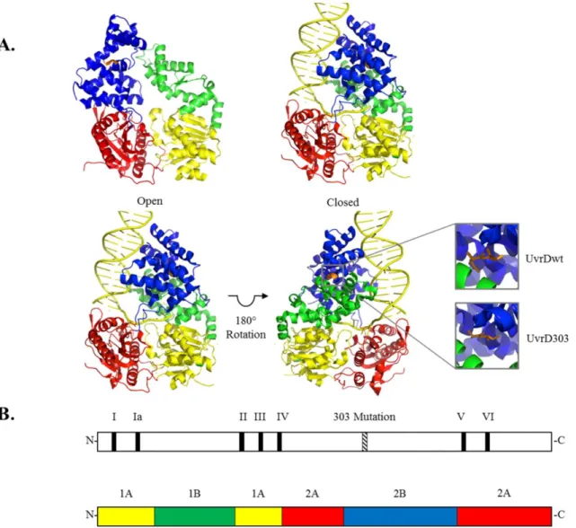
of adopting two conformations – open and closed – with the major difference between the two conformations being the orientation of the 2B subdomain. In the ‘open’ conformation the 2B subdomain is stacked above the 2A subdomain and no contacts are made between the 2B and 1B subdomains. To adopt the alternate ‘closed’ structure, the 2B subdomain rotates about a flexible hinge connected to the 2A subdomain and closes onto the 1B subdomain. The magnitude of the 2B subdomain rotation as the helicases modulate between the open and closed conformations has been reported to be 130° in Rep protein and 160° in UvrD. Although it is known that these SF1 helicases can adopt these alternate conformations, it is not clear why there are two alternate conformations. The related SF2 helicases (e.g. the hepatitis NS3 helicase) lack the equivalent of the 2B subdomain (23, 24). Consequently, the role of the 2B subdomain in SF1 helicases is not well understood.
Previous studies (25, 26) have examined what role, if any, the 2B subdomain of Rep protein plays in the activity of this helicase. It was concluded that the 2B subdomain is most likely a regulatory domain since deletion of this subdomain stimulates the unwinding and translocase activity of the protein in addition to activating the helicase activity of a Rep2B monomer. However, the role of the 2B subdomain in UvrD continues to be debated (14). The two prevailing hypotheses suggest that either the 2B subdomain is required to make direct double-stranded DNA interactions which allow the helicase to bind various substrates or that the 2B subdomain functions in a regulatory role, modulating helicase activity. Direct studies of a corresponding deletion of the 2B subdomain in UvrD have not been reported due to the apparent lethality of the expression of UvrD2B (25).
30
native amino acids at positions 403 and 404 (both aspartic acids) were mutated to alanines (Fig. 4). When UvrD303 was expressed in place of the wild-type protein, from either the chromosome or a high copy number plasmid, cells exhibited antimutator, UV sensitive and hypo-recombinant phenotypes (27, 28). When purified and analyzed in vitro, UvrD303 exhibited a ten-fold increase in unwinding activity compared to its wild-type counterpart. This increased activity led to UvrD303 being referred to as a “hyper-helicase”. Interestingly, neither D403A nor D404A alone were sufficient to convey this hyper-helicase activity (27).
This chapter presents a biochemical characterization of the UvrD303 mutant including a possible explanation for how the 2B subdomain mutation increases the unwinding activity of the helicase. Using single turnover, rapid quench helicase assays we have shown that the processivity of UvrD303, as a DNA helicase, is approximately ten -fold higher than that of wild-type UvrD. We speculate that the mutation alters the ability of the 2B subdomain to properly adopt a closed conformation, as referred to above. The closed conformation appears to be important for normal endogenous helicase activity of wild-type UvrD as evidenced by the phenotype of cells expressing UvrD303.
Materials and Methods
Helicase purification –The uvrD gene was amplified via polymerase chain reaction from genomic K12 +
F+ DNA using the following primers: forward primer 5ʹ-CGGC
31
32
electrophoresis and excision of the DNA followed by use of a Qiagen quick spin purification column. The purified digested vector was treated with shrimp alkaline phosphatase to reduce the likelihood of vector re-ligation in subsequent cloning reactions. The uvrD containing PCR product was digested with the restriction enzyme NcoI and purified in a manner similar to the vector. The resultant digested vector and PCR insert were ligated using T4 ligase. Using this cloning technique, the SmaI site in pTYB4-His is destroyed, but will ensure that only a C-terminal glycine will be added to the protein after purification, yielding essentially native protein. This construct enabled the rapid purification of the helicase as previously described (29). The uvrD303 allele was constructed using site-directed mutagenesis. A quickchange PCR was performed using primers of the following sequence: forward CTGATTGCCAACCGCAACGACGACGCGGCCTTTGAGCGTGTG-3ʹ and reverse 5ʹ-CACACGCTCAAAGGCCGCGTCGTCGTTGCGGTTGGCAATCAG-3ʹ to change the 403rd and 404th codons from -GACGAC- to -GCGGCC-, thus converting both the aspartic acids to alanines. This mutagenesis also introduced a silent NotI restriction site which was used to screen potential clones for the uvrD303 allele. The resulting plasmid was sequenced to ensure no additional mutations had been introduced.
33
(Clone-tech) and Chitin (NEB) resins to take advantage of the two affinity tags present on the overexpressed fusion protein (29). Protein that eluted from the Chitin column was dialyzed against UvrD storage buffer (31) and stored at -20oC. The purified protein was greater than 95% homogeneous as determined by polyacrylamide gel electrophoresis in the presence of sodium dodecyl sulfate (SDS).
Wild-type UvrD was purified as previously described (31).
DNA Substrates – Partial duplex substrates were prepared by radiolabeling the 5'-end of oligonucleotides obtained from Integrated DNA Technologies (Coralville, Iowa) using [
-32
P]ATP and T4 polynucleotide kinase (see Table 1 for sequences). A 1.1 fold excess of an unlabeled complementary oligonucleotide was annealed to the labeled strand by heating the two strands to 95°C for 5 minutes and allowing them to slow cool to 25°C overnight. The partial duplex substrate was purified from free nucleotide using a Sephadex G50 spin column and dialyzed into TEN buffer (10 mM Tris-HCl (pH 7.5), 1 mM EDTA, 50 mM NaCl). This process was used to generate the 24/64 and 90/130 partial duplex substrates. In each case the shorter DNA strand was radioactively labeled. The longer DNA strand contains a 40 base 3'-tail ssDNA to facilitate loading of UvrD.
34 Table 1. Oligonucleotides used in the UvrD303 study
Oligonucleotide Sequence
RQ24: 5ʹ -GCCCTGCTGCCGACCAACGAAGGT- 3ʹ
RQ64: 5ʹ -ACCTTCGTTGGTCGGCAGCAGGGC (T40) -3ʹ
RQ90: 5ʹ -GCCCTGCTGCCGACCAACGAAGGTTACATTCCCCGTGCTGGCCGT TTG
CGGTTGTCCTGTACCACTCGAAGTAGGAGGGGTGCTCACCGA -3ʹ
RQ130: 5ʹ -TCGGTGAGCACCCCTCCTACTTCGAGTGGTACAGGACAACCGCAA
ACGGCCAGCACGGGGAATGTAACCTTCGTTGGTCGGCAGCAGGGC (T40) -3ʹ
Hairpin Trap: 5ʹ -CCTCGCTGCTTTTTGCAGCGAGGC (T30) -3ʹ
Fluorescence Anisotropy Labeled: 5ʹ -TATCGGCACGTCTCGAGATG-Cy5 -3ʹ
35
elongated version of pUC19 was then used in a site directed mutagenic PCR (with the following primers: forward 5ʹ-GGATCCTCAGCAGTCGACCTCAG CGCATGC-3ʹ and reverse 5ʹ-GCATGCGCT GAGGTCGACTGCTGAGGATCC-3ʹ to create pUC19-TS, which contains a 67 bp stretch of DNA between the EcoRI and HindIII restriction sites (5ʹ-CCTCAGCAATCCTCAGCCAGGCC
TCAGCTGGCCTCAGCGGATCCTCAGCAGTCGACCTCAGCGCATG-3ʹ) containing
multiple Nt.BbvC1 nicking sites.
To produce the 243 bp partial duplex substrate 50 pmols of plasmid DNA were digested with EcoRI and SapI to completion to produce a 297 bp DNA fragment. The enzymes were heat killed at 65°C for 20 minutes and the resulting DNA was purified by gel extraction using a 1.5% (w/v) (0.75% (w/v) agarose, 0.75% (w/v) low melting agarose) agarose gel. The three nucleotide overhang generated by the SapI digest was filled in using Klenow fragment polymerase and [-32
P]dCTP, dGTP and dTTP. The fill-in reaction was incubated at 37oC for 30 minutes. The purified DNA fragment, which contains seven Nt.BbvCI nickase sites (CCTCAGC), was nicked with Nt.BbvCI followed by heating to 80°C for 20 minutes to denature the on-average 11 nucleotide fragments generated by the nicking reaction. The resulting DNA molecule contains a 45 base 3'-ssDNA tail preceding a 243 bp duplex region. The nicked DNA was immediately applied to Qiagen QIAquick PCR Purification spin columns for purification (1 column used per 8 pmols of DNA) and eluted in TEN buffer. The final DNA concentration was determined by scintillation counting.
DNA-dependent ATPase assays – The standard reaction mixture (40 l) contained 25
mM Tris-HCl (pH 7.5), 3 mM MgCl2, 20 mM NaCl, 5 mM 2-mercaptoethanol, 50 g/mL
36
or UvrD303 helicase. For kcat determinations, the ATP concentration was 400 M. For Km determinations the ATP concentration ranged from 50 to 500 M. All reagents except ATP were mixed and allowed to incubate on ice for 5 minutes. Then ATP was added and the reaction was incubated at 37°C for 10 minutes. Aliquots (5 l) were removed every two minutes and 2 L of 5 M formic acid was added to stop the reaction. 2.5 L of this mixture was spotted onto a thin layer chromatography plate and developed in a 0.45 M ammonium sulfate solution. Results were visualized by PhosphorImaging (Molecular Dynamics).
Helicase Assays – Helicase reaction mixtures (16 L) for steady-state experiments contained 25 mM Tris-HCl (pH7.5), 3 mM MgCl2, 20 mM NaCl, 5 mM 2-mercaptoethanol
(ME), 50 g/mL bovine serum albumin, 3 mM ATP, helicase concentration as described, and approximately 0.2 nM radiolabeled partial duplex DNA substrate. The reactions were assembled on ice and the helicase was allowed to preincubate with the DNA substrate for several minutes. The reactions were initiated by the addition of ATP and incubated at 37°C for 5 minutes before addition of 8 L of stop solution, 37.5% (v/v) glycerol, 50 mM EDTA, 0.3% (w/v) SDS, 0.5x TBE and dyes. The reactions were resolved on 12% (w/v) or 8% (w/v) non-denaturing polyacrylamide gels (19:1 crosslinking ratio), or 3% (w/v) nusieve agarose gels depending on substrate size and visualized by PhosphorImaging (Molecular Dynamics).
37
mM ATP on ice for 15 min, and then loaded into the D loop. The opposite E loop was loaded with 6 mM MgCl2 and 3 M DNA hairpin trap in buffer M (25 mM Tris-HCl (pH 7.5), 25
mM NaCl, 10% (v/v) glycerol, 5 mM ME). Reactions were initiated by rapidly mixing equal volume aliquots of the solutions from loops D and E, and were quenched with 200 mM EDTA and 0.2% (w/v) SDS. Samples were resolved on non-denaturing polyacrylamide gels or 3% (w/v) nusieve agarose gels as described above and visualized by PhosphorImaging (Molecular Dynamics).
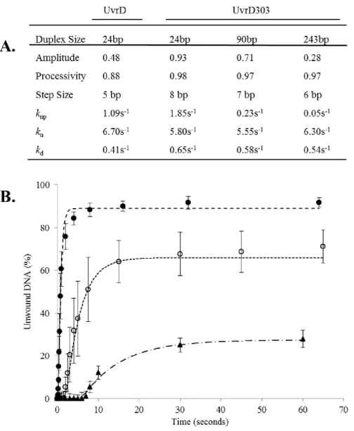
The modeled unwinding curves were fit to the data collected with each partial duplex DNA substrate using a linear regression model and the kinetic simulator Tenua (Bililite). Using the previously published step size as a starting reference, the pre-steady state kinetics were modeled using the n-step kinetic scheme (see Scheme 1). The modeled data were compared to the collected data by calculating the residuals for each collected data point at the corresponding point generated by the model. The number of steps, knp, kd and ku were manually adjusted until a best fit was obtained. The fits which produced the smallest residuals compared to model were defined as best fits. The modeled curves were visually inspected to ensure the quality of the fit. The data for each substrate length was ana lyzed in this manner and best fits calculated.
38 Results
The uvrD303 allele, containing amino acid substitutions at positions 403 and 404 (D403A, D404A), was originally isolated by Kushner and colleagues (27) in a screen for mutations in regions outside the conserved helicase motifs that impact biological function. Cells expressing this mutant form of UvrD from a plasmid exhibit increased sensitivity to ultraviolet light and methyl methane sulfonate as do cells containing a deletion of the uvrD gene. Interestingly, cells expressing uvrD303 also exhibit an antimutator phenotype (27) whereas a uvrD strain has a mutator phenotype (36). A strain containing uvrD303 in the chromosome exhibits similar phenotypes (28) indicating that the phenotypes observed are not simply due to increased expression of the mutant protein. Importantly, the purified UvrD303 protein was characterized as having a slightly increased specific activity as an ATPase and unwinds partial duplex substrates 10-fold better than the wild-type enzyme (27) leading to its designation as a hyper-helicase. We have used biochemical assays, including pre-steady state, single turnover kinetic experiments, to further understand the mechanistic basis of the increased helicase activity associated with this mutant protein.
39
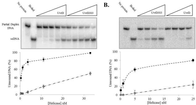
Either UvrD or UvrD303 was incubated with the DNA substrate and unwinding was initiated by the addition of ATP. Using a 24 base pair (bp) partial duplex, the wild-type helicase unwound approximately 6% of the substrate at a concentration of 3.7 nM as compared to unwinding of 77% of the substrate at an equal concentration of UvrD303 (Fig. 5A). This represents nearly a 13-fold increase in unwinding activity. The significant increase in unwinding activity observed with UvrD303 was also demonstrated using a 90 bp partial duplex substrate. On this longer substrate only about 4% of the DNA was unwound by the wild-type helicase at a concentration of 5 nM as compared to 59% unwound by UvrD303 at the same concentration (Fig. 5B). These data support previous work showing that UvrD303 exhibits a marked increase in helicase activity under multiple turnover conditions (27).
The multiple turnover unwinding experiments, while informative, do not yield significant new information about the mechanism by which the helicase activity is increased. The observed increase could be due to one or more of any number of properties being altered in the mutant including binding affinity for the substrate, an increase in the rate of unwinding or an increase in the processivity of the unwinding reaction.
40
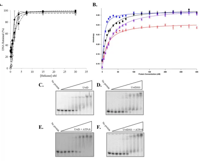
ligand with 20 bp of duplex DNA and a 40 nucleotide 3'-ssDNA tail to mimic the substrate used in unwinding experiments. Ten individual measurements were made at each protein concentration and the average was plotted as a function of protein concentration (Fig. 6B). These experiments yielded a Kd for UvrD of 23.5 ± 1.4 nM in the absence of nucleotide and 14.6 ± 1.4 nM in the presence of the poorly hydrolyzed ATP analog, ATPS. These values are somewhat higher than previously reported Kd values for UvrD (37) which were determined using a different technique and a different DNA ligand. The Kd measured for UvrD303 was 15.7 ± 2.3 nM in the absence of nucleotide and 3.9 ± 0.6 nM in the presence of ATPS. These data suggest that UvrD303 has a somewhat higher initial binding affinity for the partial duplex substrate than UvrD. However, this difference in initial DNA binding affinity alone is not likely to account for the dramatic increase in unwinding observed using UvrD303.
To investigate further the increase in UvrD303-catalzyed unwinding, single turnover pre-steady state kinetic experiments were performed. Using a rapid chemical quenched flow protocol, excess helicase (50 nM final concentration), the partial duplex substrate (1 nM final concentration) and ATP were pre-incubated to allow formation of the enzyme-substrate complex, and then rapidly mixed with MgCl2 and a 1500-fold excess of a DNA hairpin trap
41
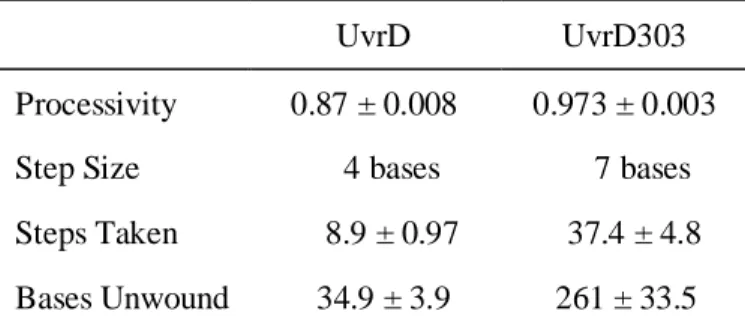
partial duplex substrate generated by digestion of pUC19-TS plasmid as described under “Materials and Methods”. It is important to note that these unwinding assays are ‘all or none’ and do not detect partially unwound DNA molecules. Using the 24 bp partial duplex DNA, both UvrD and UvrD303 were capable of converting the duplex substrate to ssDNA (Fig. 7A). Under these conditions UvrD unwound approximately 40% of the substrate while UvrD303 unwound nearly 95% of the substrate. It also appears that the exponential rise trends generated by the unwinding reactions level off at very similar time points. If these trends are compared with respect to the total amount of partial duplex substrate, a simple rate describing the approximate number of base pairs unwound per second per UvrD molecule can be estimated. These rates on this 24 bp partial duplex substrate are estimated to be 0.13 bp unwound per second per UvrD and 0.19 bp unwound per second per UvrD303. These data suggest that the unwinding reaction catalyzed by UvrD and UvrD303 occurs at a similar rate but UvrD303 is able to unwind a much larger fraction of the substrate.
The similarity of the rates of unwinding prompted us to also measure the rate of ATP hydrolysis by the wild-type and mutant protein. Previous data (27) suggested there was a modest increase in ATP hydrolysis activity conveyed by the 303 mutation. However, the Km
for ATP was nearly identical for each helicase. We performed similar ATPase assays and observed the same result. UvrD303 does have a somewhat higher ATPase activity and the Km
42
43
44
Therefore, UvrD is not expected to unwind a 90 bp partial duplex substrate in a single turnover experiment. UvrD303, however, was able to unwind nearly 70% of the of the 90 bp substrate (Fig. 7B).
45
Figure 7. UvrD303 exhibits a stimulated helicase activity in single turnover rapid quench reactions. Panel A – DNA helicase single turnover rapid quench reactions were performed as described under “Materials and Methods” using a 24 bp partial duplex substrate and increasing time. The first two lanes are controls for heat denatured substrate and for no magnesium chloride (MgCl2). The remaining lanes depict a time course from 0.25 to 64
seconds for both UvrD and UvrD303. Quantitative data from 3 experiments for UvrD (open circles) and UvrD303 (filled squares) were plotted as an average. Panel B – DNA helicase single turnover rapid quench reactions were performed as described under “Materials and Methods” using a 90 bp partial duplex substrate and increasing time. The first two lanes are controls for heat denatured substrate and no magnesium chloride (MgCl2). The remaining
lanes depict a time course from 0.075 to 65 seconds for UvrD303. Quantitative data represent the average of 3 experiments using UvrD303. Panel C – DNA helicase single turnover rapid quench reactions were performed as described in “Materials and Methods” using a 243 bp partial duplex substrate and increasing time. The first two lanes are controls for heat denatured substrate and no magnesium chloride (MgCl2). The remaining lanes depict a time
46
47
unwound per binding event is an approximate eight-fold increase and accounts for the previously reported ten-fold increase in helicase activity.
Discussion
The uvrD303 allele was discovered in a screen for mutations outside the conserved helicase motifs of UvrD that had a clear impact on the activity of the protein as measured in genetic assays (27). UvrD303 contains two amino acid substitutions (D403A, D404A) in the 2B subdomain of the protein between conserved motifs IV and V (see Fig. 4B). The impact of this mutation is remarkable both for its alteration of the biochemistry of the protein and the significantly different phenotype of cells harboring this mutation. The purified protein has been described as a “hyper-helicase” based on a steady state analysis of helicase activity.