DORSAL HIPPOCAMPAL ASTROCYTE SIGNALING REGULATES HEROIN-CONDITIONED IMMUNOMODULATION BUT NOT HEROIN-HEROIN-CONDITIONED
PLACE PREFERENCE
Jacqueline E. Paniccia
A thesis submitted to the faculty at the University of North Carolina at Chapel Hill in partial fulfillment of the requirements for the degree of Master of Psychology and Neuroscience in the
Behavioral Neuroscience program in the College of Arts and Sciences.
Chapel Hill 2018
Approved by:
Donald T. Lysle
Kathryn J. Reissner
ii © 2018
ABSTRACT
Jacqueline E. Paniccia: Dorsal hippocampal astrocyte signaling regulates heroin-conditioned immunomodulation but not heroin-conditioned place preference
(Under the direction of Donald T. Lysle)
Repeated context-heroin pairings result in Pavlovian associations that manifest as
conditioned appetitive responses or peripheral immunomodulation upon re-exposure to
heroin-conditioned stimuli. The dorsal hippocampus (DH) is a critical neural substrate governing these
context-heroin associations. Within the DH, there appears to be divergent mechanisms mediating
heroin-conditioned Pavlovian responses. Evidence suggests that DH interleukin-1 signaling
regulates heroin-conditioned immunomodulation but not heroin-conditioned place preference
(CPP). The present study sought to further investigate the role of DH neuroimmune signaling in
heroin-conditioned Pavlovian responses. Astroglial activity has been implicated in both drug
addiction and mechanisms of learning and memory. As such, we employed chemogenetic tools to
examine the involvement of DH astrocytes in the expression of both heroin-conditioned
immunomodulatory and appetitive responses. Interestingly, chemogenetic stimulation of DH
astroglial Gi-signaling disrupted conditioned immunomodulation but did not alter CPP. These data provide further evidence that differential DH mechanisms regulate
iv
To my parents:
ACKNOWLEDGEMENTS
Many thanks to the Lysle Laboratory, Christina L. Lebonville, Meghan E. Jones, Shveta
V. Parekh, and to my advisor, Donald T. Lysle, whose guidance, support, and editing were
vi
TABLE OF CONTENTS
LIST OF FIGURES...viii
LIST OF ABBREVIATIONS...ix
CHAPTER 1: DORSAL HIPPOCAMPAL ASTROCYTE SIGNALING REGULATES CONDITIONED IMMUNOMODULATION BUT NOT HEROIN-CONDITIONED PLACE PREFERENCE...1
Introduction...1
Methods and Materials...4
Animals...4
Drug Administration...4
Surgical Procedures...4
Heroin-conditioned immunomodulation...5
Heroin-conditioned place preference...6
Tissue Collection and Histology...7
Immunohistochemistry...7
Microscopy...8
RNA Extraction and cDNA Synthesis...8
qPCR Quantification of Splenic iNOS Gene Expression...9
Nitrate/nitrite Assay...10
Statistical Analysis...11
Experiment 1: Stimulation of astroglial Gi-signaling disrupts
heroin-conditioned immunomodulation...11
Experiment 2: Stimulation of astroglial Gi-signaling does not alter heroin-conditioned place preference...13
Discussion...15
Acknowledgement...20
viii
LIST OF FIGURES
Figure 1 – Stimulation of GFAP-hM4D(Gi) blocks heroin-conditioned immunomodulation...12 Figure 2 – Expression of GFAP-hM4D(Gi) is specific to DH astrocytes...13 Figure 3 – Stimulation of GFAP-hM4D(Gi) does not alter heroin-conditioned place
LIST OF ABBREVIATIONS
AAV adeno-associated virus
ANOVA analysis of variance
cAMP cyclic adenosine monophosphate
CNO clozapine-N-oxide
CS conditioned stimulus
CPP conditioned place preference
ΔΔCT Comparative CT
DH dorsal hippocampus
DMSO dimethyl sulfoxide
DREADD designer receptors exclusively activated by designer drugs
GFAP glial fibrillary acidic protein
IHC immunohistochemistry
IL-1 interleukin-1
IL-1β interleukin-1β
IL-1R1 IL-1 receptor type 1
IL-1RA interleukin-1 receptor antagonist
iNOS inducible nitric oxide synthase
LPS lipopolysaccharide
mRNA messenger RNA
NAc nucleus accumbens
NF-κB nuclear factor-κB
x
PB phosphate buffer
Rpl13a 60S ribosomal protein L13a
CHAPTER 1: DORSAL HIPPOCAMPAL ASTROCYTE SIGNALING REGULATES CONDITIONED IMMUNOMODULATION BUT NOT
HEROIN-CONDITIONED PLACE PREFERENCE 1
Introduction
Repeated pairings between environmental stimuli and the subjective and physiological
effects of heroin result in robust associative learning. The consequent stimulus control over
physiology and behavior is integral to heroin addiction, and has detrimental health consequences
that represent a growing public health concern. Heroin-associated contextual stimuli can act as
conditioned stimuli (CS) that trigger Pavlovian appetitive conditioned responses, including
conditioned place preference (CPP) (Tzschentke, 1998). Additionally, drug-paired contextual
stimuli can act as discriminative stimuli or occasion setters that signal drug availability and thus
engender drug-seeking behavior in instrumental paradigms (Crombag et al., 2008; Fuchs et al.,
2008). Regardless of the specific role of the contextual stimulus, the hippocampus is essential for
context-drug associative learning (Kutlu and Gould, 2016). In particular, the dorsal hippocampus
(DH) plays a critical role in drug-induced CPP (Corrigall and Linseman, 1988; Meyers et al.,
2003; Xia et al., 2017) as well as context-induced drug-seeking behaviors (Fuchs et al., 2007;
Fuchs et al., 2005; Ge et al., 2017; Xie et al., 2010).
In addition to heroin-conditioned appetitive responses, heroin-associated contextual
1This chapter previously appeared as an article in the journal Brain, Behavior, and Immunity. The original citation is
as follows: Paniccia, J.E. et. al. 2018. Dorsal hippocampal neural immune signaling regulates heroin-conditioned
2
stimuli can elicit the immunomodulatory effects induced by opioids (Lysle and Ijames, 2002).
Heroin and other opioids negatively alter host immunity (McCarthy et al., 2001; Wang et al.,
2011). Following repeated context-heroin pairings, exposure to the heroin-paired CS is sufficient
to evoke heroin-conditioned suppression of lipopolysaccharide (LPS)-induced peripheral
immune parameters (Lysle and Ijames, 2002). We have characterized heroin-conditioned
immunomodulation as classically conditioned response that follows the principles of learning
(Szczytkowski and Lysle, 2007), and found it is mediated through DH-dependent processes.
GABA agonist-induced DH inactivation during CS exposure significantly disrupts
heroin-conditioned suppression of LPS-induced peripheral indices of nitric oxide (NO) production
(Szczytkowski et al., 2013). Thus, the DH is an essential component of the neural circuitry
governing the retrieval or utilization of the context-heroin association that controls host
immunity.
Within the DH, we have discovered a distinct role of the neuroimmune system in
governing heroin-conditioned immunomodulation. The role of the pro-inflammatory cytokine
interleukin-1β (IL-1β) is well established in hippocampal-dependent memory processes (Goshen
et al., 2007; Jones et al., 2015), and there is evidence to suggest its involvement in the
development and maintenance of long-term potentiation (Donzis and Tronson, 2014; Yirmiya
and Goshen, 2011). Furthermore, signaling through the active IL-1 receptor, IL-1 receptor type 1
(IL-1R1), is vital in hippocampal-dependent learning (Ben Menachem-Zidon et al., 2011). We
have determined that both expression of DH IL-1β and signaling through IL-1R1 is required
during presentation of heroin-paired cues for heroin-conditioned immunomodulation to occur
(Paniccia et al., 2018; Szczytkowski et al., 2013). Interestingly, this involvement of IL-1
signaling in heroin-conditioned responses does not extend to heroin-conditioned appetitive
The neuroimmune system is a vastly complex network involving multiple cell types and
signaling molecules. Relevant to our model, astrocyte activity has been implicated in both
mechanisms of learning and memory (Ben Achour and Pascual, 2010; Jones et al., 2018b; Ota et
al., 2013), and substance use disorders (Lacagnina et al., 2018; Miguel-Hidalgo, 2009; Scofield
and Kalivas, 2014). Astrocytes can directly alter neuronal function and synaptic plasticity
through the release of gliotransmitters (Haydon and Carmignoto, 2006) and cytokines
(Lacagnina et al., 2018; Santello and Volterra, 2012). Interestingly, astroglia have been shown to
support hippocampal-dependent learning and memory through the expression of IL-1β (Jones et
al., 2018a) and IL-1R1 (Ben Menachem-Zidon et al., 2011). While a mechanistic link between
astrocyte activity and subsequent IL-1β release has not yet been confirmed, astrocytes may be a
critical cell population involved in mediating heroin-conditioned immunomodulation. Moreover,
the role of hippocampal astroglia in heroin-conditioned appetitive responses is presently
unknown. Thus, the current study is aimed at extending our knowledge of neuroimmune
regulation of heroin-conditioned Pavlovian responses, and examining the role of astrocyte
activity in heroin-condition immunomodulation and heroin-CPP. We employed chemogenetic
techniques to evaluate the importance of DH astroglial signaling during exposure to
heroin-associated contextual stimuli. An adeno-heroin-associated viral construct was used to selectively target
DH astroglia and express Gi-coupled designer receptors exclusively activated by designer drugs (DREADDs) in this cell population. DREADDs are mutated muscarinic receptors that no longer
respond to endogenous ligands and instead are activated by clozapine-N-oxide (CNO) (Roth,
2016). CNO-induced stimulation of astroglial Gi-signaling will attenuate induction of cyclic adenosine monophosphate (cAMP) (Jones et al., 2018b) and have distinct functional outcomes
4
astroglial Gi-signaling, in two Pavlovian procedures: heroin-conditioned immunomodulation and heroin-CPP.
Materials and Methods
Animals. Adult, male Lewis rats (~225-250 g) were purchased from Charles River
Laboratories (Kingston, NY). All rats were individually housed on a 12-hour reversed light-dark
cycle. Animals were handled regularly prior to and throughout experimental procedures.
Animals received ad libitum home cage access to food and water. All procedures were conducted
in compliance with regulations by the University of North Carolina at Chapel Hill Institutional
Animal Care and Use Committee.
Drug Administration. Heroin (diacetylmorphine, National Institute on Drug Abuse
Drug Supply Program, Bethesda, MD) was dissolved in 0.9% sterile saline. Heroin was stored at
4°C until use at room temperature. In all experiments, heroin was administered subcutaneously at
a dose of 1 mg/kg. This dose was selected based on prior research showing that it induces
conditioning and alters endotoxin-induced indices of NO production (Lysle and How, 2000;
Lysle and Ijames, 2002; Szczytkowski and Lysle, 2007). Clozapine-N-oxide (CNO; Sigma, St.
Louis, MO or the National Institutes of Health, Bethesda, MD) was dissolved in a vehicle of
0.9% sterile saline with 0.5% dimethyl sulfoxide (DMSO). In both experiments, CNO (3 mg/kg)
or vehicle was administered subcutaneously. Lipopolysaccharide (LPS; derived from E. coli,
serotype O55:B5, Sigma) was dissolved in 0.9% sterile, pyrogen-free saline. In Experiments 1,
LPS (1 mg/kg) was administered subcutaneously. This LPS dose produces sickness behavior and
induces measures of NO production.
Surgical Procedures. Animals were fully anesthetized with a 1 mg/kg intraperitoneal
injection of ketamine hydrochloride (100 mg/mL) mixed with xylazine (100 mg/mL) in a 9:1
was infused into the DH. The DREADD construct was packaged into an adeno-associated virus
(AAV) by the University of North Carolina at Chapel Hill Vector Core (Chapel Hill, North
Carolina). Injectors (33 Gauge, Plastics One) were directed bilaterally at the DH (AP -3.4 mm,
ML ± 3.1 mm, DV -3.2 mm, relative to bregma, 15° angle laterally, (Paxinos and Watson,
2006)). Purified viruses were obtained pre-dialyzed (350 mM NaCl, 5% D-sorbitol in PBS) and
were microinjected at a viral titer of 2.0 x 10¹² particles/mL (Experiment 1) or 9.8 x 10¹²
particles/mL (Experiment 2). Virus infusions of 0.7 µL per hemisphere were delivered bilaterally
at a rate of 0.05-0.1 μL/min. At the end of the infusion, injectors were left in place for 10-15 min
to allow for diffusion away from the injection site. Following virus infusion surgeries, animals
remained in their home cage for three weeks to allow for post-operative recovery and astroglial
DREADD expression.
Heroin-conditioned Immunomodulation. The heroin-conditioning paradigm employed
here has been described previously (Szczytkowski et al., 2011; Szczytkowski et al., 2013).
Briefly, all animals received five 1-h pairings of heroin with a conditioning chamber
(conditioned stimulus, CS). The conditioning chambers (BRS/LVE, Laurel, MD; H 26.7 cm × D
24.1 cm × W 30.5 cm) were located in a room separate from the vivarium. The chambers
contained metal grid flooring and cedar bedding to create an environment with different
olfactory, tactile, and visual characteristics relative to the home cage. The chambers were
enclosed within sound- and light-attenuating chambers (H 36.8 cm x D 34.3 cm x W 50.8 cm)
with a house fan to mask background noise. Heroin-conditioning sessions took place during the
dark phase of the light cycle and were separated by 48 h. Following the last conditioning session,
animals remained undisturbed in their home cage for 6 days. Animals were randomly assigned to
four groups according to a 2 (CS or home cage) x 2 (drug or vehicle) between-subjects design. In
6
treatment, the animals were re-exposed to the heroin-paired context (CS) for 1 h in the absence
of heroin or remained in their home cage. Immediately after the CS exposure or equivalent home
cage stay, the animals were injected with LPS and placed into their home cages until tissue
collection, 6 h later.
Heroin-conditioned Place Preference. The conditioned place preference (CPP)
apparatus was located in a room separate from the vivarium. A three-chambered apparatus was
used, with the two large chambers containing distinct olfactory, visual, and tactile cues from
home cage, as well as each other. Animals were habituated to the CPP apparatus. During
habituation to the CPP apparatus, baseline test, and each subsequent CPP test, animals were
given free access to all three chambers for 15 min in a heroin-free state. Behavior within the
apparatus during test sessions was video recorded using a Sony Handycam (HDR-CX455, 9.2
megapixels). The time spent in each side of the apparatus was scored manually by an
experimenter blind to treatment assignment. Twenty-four hours after habituation, a
pre-conditioning baseline CPP test was conducted to determine unconditioned side preferences.
Using a biased conditioning procedure, heroin was paired with the initially non-preferred side of
the apparatus. Saline-conditioned controls were included to test for unconditioned drift in side
preference that might occur with repeated exposure to the apparatus.
Assignment to heroin- and saline-conditioned groups, as well as to the order of heroin
and saline conditioning sessions, was counterbalanced based on unconditioned side preferences.
Animals received a heroin or saline injection and were confined to one side for 30 min. The next
day animals were injected with the opposite treatment and confined to the opposite side for 30
min. Conditioning continued as an alternating regimen across a total of 10 daily sessions.
Animals then received a CPP test. After heroin CPP was confirmed, as indicated by significantly
animals were assigned to treatment groups, counterbalanced based by initial and post-training
preferences. In Experiment 2, animals received two CNO test sessions, 24 h apart, with CNO or
vehicle administered 30 min prior to Test 1, and the opposite treatment administered prior to Test
2. There were no statistical differences between these two tests, thus data across CNO test days
were combined to increase power. Data are presented for both experiments as time (sec) spent in
the heroin-paired side during CNO test and as change in time spent in the heroin-paired side
during CNO test relative to baseline Additionally, CPP score is reported and is defined as the
time spent in the heroin-paired side minus that in the saline-paired side.
Tissue Collection and Histology. Animals were sacrificed via cervical dislocation
(Experiments 1) or transcardial perfusion (Experiment 2). In studies examining the effects of
heroin-conditioned immunomodulation (Experiments 1), samples of spleen and blood plasma
were collected 6 h following LPS injection to assess indices of NO production. Spleen tissue for
RNA extraction was divided into ~100 mg samples which were stored in RNAlater (Ambion,
ThermoFisher Scientific, Waltham, MA). Brain tissue from both experiments was post-fixed in
4% paraformaldehyde for 48 h, cryoprotected in 30% sucrose in 0.1 M phosphate buffer (PB; pH
= 7.4), and stored at 4°C until sectioned. All brain tissue was frozen and sectioned into 40 µm
coronal slices via cryostat (CM3050 S, Leica, Buffalo Grove, IL) or freezing microtome (SM
2000R, Leica). To ensure DREADD specificity, sections were labeled using standard
immunohistochemistry (IHC) methods as described below. All tissue sections were analyzed by
an experimenter blind to treatment group.
Immunohistochemistry. To verify cell-type specificity of GFAP-hM4Di(Gi)-mCherry
expression in Experiments 1 and 2, sections were washed three times for 10 min in 0.1 M PB
(pH = 7.4) and incubated in 5% Normal Goat Serum (NGS; Vector Laboratories, Burlingame,
8
at 4°C in 5% NGS, 0.5% Triton-X100, and primary antibody, mouse anti-GFAP (1:1000,
ThermoFisher Scientific, Waltham, MA, Cat# MS-1376P) or mouse anti-NeuN (1:1000,
Millipore, Burlington, MA, Cat# MAB377). The next day, tissue was washed three times for 10
min in 0.1 M PB (pH = 7.4) and then incubated at room temperature in 5% NGS, 0.5%
Triton-X100, and secondary antibody for 2 h. Secondary antibodies used for visualization were
conjugated with Alexa-Fluor dyes (Alexa-488, 1:1000, Invitrogen, ThermoFisher Scientific,
Cat#A-11001). Tissue was then washed three times for 10 min in 0.1 M PB (pH = 7.4), mounted
onto SuperFrost Plus slides (ThermoFisher Scientific), and coverslipped using Vectashield
HardSet mounting medium (Vector Laboratories). Slides were stored at 4C until time of
analysis. Specificity of each primary antibody was verified in control experiments.
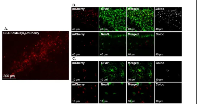
Microscopy. In order to verify DREADD DH- and astroglial-specificity, mCherry
expression was carefully examined by an experimenter blind to treatment group. DH sections
were visualized using confocal microscopy (Zeiss LSM800, Jena, Germany) and representative
images for publication were acquired using 1024 x 1024 frame size, 16-bit image resolution, and
frame average of 4. Laser lines that excite at 488 nm and 561 nm were used to visualize
AlexaFluor-488 and mCherry respectively. Images were deconvolved using Bitplane AutoQuant
X3 (10 iterations), and exported to Biplane Imaris Software (Zurich, Switzerland). mCherry was
expected to be expressed bilaterally throughout the DH, selectively within the DH, and
specifically in DH astrocytes. Animals with non-DH and/or non–astrocyte specific mCherry
expression were removed from data analysis.
RNA Extraction and cDNA synthesis. Messenger RNA (mRNA) was extracted to
assess measures of NO in the spleen. Spleen tissue was homogenized in 1 mL of cold TriReagent
(Molecular Research Center, Cincinnati, OH) using a bead homogenizer (Precellys Instruments,
second tube. Next, the samples were shaken and incubated with BCP at room temperature and
centrifuged for phase separation. The aqueous layer was thoroughly mixed with isopropanol,
incubated at room temperature, and samples were centrifuged to form the RNA pellet. The pellet
was then washed three times in 75% ethanol and air dried to remove residual ethanol. The RNA
pellet was reconstituted in warm RNase-free water. Absorbance for samples diluted (1:20) in
1xTE (pH = 7.5) was assessed using spectrophotometer (Epoch™, BioTek Instruments Inc.,
Winooski, VT). Sample mRNA concentrations were read using the Take3 Application and Gen5
Software for Nucleic Acid Quantification (BioTek Instruments Inc.), and A260/280 ratios were
assessed to ensure purity.
Sample mRNA input concentration was equalized using PCR-grade water. cDNA was
synthesized using the Advantage for RT-PCR Kit (ClonTech, Takara, Mountain View, CA)
following the manufacturer’s protocol and using the Veriti 96 Well Fast Thermal Cycler
(Applied Biosystems, ThermoFisher Scientific). A subset of undiluted cDNA samples were
pooled together, and five serial 1:10 dilutions were made to test qPCR reaction efficency. The
remaining original sample was then diluted 1:5 in PCR-grade water for qPCR.
qPCR Quantification of Splenic iNOS Gene Expression. qPCR was performed using
the TaqMan™ Fast Advanced Master Mix Kit (Applied Biosystems, ThermoFisher Scientific)
according to the manufacturer’s instructions. Reactions were carried out in triplicate on a
384-well plate, with each individual reaction containing 1.5 µL of cDNA pooled or sample cDNA. In
order to assess indices of NO production, levels of splenic inducible nitric oxide synthase (iNOS)
gene expression were analyzed. NO is produced by iNOS in response to inflammatory stimuli
(Nathan and Shiloh, 2000). Thus, two different genes were analyzed by using the TaqMan™
Gene Expression Assays (FAM): inducible nitric oxide synthase 2 (iNOS/NOS2, Assay ID:
10
gene, Assay ID: Rn01475911_g1; ThermoFisher Scientific). A no template control was run to
ensure purity of these reactions. Plates were run in the QuantStudioTM 6 Flex RealTime PCR
System (Applied Biosystems, ThermoFisher Scientific). Data were collected using the
QuantStudioTM RealTime PCR Software with a PCR Run Method as follows: 50°C for 2 min
for PCR product contamination degradation, hold at 95°C for 20 sec for polymerase activation,
and 45 PCR cycles of 95° C for 1 sec and 60° C for 20 sec with data collection at the end of each
cycle. Data were analyzed using the Comparative CT (ΔΔCT) Method. iNOS CT data were
normalized to the reference gene (Rpl13a), and then normalized to the overall average of
reference normalized values.
Nitrate/nitrite Assay. As NO is degraded quickly, degradation products in plasma can be
analyzed in combination with iNOS expression as indices of NO production. Plasma
nitrate/nitrite concentrations were assessed using the Griess reagent assay as described
previously (Szczytkowski and Lysle, 2007). Briefly, plasma was diluted in dH2O and incubated
with nitrate reductase (1.0 U/mL), 0.31 M PB (pH = 7.5), 0.86 mM NADPH (Sigma-Aldrich
Inc., Milwaukee, WI), and 0.11 mM flavin adenine dinucleotide in a 96-well plate for 90 min at
room temperature in the dark. Following incubation, Griess reagent (1:1 (vol:vol) solution 1%
sulfanilamide in 5% phosphoric acid and 0.1% N-(1-napthyl)ethylenediamine dihydrochloride in
distilled H2O) was added to the samples and allowed to develop at room temperature.
Absorbance was assessed at 550 nm using a spectrophotometer (Epoch™, BioTek Instruments
Inc). Reactions were carried out in triplicate. The total micromolar concentration of nitrite was
determined for each sample based on a concurrently run standard curve.
Statistical Analysis. Data for each experiment herein was analyzed using 2x2 analysis of
variance (ANOVA) in SPSS Statistics (IBM, Armonk, NY). Planned contrasts were made using
Test. For Experiments 1, we tested planned comparisons between CS-exposed and corresponding
home cage control groups, as well as differences between CS-exposed groups themselves. For
analysis of RT-qPCR, ΔΔCT values were analyzed, although the linearly transformed were used
to display the data graphically. For Experiments 2, we tested a planned contrast between the
heroin-conditioned groups at CNO test for time spent in heroin-paired side, change in time spent
in heroin-paired side relative to baseline, and CPP score. Initial verification of acquired CPP was
performed using an independent t-test comparing heroin-conditioned to saline-conditioned
animals. Statistically significant outliers were detected using Grubb’s test and removed from
analysis. Alpha was set at p = 0.05.
Results
Experiment 1: Stimulation of astroglial Gi-signaling in the DH disrupts heroin-conditioned
immunomodulation. Experiment 1 examined the role of DH astrocyte signaling in the
expression of heroin-conditioned suppression of LPS-induced indices of NO production (see
timeline in Fig. 1A). DREADD expression, as indicated by mCherry, was observed throughout
the DH (Fig.1B). Furthermore, hM4Di-mCherry expression was restricted to astrocytes (Fig. 2).
CNO-induced stimulation of DH astroglial Gi-signaling attenuated heroin-conditioned
splenic iNOS mRNA suppression (Fig. 1C). A 2 x 2 ANOVA of splenic Rpl13A mRNA levels
revealed no significant differences between the groups (F(3,18) = 1.48, p = 0.252), validating
Rpl13A as a reference gene. A 2 x 2 ANOVA of splenic iNOS mRNA levels revealed significant
main effects of CS exposure (F(1,18) = 30.96, p < 0.05) and CNO treatment (F(1,18) = 6.05, p <
0.05), but no CS exposure by CNO treatment interaction (F(1,18) = 2.57, p = 0.127). Planned
contrasts revealed that CS exposure significantly reduced splenic iNOS mRNA levels relative to
home cage controls in the vehicle-treated (p < 0.05). CNO treatment partially attenuated
12
iNOS mRNA expression was reduced relative to CNO-treated home cage controls (p < 0.05), but
was higher than vehicle-treated CS-exposed animals (p < 0.05). Thus, stimulation of DH
astroglial Gi-signaling significantly increased splenic iNOS gene expression, yet does not completely restore mRNA levels to those of control animals.
In contrast to splenic iNOS mRNA levels, CNO-induced stimulation of DH astroglial
Gi-signaling completely inhibited heroin-conditioned suppression of plasma nitrate/nitrite
concentration (Fig. 1D). A 2 x 2 ANOVA of nitrate/nitrite concentration revealed a significant
CNO treatment x CS exposure interaction (F(1,18) = 8.05, p < 0.05). Planned contrasts revealed
that, in vehicle-treated groups, CS exposure reduced plasma nitrate/nitrite concentrations relative
to home cage controls (p < 0.05), indicating expression of heroin-conditioned
immunomodulation. CNO-induced stimulation of Gi-signaling in DH astrocytes restored plasma
Figure 1: Activation of astroglial Gi-coupled signaling in the DH disrupts heroin-conditioned suppression of
peripheral indices of NO production. For Experiment 1, the timeline is depicted (A) as well as the spread of
GFAP-hM4D(Gi) as indicated by mCherry expression throughout the DH (B). Darker red areas are indicative
of denser mCherry expression, with coordinates indicating distance from bregma based on Paxinos and Watson (2006). CNO administration significantly attenuated heroin-conditioned LPS-induced splenic iNOS mRNA
expression (C) and completely blocked heroin-conditioned LPS-induced plasma nitrate/nitrite concentration
(D). Group sizes were n = 5-6 in the final analysis for splenic iNOS mRNA expression and plasma
nitrate/nitrite concentration. * represents statistically significant differences relative to respective home cage
nitrate/nitrite concentrations, such that concentrations for the CS-exposed group did not
significantly differ from CNO-treated home cage controls (p = 0.646) and were higher than
vehicle-treated CS-exposed animals (p < 0.05).
Experiment 2: Stimulation of astroglial Gi-signaling in the DH does not alter
heroin-conditioned place preference. Experiment 2 investigated the role of astrocyte signaling in the
expression of heroin-CPP (see timeline in Fig. 3A). DREADD expression, as indicated by
mCherry, was observed throughout the DH (Fig.3B). All animals acquired CPP.
Heroin-conditioned animals spent significantly more time in the heroin-paired side than
saline-conditioned animals during the CPP test session (t(12.47) = -3.22, p < 0.05), verifying the
effectiveness of the biased conditioning procedure. CPP data were collapsed across experimental
Figure 2: GFAP-hM4Di-mCherry is selectively expressed in DH astrocytes. A representative confocal 10X tile
14
CNO-induced stimulation of DH astroglial Gi-signaling failed to alter heroin-CPP at CNO test relative to controls (Fig. 3C and 3D). Specifically, a 2 x 2 ANOVA for total time
spent in the heroin-paired chamber at CNO test revealed a significant main effect of conditioning
(F(1, 26) = 17.63, p < 0.05) with no significant main effect of CNO treatment (F(1, 26) = 0.44, p
= 0.511) nor interaction (F(1, 26) = 1.37, p = 0.253). Similarly, the 2 x 2 ANOVA for change in
time spent in the heroin-paired side relative to baseline indicated a significant main effect of
heroin conditioning (F(1, 24) = 23.28, p < 0.05) with no significant main effect of CNO
treatment (F(1, 24) = 0.12, p = 0.729) nor interaction (F(1, 24) = 1.88, p = 0.183). Finally, a 2 x
2 ANOVA of CPP scores on CNO test day (data not shown) also revealed a significant main
effect of conditioning (F(1, 26) = 16.18, p < 0.05), with no significant main effect of CNO
treatment (F(1, 26) = 0.12, p = 0.736) nor interaction (F(1, 26) = 3.91, p = 0.059). Thus,
heroin-conditioned animals spent significantly more total time, time relative to baseline, and time
relative to saline-paired side in the heroin-paired side independent of CNO treatment. Planned
contrasts between the heroin-conditioned groups revealed no differences between these groups
regardless of CNO treatment for total time spent in the heroin-paired side (p = 0.694), for change
Discussion
Through associative learning, contextual stimuli can come to elicit heroin-conditioned
responses, including CPP and immunomodulation. The DH plays a critical role in contextual
learning and memory, and has been implicated in both opioid-conditioned reward (Corrigall and
Linseman, 1988) and -conditioned immunomodulation (Szczytkowski et al., 2013). In addition,
neuroimmune signaling, in terms of both gliotransmission and cytokine signaling, is essential in
learning and memory processes (Ben Achour and Pascual, 2010; Donzis and Tronson, 2014;
Santello and Volterra, 2012; Yirmiya and Goshen, 2011) and in some drug-conditioned
responses and instrumental behaviors relevant for drug addiction (Haydon et al., 2009;
Figure 3:Activation of astroglial Gi-coupled signaling in the DH fails to alter heroin-CPP. For Experiment 2,
the experimental timeline is shown (A) as well as the spread of GFAP-hM4D(Gi) as indicated by mCherry
expression throughout the DH (B). Darker red areas are indicative of denser mCherry expression with
coordinates indicating distance from bregma based on Paxinos and Watson (2006). CNO administration fails to disrupt total time spent in the heroin-paired side (C) or change in time spent in the heroin-paired side relative to a pre-conditioning baseline (D). Group sizes were n = 6 for each saline-conditioned group and n = 8-9 for each conditioned group in the final analysis of CPP measures. * represents a main effect of
16
Lacagnina et al., 2018; Scofield and Kalivas, 2014). Astrocytes, for example, have an established
involvement in the IL-1R1 signaling required for some forms of learning and memory (Ben
Menachem-Zidon et al., 2011). Findings in the present study significantly extend these lines of
research by demonstrating that DH neuroimmune signaling plays a causal and selective role in
heroin-conditioned immunomodulation, but not in heroin-CPP. We have demonstrated that IL-1
signaling is necessary for heroin-conditioned immunomodulation but not heroin-conditioned
appetitive responses (Paniccia et al., 2018). The present study complements these findings such
that undisturbed astroglial signaling during CS exposure is necessary for heroin-conditioned
suppression LPS-induced of indices of NO production. Conversely, manipulations of the same
signaling pathways failed to disrupt measures of heroin-CPP under the present experimental
parameters. Together, our data suggest that divergent mechanisms within the DH govern
heroin-conditioned peripheral immunomodulation and heroin-heroin-conditioned appetitive behavior.
Findings from our laboratory have furthered our understanding into the role of DH IL-1
signaling in heroin-conditioned Pavlovian responses. We have established sustained, inducible
knockdown of DH IL-1β mRNA expression prior to CS exposure disrupts heroin-conditioned
suppression of peripheral modulators, including indices of NO production (Szczytkowski et al.,
2013). Additionally, signaling of DH IL-1R1 mediates the expression of heroin-conditioned
immunomodulation, but antagonism of IL-1R1 does not alter heroin-CPP. Within the
hippocampus, both astrocytes and microglia are capable of producing and responding to IL-1β
signaling (Friedman, 2001; Hanisch, 2002), indicating either or both of these cell types could
facilitate the IL-1 signaling required for heroin-conditioned immunomodulation. While the
experiments in the present study strongly suggest astroglia mediate heroin-conditioned
immunomodulation, the additional role of DH microglial involvement in this conditioned
Hippocampal astrocytes are capable of expressing IL-1R1, and IL-1β administration
triggers receptor upregulation of this receptor (Friedman, 2001). 1β action at astroglial
IL-1R1 evokes nuclear factor-κB (NF-κB) signaling cascades (Srinivasan et al., 2004) and thus
elicits the transcription of pro-inflammatory factors, including IL-1β and other cytokines, serving
as a potential positive feedback loop for IL-1β expression. Presently, we establish a role for
hippocampal astrocyte activity in mediating heroin-conditioned immunomodulation. The same
chemogenetic stimulation of astroglial Gi-signaling used herein attenuates cAMP induction in DREADD-positive astrocytes (Jones et al., 2018b). As converging evidence suggests that
activity of NF-κB is modulated by cAMP induction (Gerlo et al., 2011), it is possible astroglial
Gi-signaling attenuates IL-1β production in hippocampal astrocytes. Future experiments should be aimed at testing the relationship between astrocyte activity and subsequent IL-1 signaling in
heroin-conditioned immunomodulation.
The current study strongly suggests that DH astroglial signaling is a critical component in
the expression of heroin-conditioned immunomodulation, but not heroin-CPP. The absence of
effects on heroin-CPP were surprising given the established role of astroglial activity in
addiction (Scofield and Kalivas, 2014). Specifically, prior research has shown that chemogenetic
manipulation of astroglial Gq-signaling in the nucleus accumbens (NAc) core ameliorates the ability of cocaine-conditioned stimuli to elicit drug-seeking behaviors (Scofield et al., 2015).
Although there is a functional projection from the DH to the NAc core (Peleg-Raibstein and
Feldon, 2006), the current study targeted DH astroglial Gi-signaling in vivo during exposure to heroin-paired stimuli. While the DH is critical for encoding context-drug associations (Xia et al.,
2017), it is the connection from the ventral hippocampus to the NAc shell that drives
context-induced heroin-seeking behaviors (Bossert et al., 2016). It is possible that chemogenetic
heroin-18
conditioned appetitive responses. The current data suggest astroglial involvement varies across
conditioned appetitive behaviors as a function of evoked signaling pathway, target brain region,
animal model, and drug of abuse.
The neuroimmune system is both impacted by opioid administration and serves as a key
regulator of opioid-induced responses. Opioids produce alterations in hippocampal GFAP and
IL-1β protein expression that are attenuated through anti-inflammatory compounds, including
ibudilast (Hutchinson et al., 2009). At the same time, ibudilast administration reduces opioid
withdrawal and simultaneously increases antinociception (Hutchinson et al., 2009). These
findings indicate the neuroimme system differentially regulates opioid-induced responses
depending on the type of response in question. Consistent with this, the current study establishes
a divergence in mechanism governing heroin-conditioned responses.
The data demonstrating astroglial Gi-signaling disrupts heroin-conditioned
immunomodulation are in line with recent findings demonstrating that modulation of DH
astroglial signaling directly alters hippocampal-dependent mechanisms of learning and memory
(Adamsky et al., 2018). Notably, chemogenetic stimulation of astroglial Gi-signaling did not fully restore LPS-induced NO measures. It is possible that astrocytes are not the only cellular
component involved in the expression of heroin-conditioned immunomodulation. Consistent
with this, astroglial-mediated neuronal alterations improve hippocampal-dependent memory,
while neuronal activation alone impairs it (Adamsky et al., 2018). We have previously
demonstrated hippocampal neuronal involvement in heroin-conditioned immunomodulation
(Szczytkowski et al., 2013). Given the current findings that astrocyte activity mediates
heroin-conditioned immunomodulation, the possibility of astrocyte-neuron interplay and the specific
In the current set of experiments we employed a 2 x 2 statistical design in which all
animals received intra-DH infusions of AAV8-GFAP-hM4D(Gi)-mCherry. Thus, DREADD
expression was present in all animals and transfection alone could not account for group
differences in heroin-conditioned immunomodulation or heroin-CPP. Furthermore, all animals
were thoroughly examined for site- and cell-type-specific expression which did not differ across
groups. Although there have been recent concerns of CNO effects irrespective of DREADD
expression (Gomez et al., 2017), other groups report no effect of CNO administration alone
during experiments involving astroglial chemogenetic techniques (Adamsky et al., 2018; Bull et
al., 2014; Scofield et al., 2015). While we do not presently report use of a control DREADD,
CNO did not alter any of the current measures relative to vehicle in home cage controls. Thus,
effects on reported measures were likely induced by astroglial Gi-signaling pathway
manipulation, specifically. Importantly, we have recently demonstrated CNO attenuates
LPS-induced cAMP expression in mCherry-positive DH astrocytes using the same viral construct
(Jones et al., 2018b). This confirms CNO exerts its effects through the stimulation of Gi -signaling cascades and the inhibition of downstream cAMP within DH astrocytes.
In summary, the present study suggests that divergent mechanisms within the DH
regulate Pavlovian heroin-conditioned responses. The current findings suggest that astrocyte
signaling in the DH regulate conditioned immunomodulatory, but not conditioned appetitive,
effects of heroin. The immunomodulatory effects of heroin can exacerbate infectious and other
disease progression in addicts (Ninković and Roy, 2013; Wang et al., 2011). Since
immunomodulation can become conditioned to environmental stimuli over the course of chronic
heroin use, the detrimental health effects of heroin may persist in heroin-associated environments
20
substrates that maintain heroin-conditioned immunomodulation may be a promising therapeutic
target for harm reduction in heroin use disorders.
Acknowledgments
This research was supported by the National Institute on Drug Abuse grants DA034721
REFERENCES
Adamsky, A., Kol, A., Kreisel, T., Doron, A., Ozeri-Engelhard, N., Melcer, T., Refaeli, R., Horn, H., Regev, L., Groysman, M., London, M., Goshen, I., 2018. Astrocytic Activation Generates <em>De Novo</em> Neuronal Potentiation and Memory Enhancement. Cell.
Ben Achour, S., Pascual, O., 2010. Glia: The many ways to modulate synaptic plasticity. Neurochemistry International 57, 440-445.
Ben Menachem-Zidon, O., Avital, A., Ben-Menahem, Y., Goshen, I., Kreisel, T., Shmueli, E.M., Segal, M., Ben Hur, T., Yirmiya, R., 2011. Astrocytes support hippocampal-dependent memory and long-term potentiation via interleukin-1 signaling. Brain, Behavior, and Immunity 25, 1008-1016.
Bossert, J.M., Adhikary, S., St Laurent, R., Marchant, N.J., Wang, H.-L., Morales, M., Shaham, Y., 2016. Role of projections from ventral subiculum to nucleus accumbens shell in context-induced reinstatement of heroin seeking in rats. Psychopharmacology 233, 1991-2004.
Bull, C., Freitas, K.C.C., Zou, S., Poland, R.S., Syed, W.A., Urban, D.J., Minter, S.C., Shelton, K.L., Hauser, K.F., Negus, S.S., Knapp, P.E., Bowers, M.S., 2014. Rat Nucleus Accumbens Core Astrocytes Modulate Reward and the Motivation to Self-Administer Ethanol after Abstinence. Neuropsychopharmacology 39, 2835-2845.
Corrigall, W.A., Linseman, M.A., 1988. Conditioned place preference produced by intra-hippocampal morphine. Pharmacology Biochemistry and Behavior 30, 787-789.
Crombag, H.S., Bossert, J.M., Koya, E., Shaham, Y., 2008. Context-induced relapse to drug seeking: a review. Philosophical Transactions of the Royal Society B: Biological Sciences 363, 3233-3243.
Donzis, E.J., Tronson, N.C., 2014. Modulation of learning and memory by cytokines: signaling mechanisms and long term consequences. Neurobiology of learning and memory 0, 68-77.
Friedman, W.J., 2001. Cytokines Regulate Expression of the Type 1 Interleukin-1 Receptor in Rat Hippocampal Neurons and Glia. Experimental Neurology 168, 23-31.
Fuchs, R.A., Eaddy, J.L., Su, Z.I., Bell, G.H., 2007. Interactions of the basolateral amygdala with the dorsal hippocampus and dorsomedial prefrontal cortex regulate drug context‐induced
reinstatement of cocaine‐seeking in rats. European Journal of Neuroscience 26, 487-498.
Fuchs, R.A., Evans, K.A., Ledford, C.C., Parker, M.P., Case, J.M., Mehta, R.H., See, R.E., 2005. The Role of the Dorsomedial Prefrontal Cortex, Basolateral Amygdala, and Dorsal Hippocampus in Contextual Reinstatement of Cocaine Seeking in Rats. Neuropsychopharmacology 30, 296.
22
Ge, F., Wang, N., Cui, C., Li, Y., Liu, Y., Ma, Y., Liu, S., Zhang, H., Sun, X., 2017.
Glutamatergic Projections from the Entorhinal Cortex to Dorsal Dentate Gyrus Mediate Context-Induced Reinstatement of Heroin Seeking. Neuropsychopharmacology 42, 1860-1870.
Gomez, J.L., Bonaventura, J., Lesniak, W., Mathews, W.B., Sysa-Shah, P., Rodriguez, L.A., Ellis, R.J., Richie, C.T., Harvey, B.K., Dannals, R.F., Pomper, M.G., Bonci, A., Michaelides, M., 2017. Chemogenetics revealed: DREADD occupancy and activation via converted clozapine. Science 357, 503-507.
Goshen, I., Kreisel, T., Ounallah-Saad, H., Renbaum, P., Zalzstein, Y., Ben-Hur, T., Levy-Lahad, E., Yirmiya, R., 2007. A dual role for interleukin-1 in hippocampal-dependent memory processes. Psychoneuroendocrinology 32, 1106-1115.
Hanisch, U.-K., 2002. Microglia as a source and target of cytokines. Glia 40, 140-155.
Haydon, P.G., Blendy, J., Moss, S.J., Jackson, F.R., 2009. Astrocytic control of synaptic transmission and plasticity: a target for drugs of abuse? Neuropharmacology 56, 83-90.
Haydon, P.G., Carmignoto, G., 2006. Astrocyte Control of Synaptic Transmission and Neurovascular Coupling. Physiological Reviews 86, 1009-1031.
Jones, M.E., Lebonville, C.L., Barrus, D., Lysle, D.T., 2015. The role of brain interleukin-1 in stress-enhanced fear learning. Neuropsychopharmacology 40, 1289-1296.
Jones, M.E., Lebonville, C.L., Paniccia, J.E., Balentine, M.E., Reissner, K.J., Lysle, D.T., 2018a. Hippocampal interleukin-1 mediates stress-enhanced fear learning: A potential role for astrocyte-derived interleukin-1β. Brain, Behavior, and Immunity 67, 355-363.
Jones, M.E., Paniccia, J.E., Lebonville, C.L., Reissner, K.J., Lysle, D.T., 2018b. Chemogenetic Manipulation of Dorsal Hippocampal Astrocytes Protects Against the Development of Stress-enhanced Fear Learning. Neuroscience 388, 45-56.
Kutlu, M.G., Gould, T.J., 2016. Effects of drugs of abuse on hippocampal plasticity and
hippocampus-dependent learning and memory: contributions to development and maintenance of addiction. Learning & Memory 23, 515-533.
Lacagnina, M.J., Rivera, P.D., Bilbo, S.D., 2018. Glial and Neuroimmune Mechanisms as Critical Modulators of Drug Use and Abuse. Neuropsychopharmacology 42, 156-177.
Lysle, D.T., How, T., 2000. Heroin modulates the expression of inducible nitric oxide synthase. Immunopharmacology 46, 181-192.
Lysle, D.T., Ijames, S.G., 2002. Heroin-associated environmental stimuli modulate the expression of inducible nitric oxide synthase in the rat. Psychopharmacology (Berl) 164, 416-422.
Meyers, R.A., Zavala, A.R., Neisewander, J.L., 2003. Dorsal, but not ventral, hippocampal lesions disrupt cocaine place conditioning. NeuroReport 14, 2127-2131.
Miguel-Hidalgo, J.J., 2009. The Role of Glial Cells in Drug Abuse. Current drug abuse reviews 2, 76-82.
Nathan, C., Shiloh, M.U., 2000. Reactive oxygen and nitrogen intermediates in the relationship between mammalian hosts and microbial pathogens. Proceedings of the National Academy of Sciences 97, 8841-8848.
Ninković, J., Roy, S., 2013. Role of the mu opioid receptor in opioid modulation of immune function. Amino acids 45, 9-24.
Ota, Y., Zanetti, A.T., Hallock, R.M., 2013. The Role of Astrocytes in the Regulation of Synaptic Plasticity and Memory Formation. Neural Plasticity 2013, 11.
Paniccia, J.E., Lebonville, C.L., Jones, M.E., Parekh, S.V., Fuchs, R.A., Lysle, D.T., 2018. Dorsal hippocampal neural immune signaling regulates heroin-conditioned immunomodulation but not heroin-conditioned place preference. Brain, Behavior, and Immunity.
Peleg-Raibstein, D., Feldon, J., 2006. Effects of dorsal and ventral hippocampal NMDA stimulation on nucleus accumbens core and shell dopamine release. Neuropharmacology 51, 947-957.
Roth, B.L., 2016. DREADDs for Neuroscientists. Neuron 89, 683-694.
Santello, M., Volterra, A., 2012. TNFα in synaptic function: switching gears. Trends in Neurosciences 35, 638-647.
Scofield, M.D., Boger, H.A., Smith, R.J., Li, H., Haydon, P.G., Kalivas, P.W., 2015. Gq-DREADD Selectively Initiates Glial Glutamate Release and Inhibits Cue-induced Cocaine Seeking. Biological psychiatry 78, 441-451.
Scofield, M.D., Kalivas, P.W., 2014. Astrocytic Dysfunction and Addiction: Consequences of Impaired Glutamate Homeostasis. The Neuroscientist : a review journal bringing neurobiology, neurology and psychiatry 20, 610-622.
Srinivasan, D., Yen, J.-H., Joseph, D.J., Friedman, W., 2004. Cell Type-Specific Interleukin-1β Signaling in the CNS. The Journal of Neuroscience 24, 6482-6488.
Szczytkowski, J.L., Fuchs, R.A., Lysle, D.T., 2011. Ventral tegmental area-basolateral
amygdala-nucleus accumbens shell neurocircuitry controls the expression of heroin-conditioned immunomodulation. J Neuroimmunol 237, 47-56.
24
Szczytkowski, J.L., Lysle, D.T., 2007. Conditioned effects of heroin on the expression of inducible nitric oxide synthase in the rat are susceptible to extinction and latent inhibition. Psychopharmacology 191, 879-889.
Tzschentke, T.M., 1998. Measuring reward with the conditioned place preference paradigm: a comprehensive review of drug effects, recent progress and new issues. Progress in Neurobiology 56, 613-672.
Wang, X., Zhang, T., Ho, W.-Z., 2011. Opioids and HIV/HCV Infection. Journal of
neuroimmune pharmacology : the official journal of the Society on NeuroImmune Pharmacology 6, 477-489.
Xia, L., Nygard, S.K., Sobczak, G.G., Hourguettes, N.J., Bruchas, M.R., 2017. Dorsal-CA1 Hippocampal Neuronal Ensembles Encode Nicotine-Reward Contextual Associations. Cell Reports 19, 2143-2156.
Xie, X., Ramirez, D.R., Lasseter, H.C., Fuchs, R.A., 2010. Effects of mGluR1 Antagonism in the Dorsal Hippocampus on Drug Context-induced Reinstatement of Cocaine-seeking Behavior in Rats. Psychopharmacology 208, 1-11.