A CONTEMPORARY PERSPECTIVE ON TOOTH EXTRACTION IN ORTHODONTICS
Camille Guez
A thesis submitted to the faculty at the University of North Carolina at Chapel Hill in partial fulfillment of the requirements for the degree of Master of Science in the School of Dentistry
(Orthodontics).
Chapel Hill 2015
Approved by:
Ching-Chang Ko
Feng-Chang Lin
© 2015 Camille Guez
ABSTRACT
Camille Guez: A contemporary perspective on tooth extraction in orthodontics (Under the direction of Ching-Chang Ko)
Introduction: The decision to extract teeth for orthodontic purposes is one of the most
complex and debated topics in the specialty. The profession’s understanding of factors affecting
the extraction decision (e.g., outcome stability and facial appearance) has evolved over time, and
estimated extraction rates have varied from 10% in 1953 to 78% in 1968 to 28% in 1994. A
contemporary perspective on the rate of orthodontic extractions is needed to help clinicians
understand this treatment choice in light of 21st century philosophies, techniques, and
appliances. To this end, we investigated changes in orthodontic extraction rates at the University
of North Carolina from 2000 to 2011, as well as factors that may have affected those rates. We
hypothesize that extraction rates have changed as a result of evolving diagnostic methodology
and appliance selection (e.g., self-ligating brackets). Methods: Pre- and post- treatment records
were analyzed to determine extraction rates over time, and different factors (Angle classification,
skeletal relationship, use of self-ligation, etc.) were investigated to evaluate potential impact on
the extraction decision. The sample consisted of 2,184 patients, with 1,263 females (58%). Age
at the start of treatment ranged from 7 to 67 years. Third molar extractions were excluded from
analyses. Student t-test and chi square were used to evaluate the extraction rate over time.
Logistic regression was used to investigate diagnostic and treatment factors that might affect
extraction rates over time. Results: The extraction rate decreased significantly from 2000 to
explanatory variables were found to influence the overall extraction rate but none of them could
explain the decrease of the extraction rate over time. Conclusions: The extraction rate
fluctuated greatly since 2000 and has decreased linearly over the years (p<0.05). Several
explanatory variables were found to influence the overall extraction decision, but none of them
could explain the decreasing trend over time. Irrational cognition with medical concerns about
ACKNOWLEDGEMENTS
Thank you to my committee members, Dr. Ko, Dr. Lin and Dr. Proffit for your expertise,
guidance, and advice throughout my project. Thank you to Tate Jackson for your ideas and your
feedback. Thank you to my fiancé Raphael and my family for your love and support. To my son
Vadim, I am so proud to be your mother. Thank you to the Southern Association of
TABLE OF CONTENTS
LIST OF TABLES ... vii
LIST OF FIGURES ... viii
LITERATURE REVIEW ...1
Introduction ...1
History...2
The extraction decision, a multi-factorial decision ...5
Previous studies ...14
Conclusions ...16
REFERENCES ...17
A CONTEMPORARY PERSPECTIVE ON TOOTH EXTRACTION IN ORTHODONTICS ..21
Introduction ...21
Materials and Methods ...23
Results ...25
Discussion ...29
Conclusions ...32
Tables ...34
LIST OF TABLES
Table 1 – Influence of explanatory variables on the overall extraction rate. ...34
Table 2 – Extraction rate versus explanatory variables from 2000 to 2011 per year ...35
LIST OF FIGURES
Figure 1 – Fauchard “bandeau” appliance30 ...2
Figure 2 – Angle School of Orthodontia, first postgraduate school for orthodontists7 ...3
Figure 3 –”Envelope of discrepancy”. Adapted from Proffit12 ...12
Figure 4 – Extraction of four first premolars percentages at UNC. Adapted from Proffit12 ...15
Figure 5 – Extraction rate per year ...25
Figure 6 – Extraction percentage and Angle classification ...26
LITERATURE REVIEW
Introduction
The first organized data on malocclusion in the United States was reported by the US
Public Health Service in the 1960s, which was collectively documented as part of the first
National Health and Nutrition Examination Survey (NHANES). According to NHANES, Kelly
et al. reported that 24.4% of the children between 6 and 11 years had normal occlusion, 22.4%
had a definite malocclusion, 8.7% had severe malocclusion, and 5.5% were classified as having a
very severe malocclusion1. This was the first epidemiological study showing that at least 35% of
American children are candidates for orthodontic treatment. More recently, the American
Association of Orthodontists stated that in 2004, there were 5,750,000 patients undergoing
orthodontic treatment, and that this statistic had undergone a steady and significant increase since
1989. Depending upon the year of treatment, 28% to 76% of orthodontic patients have
undergone tooth extraction, which represents a substantial surgical intervention in orthodontics
2-3-4
. Some patients have concerns about losing teeth and how this will affect their general health.
As evidence-based research and new technology advance the field of orthodontics, it is important
to understand what factors affect the decision to extract teeth and whether it is possible to limit
History
Pierre Fauchard, widely acknowledged as the father of modern dentistry, published “The
Surgeon Dentist” in 1728. His book contained detailed information about all areas of
contemporary dentistry and illustrated his invention of one of the first orthodontic appliances
called “bandeau” (Figure 1). This “bandeau” consisted of a horseshoe-shaped strip of precious
metal to which the teeth were ligated. This appliance became the basis for Angle’s E-arch and its
principle is still used today to unravel crowded arches.
Figure 1. Fauchard “bandeau” appliance30
Another French dentist, Etienne Bourdet, followed Fauchard’s footsteps and refined the
“bandeau” appliance. He published “The Dentist’s Art” in 1757 which contained a chapter
dedicated to orthodontics. He is the first dentist known to have recommended serial extractions
and extraction of permanent teeth, especially premolars, as a mean to alleviate crowding. He was
also the first known dentist to practice “lingual orthodontics”, expanding the arches from the
lingual. He emphasized the importance of dental hygiene and described the symptoms of tooth
eruption in children. He was the dentist of the King of France, Louis XV then Louis XVI5.
Extractions have been proven to improve alignment since early times but orthodontics as
systematic extraction of teeth: he invented the occipital traction, precursor to the Headgear
appliance, and used occlusal coverage appliances among many technical innovations. These
technics are still widely used today. He also focused on cleft patients and perfected a gold
obturator and artificial vellum of soft rubber in 18597. From a biological standpoint, John Farrar
introduced the foundation of the scientific approach of orthodontics by studying the limits of
tooth movements. He investigated the physiologic and pathologic changes occurring in animals
as a result of orthodontic tooth movement. He recommended the bodily movement of teeth as
opposed to a simple tipping movement and advised orthodontists to limit the orthodontic force to
avoid any pain to the patient5. Based on animal studies, he enunciated the theory of intermittent
force and developed a screw to deliver this force in controlled increments.
At the beginning of the twentieth century, Edward Angle (1855-1930) founded the first
orthodontic school, first orthodontic society and first orthodontic journal.
Figure 2. Angle School of Orthodontia, first postgraduate school for orthodontists7
He was the most influential figure in orthodontics and is considered as “the father of
modern orthodontics”. He created a dental classification of malocclusion which is still the most
widely used classification around the world today. His vision of orthodontic treatment was based
strongly advocated a non-extraction approach stating that jaws and bones would grow
accordingly and the adjacent tissues would adapt to their new position. Ideal occlusion is
“nature’s intended ideal form”8-9-10. His credo was that “the best balance, the best harmony, the
best proportions of the mouth in its relation to the other features require that there shall be a full
complement of teeth, and that each tooth shall be made to occupy its normal position—i.e.,
normal occlusion”. Calvin Case defended extractions as a treatment to correct facial deformities
in one of his articles and instigated the “Great Extraction Debate” in 1911 with Edward Angle7-8
.
One of Angle’s disciples, Charles Tweed, followed his teacher’s approach and realized later in
his career that many of his patients experienced relapse after the end of their non-extraction
treatment, especially when the lower incisors were overly proclined. Non-extraction
arch-expansion, originally proposed by Edward Angle, was found to be unstable after treatment11.
Tweed re-treated a number of his patients with the extraction of four premolars and obtained a
satisfactory result. Other orthodontists like Raymond Begg followed his footsteps and advocated
premolar extraction as a valid way to treat patients. Technological advances also played a major
role in that direction. As an example, the possibility to bond to enamel gave the clinician better
control over the tooth movements.
William Proffit and James Ackerman added another dimension to the debate in the 1980s
with the soft tissue paradigm12-13. In opposition to Angle’s theory, they developed the idea that
the clinician should focus on the soft tissue balance instead of focusing solely on a perfect dental
occlusion. Variation is now accepted as a natural form, perfect occlusion is more the exception
than the rule. The orthodontist has to plan his treatment based on the soft tissue limitations and
contours of his patient. They stressed the importance of the smile arc, defined by the contour of
As of today, the philosophic evolution in orthodontic treatment lies on three principles:
occlusion, stability, and soft tissue balance. Derived from these principles, it has been accepted
by the orthodontic community that some patients will need extractions and other will not.
However, in practicality, the question remains as to which patients should benefit from these
extractions and how the clinician should make that decision.
The extraction decision, a multi-factorial decision
Three main aspects have historically guided the practitioner in his treatment plan. First
the occlusion with Edward Angle, then the stability and concerns about relapse with Charles
Tweed, and finally esthetics, with the soft tissue paradigm. Occlusion, stability and esthetics are
the three goals for a successful treatment plan, but no single rule can give the orthodontist a
simple way to decide how to reach these three goals: The extraction decision is multi-factorial3.
The historical and most common extraction pattern is the extraction of four premolars,
two on the upper arch and two on the lower arch. However, different extraction patterns can be
followed depending on the type of malocclusion and are all successful if used in appropriate
patients12-14-15.
A dilemma exists: some clinicians are more inclined to extract teeth than expand the
arches; others would rather conserve the teeth if possible and try to expand the arches to relieve
the crowding. In borderline cases, both treatments offer good stability and results16-17-18-19. A
reliable criterion for extracting teeth remains elusive. Many researchers attempted to find a way
to help the decision of the clinician, like Takada who created a mathematical model to guide the
treatment plan decision and optimize orthodontic treatment outcome. His model used 25
vertical dentoskeletal relationship, transverse dental relationship and intra-arch conditions). His
model had a success rate of 90.4% at its prediction performance, but Takada recognized the
importance of the orthodontist’s elaborate thinking to make the final decision. The model will be
tested on different ethnical groups, at different times and with different groups of orthodontists to
be improved in the future20.
Several factors guide the practitioner in his/her treatment plan, among them are:
demographical factors, clinical factors, and treatment factors as well as philosophical and
psycho-social variables12-21-22-23-24-25.
Demographical factors: Age, ethnicity, gender
• Age
The age of the patient plays a role in the decision to extract teeth. One of the reasons is
that tooth movement is slower and not as predictable in adults due to the constitution of the bone
which is less vascularized than in teenagers12.
• Ethnicity
Different ethnical groups present with different facial features and different perception of
ideal/desired esthetics, which impacts the decision to extract. As an example, 42 Japanese dental
students and 42 orthodontists were asked to rank their 3 favorite profiles among 30 constructed
profiles with different lip protrusion. The study showed that the Japanese students preferred a
retrusive profile, suggesting a reduced orthodontic extraction rate, even though natural Japanese
facial features are somewhat convex26. The same scenario may be applied to the African
• Gender
Gender could also be important for the treatment plan. Using the same example, Ioi’s
study showed that the dental students favored a more retrusive lip position in women26.
Clinical factors: Crowding, Overjet, Overbite, Bolton ratio, Angle classification, skeletal
anterior-posterior classification, vertical dimension, transverse dimension, curve of Spee,
incisors angulation, impacted teeth, periodontal status, presence of root resorption, previous
trauma or heavily restored teeth, agenesis, supernumerary, malformed teeth and soft tissue
profile.
This is a non-exhaustive list of the clinical factors which guide the orthodontist in
establishing his/her treatment plan.
• Crowding and curve of Spee
Tooth extraction in orthodontics represents one major treatment option to relieve
crowding. Proffit established three categories to guide the practitioner in his decision to extract
teeth in Class I crowding cases. Extractions are rarely recommended in patients with less than
4mm of arch length discrepancy, both extraction and non-extraction treatments are possible
between 5 and 9 mm and finally extractions are almost always required with more than 10mm of
crowding12. Patients presenting with severe crowding (greater than 6-7mm) in the upper and
lower jaws have been treated successfully by extracting four premolars with satisfactory
alignment of lower incisors up to and exceeding ten years post-retention10-17.
Francisconi et al. studied a sample of 84 Class I and Class II patients and found that there
extractions had more overbite relapse27. This could be due to the fact that extractions favor a
decrease in incisors proclination, potentially increasing the overbite28. The curve of Spee also
impacts the extraction decision since its leveling will increase the lower incisor proclination. The
curve of Spee adds up to the crowding measured on the lower arch.
Nevertheless, the long-term response to mandibular anterior alignment remains
unpredictable for extraction cases29.
• Overjet
An excessive or a negative overjet is one of the major chief complaints for orthodontic
patients. Extractions have been used as a mean to improve overjet for decades, even centuries.
Recent advocate on differential growth modification using Herbst, headgear, and temporary
skeletal anchorage may reduce the numbers of extraction. Once more, the facial profile plays a
crucial role on the extraction decision.
• Overbite and vertical dimension
The skeletal vertical dimension and the overbite influence the decision to extract teeth.
We know that a non-extraction treatment will likely procline the teeth and consequently decrease
the overbite, and the opposite is true with an increase of overbite in extraction cases. Therefore it
is important to take into consideration the initial overbite in the decision to extract28.
• Bolton Ratio
Bolton in 1958 measured mesio-distal width of 12 maxillary teeth, first molar of one side
to the first molar of the opposite side, and compared with the sum derived by the same procedure
ratios help in estimating overbite, overjet relationships, and the effects of contemplated
extractions on posterior occlusion. In case of a tooth size discrepancy, inter-proximal reduction
can be an option to improve the Bolton ratio, and sometimes extraction if the discrepancy is
important30.
• Angle and skeletal anterior-posterior classifications
Extractions have historically been used to correct Class II and Class III patients. They can
be used as camouflage or to prepare for a surgical treatment, partially depending upon the soft
tissue profile23. Sometimes, the adult with compromised periodontal tissues may prefer surgery
rather than camouflage with extraction.
• Transverse dimension (midline discrepancy, facial asymmetries)
“Extraction versus expansion”, this very contemporary debate is the illustration of the
close relationship between the transverse dimension and extractions. A patient with large buccal
corridors would probably benefit more from expansion than extraction. Also, extractions can be
used as a mean to correct midlines discrepancy and mask facial asymmetries19-31.
• Incisors angulation
Patients are very sensitive to the incisors proclination. As we stated before, extractions
will impact the incisors angulation: a non-extraction approach will favor proclination whereas
extractions will cause some retroclination. The evolution of treatment planning has shifted from
planned lower incisor position to planned upper incisor position for esthetic consideration12.
The periodontal health of the patient is of major importance for any orthodontic
treatment, especially in adults who often require a multi-disciplinary approach. Reduced attached
gingiva and bone levels will impact the way teeth will move during treatment and influence the
treatment plan including extraction decisions.
• Presence of root resorption
Research shows that extractions can be a risk factor for root resorption, especially when
the treatment involves significant retraction of maxillary incisors33.
• Previous trauma / ankylosis / heavy restoration / impaction / agenesis / supernumerary
and malformed teeth
The overall dental situation is to be considered carefully when deciding which teeth if
any should be extracted.
• Soft-tissue profile
As stated previously, the treatment plan relies on the soft tissue profile and the smile
esthetics. This factor will be studied in greater details in the next paragraph.
All these variables are important and all play a role in the decision to extract. In a sample
of 542 Class I patients, Konstantonis found that the most important clinical measurements in the
extraction decision were lower crowding, lower lip to E-Plane, upper crowding and overjet34.
Treatment factors: treatment time, bracket type, growth modification appliances and use of
orthognathic surgery
Research shows that you can treat a patient with a similar outcome and stability with or
without extractions, but the treatment length will not be the same. Closing extraction spaces
takes a considerable amount of time and impacts the overall treatment length17-18. Holman
observed an additional 3 months of treatment for the patients treated with extractions, while
Vig’s findings ranged from 3 to 7.3 months added treatment length for extraction cases10-24
. In
today’s consumer mentality and with all the marketing directed at orthodontists and patients this
could influence the practitioner’s decision.
• Bracket type
Technological advances like the recent improvement of self-ligating brackets can
influence the practitioner’s treatment plan. Self-ligating brackets have been used for decades35
.
As examples, the Russel Lock was the first one on the market in 1935, followed by the Ormco
Edgelock in 1972, the SPEED bracket in 1980, and the Damon SL in 1996. Due to technical
improvement in the past decade, their use is becoming more and more frequent. The Damon
system in particular presents not only a bracket but a “system” which is believed to help the
practitioner expand the arches more than what was previously possible with other modes of
treatment. The reason behind this belief is based on the low friction between the bracket and arch
wires36-37-38. Of course this creates a debate in the orthodontic community since no
evidence-based long-term study is available39. Fleming could not find any evidence supporting the use of
self-ligating brackets over conventional appliance systems40. In her systematic review, Chen
noted a few variables that could be the only advantages of self-ligating brackets: shortened
chair-time and a slightly less lower incisors proclination. She still concluded that evidence is lacking to
prove the real advantage of self-ligating brackets over conventional bracket36-41. Tang et al.
extraction cases. Straight soft tissue profile and upright incisor position are prerequisite for
non-extraction treatment. Also, a harmonious chin and lip position is the key factor in the success of
non-extraction treatment with the Damon appliance. However, because of the small sample size
in Tang’s study, the ability of the Damon system to reduce the number of tooth extractions
remains undetermined42. Other new devices decreasing friction in the stage 1 treatment may be
categorized under the same title as the “Damon” device.
• Increased use of growth modification devices / modern surgery
Proffit created a diagram, “the envelope of discrepancy” which shows the amount of
change that could be produced by orthodontics only, growth modification and orthodontics, and
oral surgery and orthodontics to obtain an ideal position of the upper and lower incisors (Figure
1.).
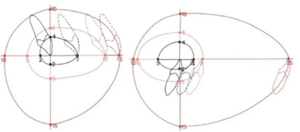
Figure 3. ”Envelope of discrepancy” shows the amount of change of tooth position that could be produced by orthodontic tooth movement alone, orthodontic tooth movement combined with growth modification, and orthognathic surgery. Adapted from Proffit12.
This diagram provides a guideline to help the clinician choose options between using a
treatment during teenage years, or waiting until the growth is over to treat the patient with
orthognathic surgery and orthodontics12.
Currently, no existing data correlate orthognathic surgery with the prevalence of tooth
extractions. It appears that the percentage of Class III patients undergoing orthognathic surgery is
increasing while the percentage of Class II patients undergoing surgery is decreasing at the
University of North Carolina. Some demographic factors could be part of the explanation (the
ethnicity of the patient population has changed with more African-American, Hispanic and Asian
patients). Also, the increased use of non-surgical appliances like the Herbst appliance or the
Forsus appliance could explain the decrease in Class II patients seeking surgery43.
Psychological- and philosophical factors:
In addition to the major variables mentioned above, the psycho-social dimension and personal
philosophy play a major role in the treatment plan.
• Esthetics
The concept of facial beauty and esthetics is of major importance for orthodontists and their
patients. Wahl wrote: “now it appears that facial esthetics is again in the forefront as we realize
why patients come to us in the first place”44
. Ker showed in his survey that an ideal and an
acceptable range for smile esthetics can be identified reliably23. As an example, Parekh showed
that both laypersons and orthodontists prefer smiles in which the smile arc parallels the lower lip
and buccal corridors are minimal45. Orthodontists and laypersons can then evaluate accurately
the smile esthetics. There is a debate about the consequence of extractions on the patient’s
profile. Some argue that extractions would cause a “dished in” profile, but this could not be
post-treatment profiles of 70 extractions and 50 non-extractions Class I and Class II Caucasian
patients. His results showed that non-extraction treatment produced very minimal effect on the
profile whereas extractions could improve the patient’s facial esthetics when they presented with
a combination of crowding and protrusion at the beginning of treatment46. Similarly, Drobocky
estimated that 90% of the patients treated with extractions saw their profile improved after
treatment22. In opposition, some studies could not find any differences in esthetic outcome
between the two treatment options47-48. Janson stated in his review of the literature that extraction
and non-extraction protocols seemed to have no predictable effect on smile esthetics32-49. In
another study, he compared Class II Division 1 patients treated with different extractions
protocols (one, three or four premolars extractions). He asked 70 orthodontists and 46 laypersons
to rate the posed-smile on a 10-point scale. His results showed that there was no significant
difference in smile attractiveness between orthodontists and laypersons or between the different
extraction patterns50.
• Personal philosophy
Individual orthodontists are influenced by their educational background, their vision of esthetics,
and their personal philosophy. As seen previously, extractions could impact the patient’s profile
1-2-51
, and some orthodontists or patients may prefer a fuller profile21-23-44-45-46. In protrusive
patients, the fullness of the lip improves after the extraction of four premolars. This improvement
is predictable but the changes are small and individual responses are very diverse.
Previous studies
Proffit14 surveyed the extraction frequency between the 1950’s and 1990’s at the
1953, reached 76% in 1968, and declined to 28% in 1993. He hypothesized that this variation
was due to considerations in outcome stability, facial esthetics, and technological changes.
However, new appliances (e.g. self-ligating brackets), development of growth modification
techniques, and improved orthognathic surgery procedures did not mature until the beginning of
the twenty-first century. It is uncertain whether these recent clinical approaches have had an
impact on the extraction frequency after 2000.
Figure 4. Extraction of four first premolars percentages in the Department of Orthodontics at the University of North Carolina over a 50-year period, from 1953-2003. Adapted from Proffit, Contemporary Orthodontics
Janson52 conducted a retrospective study at the University of Sao Paulo in Brazil to
evaluate the frequency of different extraction patterns. His sample comprised 3,413 records since
1973. He divided the sample in 10 groups depending on their extraction protocol, the first group
being the non-extraction patients. His results showed an overall increase of the non-extraction
group with a decrease of all the extraction protocols except for the two maxillary premolars
extraction groups which remained stable. There is a trend towards non-extraction treatments 31.
O’Connor found a similar trend in his survey of 814 questionnaires returned by orthodontists2
These studies of the extraction trend over time are very valuable because it can be
difficult for the practitioner to self-evaluate his extraction rate: the actual rate can differ from the
perceived extraction rate24.
Conclusions
Extractions have been performed for centuries as a treatment option in orthodontics. Their
frequency has fluctuated greatly over the years, with a very low percentage at the beginning of
the twentieth century due to the influence of Angle, a significant increase in the 1960s and 70s
and a general decrease since then. Variations in the extraction rate cannot be easily explained as
there are several criteria involved in the decision to extract teeth in addition to the practitioner
deciding based on his knowledge and philosophy of orthodontics.
The following study provides connections on how the aforementioned factors may be associated
with the likelihood of electing tooth extraction as an orthodontic treatment modality.
However, there is a consistent tendency to choose the less invasive treatment option, and some
practitioners in some occasions choose to delay the decision to extract until several months into
REFERENCES
1. Kelly JE, Sanchez M, Van Kirk LE. An assessment of the occlusion of the teeth of children 6-11 years. Washington (DC): U.S. Government Printing Office; Nov. PHS Publication. 1973; no. 74-1612
2. O'Connor BMP. Contemporary trends in orthodontic practice: A national survey. Am J Orthod Dentofacial Orthop. 1993; 103(2):163-170.
3. Peck S, Peck H. Frequency of tooth extraction in orthodontic treatment. Am J Orthod. 1979; 76(5):491-496.
4. Weintraub JA, Vig PS, Brown C, Kowalski CJ. The prevalence of orthodontic extractions. Am J Orthod Dentofacial Orthop. 1989; 96(6):462-466
5. Wahl N. Orthodontics in 3 millenia. Chapter 1: Antiquity to the mid-19th century. Am J Orthod Dentofacial Orthop. 2005; 127:255-9.
6. www.archwired.com/historyofortho.htm
7. Wahl N. Orthodontics in 3 millenia. Chapter 2: Entering the modern era. Am J Orthod Dentofacial Orthop. 2005; 127:510-5.
8. Peck S. A Biographical Portrait of Edward Hartley Angle, the First Specialist in Orthodontics, Part 1. Angle Orthod. 2009; 79:1021-1027.
9. Angle EH. Treatment of malocclusion of the teeth and fractures of the maxilla, Angle’s system. 7th edition, Philadelphia: SS White Dental Mfg. Co. 1907.
10. Holman JK, Hans MG, Nelson S, Powers MP. An assessment of extraction versus nonextraction orthodontic treatment using the peer assessment rating (PAR) index. Angle Orthod. 1998; 68(6):527-534.
11. Tweed CH. Clinical Orthodontics. 1966; Volume One pp. 1-54.
12. Proffit WR, Fields HW, Sarver D. Contemporary orthodontics. 2007; 4th ed. St. Louis: CV Mosby Co.
13. Ackerman JL, Proffit WR. Soft-tissue limitations in orthodontics: Treatment planning guidelines. Angle Orthod. 1997; 67(5):327-336.
14. Proffit WR. Forty-year review of extraction frequencies at a university orthodontic clinic. Angle Orthod. 1994; 64(6):407-414.
15. Janson G, Araki J, Estelita S, Camardella L. Stability of class II subdivision malocclusion treatment with 3 or 4 premolars extractions. Progress in Orthodontics. 2014; 15:67.
17. Boley J, Jeffrey M, Sachdeva R, Buschang P. Long-term stability of Class I premolar extraction treatment. Am J Orthod Dentofacial Orthop. 2003; 124:277-87.
18. Luppanapornlap S, Johnston LE. The effects of premolar extraction: a long-term comparison of outcomes in “clear-cut” extraction and non-extraction Class II patients. Angle Orthod. 1993; 63:257-272.
19. Anthopoulou C, Constantonis D, Makou M. Treatment outcomes after extraction and nonextraction treatment evaluated with the American Board of Orthodontics objective grading system. Am J Orthod Dentofacial Orthop. 2014; 146(6):717-723.
20. Takada K, Yagi M, Horiguchi E. Computational formulation of Orthodontic Tooth-Extraction Decision. Angle Orthod. 2009; 79:885-891.
21. Chang CA, Fields Jr. HW, Beck FM, et al. Smile esthetics from patients’ perspectives for faces of varying attractiveness. Am J Orthod Dentofacial Orthop. 2011; 140(4):e171-e180.
22. Drobocky OB, Smith RJ. Changes in facial profile during orthodontic treatment with extraction of four first premolars. Am J Orthod Dentofacial Orthop. 1989; 95(3):220-230.
23. Ker AJ, Chan R, Fields HW, et al. Esthetic and smile characteristics from the layperson’s perspective: a computer-based survey study. J Am Dent Assoc. 2008; 139:1318-1327.
24. Vig PS, Orth. D, Weintraub JA, Brown C, Kowalski CJ. The duration of orthodontic treatment with and without extractions: A pilot study of five selected practices. Am J Orthod Dentofacial Orthop. 1990; 97(1):45-51.
25. Ribarevski R, Vig P, Vig KD, Weyant R, O’Brien K. Consistency of orthodontic extraction decisions. European Journal of Orthodontics. 1996; 18:77-80.
26. Ioi H, Nakata S, Nakasima A, Counts AL. Anteroposterior lip positions of the most-favored Japanese facial profiles. Am J Orhtod Dentofacial Orthop. 2005; 128:206-11.
27. Francisconi MF, Janson G, Freitas KMS, Gobbi de Oliveira RC, Gobbi de Oliveira RC, Freitas MR, Henriques JFC. Overjet, overbite and anterior crowding relapses in extraction and nonextraction patients, and their correlations. Am J Orthod Dentofacial Orthop. 2014; 146:67-72.
28. Janson G, Valarelli FP, Beltrao RTS, Freitas MR, Henriques JFC. Stability of anterior open-bite extraction and nonextraction treatment in the permanent dentition. Am J Orthod Dentofacial Orthop. 2006; 129:768-774.
29. Little RM, Wallen TR, Riedel RA. Stability and relapse of mandibular anterior alignment –first premolar extraction cases treated by traditional edgewise orthodontics. Am J Orthod 1981; 80(4):349-365.
30. Hasija N, Bala M, Goyal V. Estimation of Tooth Size Discrepancies among Different Malocclusion Groups. Int J Clin Pediatr Dent. 2014; 7(2):82-85.
32. Janson G, Branco NC, Fernandes TMF, Sathler R, Garib D, Lauris JRP. Influence of orthodontic treatment, midline position, buccal corridor and smile arc on smile attractiveness. Angle Orthod. 2011; 81:153-161.
33. Maues CPR, Nascimento RR, Vilella OV. Severe root resorption resulting from orthodontic treatment: Prevalence and risk factors. Dental Press J Orthod. 2015; 20(1):52-8.
34. Konstantonis D, Anthopoulou C, Makou M. Extraction decision and identification of treatment predictors in Class I malocclusion. Progress in Orthodontics. 2013; 14:47.
35. Chen SS, Greenlee GM, Kim J, Smith CL, Huang GJ. Systematic review of self-ligating brackets. Am J Orthod Dentofacial Orthop. 2010; 137(6):726.e1-726.e18.
36. Shivapuja PK, Berger J. A comparative study of conventional ligation and self-ligation bracket systems. Am J Orthod Dentofacial Orthop. 1994; 106(5):472-480.
37. Harradine NWT. Current Products and Practices. Self-ligating Brackets: Where are we now? Journal of Othodontics. 2003; 30:262-273.
38. Harradine NWT. Self-ligation: Past, Present and Future. Journal of Orthodontics. 2009; 36:260-271.
39. Burrow SJ. To extract or not to extract: A diagnostic decision, not a marketing decision. Am J Orthod Dentofacial Orthop. 2008; 133:341-2.
40. Fleming PS, Johal A. Self-ligating Brackets in Orthodontics. A systematic review. Angle Orthod. 2010; 80:575-584.
41. Chen SSH, Greenlee GM, Kim JE, Smith CL, Huang GJ. Systematic review of self-ligating brackets. Am J Orthod Dentofacial Orthop. 2010; 137:726.e1-726.e18.
42. Tang GH, Zhang L, Xu XC, Li X, Chu FT. Indications for non-extraction treatment of dental crowding with Damon appliance. Shanghai Kou Qiang Yi Xue. 2008; 17(4):364-71.
43. Proffit WR, Jackson TH, Turvey TA. Changes in the pattern of patients receiving surgical-orthodontic treatment. 2013; 143(6)793-798.
44. Naini FB, Moss JP, Gill DS. The enigma of facial beauty: Esthetics, proportions, deformity, and controversy Am J Orthod Dentofacial Orthop. 2006; 130(3):277-282.
45. Parekh S, Fields HJW, Rosenstiel S, et al. Attractiveness of variations in the smile arc and buccal corridors space as judged by orthodontists and laymen. Angle Orthod. 2006; 76:612-618.
46. Bowman SJ, Johnston LE. The esthetic impact of extraction and non-extraction treatments on Caucasian patients. Angle Orthod. 2000; 70:3-10.
48. Meyer AH, Woods MG, Manton DJ. Maxillary arch width and buccal corridor changes with orthodontic treatment. Part 2: Attractiveness of the frontal facial smile in extraction and nonextraction outcomes. Am J Orthod Dentofacial Orthop. 2014; 145:296-304.
49. Johnson DK, Smith RJ. Smile esthetics after orthodontic treatment with and without extraction of four premolars. Am J Orthod Dentofacial Orthop. 1995; 108:162-7.
50. Janson G, Branco NC, Morais JF, Freitas MR. Smile attractiveness in patients with Class II Division 1 subdivision malocclusions treated with different tooth extraction protocols. The European Journal of Orthodontic. 2011; July:1-8.
51. Leonardi R, Annunziata A, Licciardello V, Barbato E. Soft Tissue Changes Following the Extraction of Premolars in Nongrowing Patients with Bimaxillary Protrusion. Angle Orthod. 2010; 80:211-216.
A CONTEMPORARY PERSPECTIVE ON TOOTH EXTRACTION IN ORTHODONTICS
Introduction
The US Public Health Service collectively documented the first organized data on
malocclusion in the United States as part of the National Health and Nutrition Examination
Survey (NHANES) in the 1960s. According to the NHANES study, Kelly et al reported that
24.4% of the children between 6 and 11 years had normal occlusion, 22.4% had a definite
malocclusion, 8.7% had severe malocclusion, and 5.5% were classified as having a very severe
malocclusion1. Kelly’s study was the first epidemiological investigation showing that at least
35% of American children are candidates for orthodontic treatment. More recently, the American
Association of Orthodontists stated that in 2004, there were 5,750,000 patients undergoing
orthodontic treatment, and that this statistic had undergone a steady and significant increase since
1989. For those individuals who may benefit from orthodontic therapy, tooth extraction
represents one major treatment option to relieve crowding or reduce protrusion. Dogmatic
non-extraction arch-expansion, originally proposed by Edward Angle, was found to be unstable and
unaesthetic in many cases2. The work of Angle’s student, Charles Tweed, who re-treated an
impressive series of patients with the extraction of four premolars, serves as a classic
counterpoint to the notion that in all cases it is best to align a full complement of teeth within the
arches2.
Contemporary guidelines suggest that patients presenting with severe crowding (greater
premolars with satisfactory alignment of lower incisors up to ten years and more post-retention3.
In many cases however, either extraction or non-extraction treatment may offer good stability
and results4-5-6. The factors that dictate the choice to extract are multi-faceted, and different
approaches have been attempted to objectify and improve the extraction decision. Takada created
a mathematical model to guide the treatment plan decision and optimize orthodontic treatment
outcome: His model used 25 morphologic traits with four major categories (sagittal dentoskeletal
and soft tissue relationship, vertical dentoskeletal relationship, transverse dental relationship and
intra-arch conditions)7. Nonetheless, a reliable criterion for extracting teeth remains elusive and
the topic continues to be one of controversy. Accordingly, extraction rates have varied over the
decades. Depending upon the year of treatment, it has been estimated that anywhere from 28% to
76% of orthodontic patients have undergone permanent tooth extraction, other than third molars
8-9-10
.
Proffit11 surveyed the extraction frequency between the 1950’s and 1990’s in orthodontic
patients cases treated at the University of North Carolina. He found that the extraction rate for
four premolars was 30% in 1953, reached a peak of 76% in 1968, and declined to 28% in 1993.
He hypothesized that this variation was due to considerations in outcome stability, facial
esthetics, and changes in technique. In addition to this study, recent research supports the notion
that the choice to extract teeth has to be made after consideration of three main aspects: esthetics,
stability, and occlusion12. Appliance design, surgical intervention and patient-related factors are
additional confounding factors. Individual orthodontists are influenced by their educational
background, the difference in treatment length13, and their vision of esthetics. Extractions can
impact the patient’s soft tissue profile1-8
, and orthodontists like patients have individual esthetic
demographic background, and diagnostic information (e.g., crowding, overjet, and overbite) also
contribute to the decision19. In short, the extraction decision is not a simple one, and some of
today’s clinicians are more inclined to extract teeth than expand the arches in borderline cases
while others would rather conserve the teeth if possible and rely on arch expansion to relieve
crowding.
With the increased access to information and esthetic demands of orthodontic patients
today, clinicians are facing ever greater pressures to perform evidence-based decision-making.
To keep pace with advances in the field of orthodontics, it is important to understand what
factors affect the extraction decision for orthodontic purposes.
We hypothesize that the chronological trend for tooth extraction is non-constant and
depends upon a multifactorial decision aforementioned. As a follow-up of Proffit’s 40-year
review, we analyzed the extraction rate at the University of North Carolina from 2000 to 2013.
Specifically, contemporary considerations for extraction, including appliances type (e.g.,
self-ligating brackets) and patient-related diagnostic factors were examined.
Materials and Methods
The Orthodontic Graduate Clinic at the University of North Carolina maintains pre- and
post-treatment records for each patient using standardized forms that record diagnostic
information as well as information capturing treatment approach and outcome. This data is
organized within a centralized database. Our study sample included all the patients who started
treatment on January 1, 2000 or later and completed treatment before December 31, 2011.
Inclusion criteria are that both the pre- and post- treatment forms are present and complete.
2,184 patients, with 1,263 females (58%) and 913 males (42%). The age at the start of treatment
ranges from 7 to 67 years old. The primary outcome measure was extraction of permanent teeth
for orthodontic purposes; third molars extractions were not included. Explanatory measures
included demographic, clinical and treatment factors. The demographic measures were the age at
start of treatment, the gender and the ethnicity of the patient. Clinical measures included the
initial overjet and overbite in millimeters, the crowding of the maxillary and mandibular arches,
the initial Angle classification, the initial skeletal anterior/posterior classification, the periodontal
health, the curve of Spee and the presence of root resorption at the end of treatment.
Additionally, the patient pool was divided in two groups based on the use of conventional
brackets or self-ligation brackets. The extraction rate in the self-ligation user group was
compared to the conventional (non-self-ligation) bracket user group.
Demographic and clinical characteristics of patients were described by mean and
standard deviation for continuous variables and proportions for categorical variables. Group
difference between patients with and without extraction was compared either based on
two-sample t-test or chi-square tests when appropriate. Characteristics that are significantly different
between extraction and non-extraction groups were considered potential risk factors for
extraction. Year by year extraction rate was explored in a time series plot and smoothed by
three-year moving average. Due to a significant lower extraction in 2005, we applied both linear and
quadratic term of (year - 2005) in the logistic model (Model I) for extraction probability. The
model was further adjusted by potential risk factors (Model II, III, and IV, Table 2) to investigate
the adjusted time effects. All of the statistical tests were 2-sided and p-value smaller than 0.05
Results
Patient Distribution: The sample composition varied over time as shown in Table 1. The
male to female ratio remained quite constant over time with female representing 52.9% to 63.9%
of the patients’ pool. The age of the patient at start of treatment ranged from 7 to 67 years old.
Our sample became more diverse in ethnicity between 2000 and 2011: The proportion of
Caucasian patients decreased from 82.3% in 2000 to 62.9% in 2011 while the African American
patients and other ethnicities increased from 11.5% to 13.3% and from 6.2% to 23.8%,
respectively.
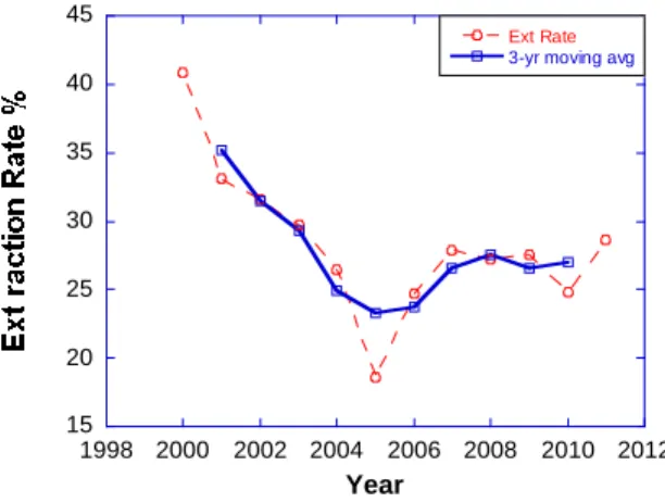
Year Effect: The year effect on extraction rate was found to be significant (p value of
0.048). The extraction rate at the University of North Carolina fluctuated significantly
throughout the years and peaked at 40.9% in the first year studied. This peak rate represents a
choice of extraction in almost half of the patients treated in the Graduate Clinic (Figure 1). This
percentage decreased significantly after 2000, going from 40.9% to 18.6% in 2005. The
extraction rate then increased to 27.9% in 2007 and remained relatively stable until 2011.
Figure 5. Extraction rate per year.
15 20 25 30 35 40 45
1998 2000 2002 2004 2006 2008 2010 2012
Ext Rate 3-yr moving avg
Several variables were found to exert a significant influence on the overall extraction rate
(p-value less than 0.05): the year of treatment, the ethnicity of the patient, overjet and overbite,
crowding, the Angle and skeletal classification, root resorption, and the bracket used for the
patient (Table 1 and Table 2).
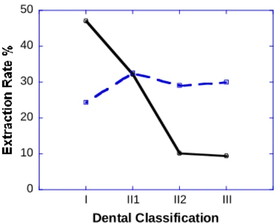
Effect of Angle Classification: Extraction rates were examined for each dental
antero-posterior diagnostic (Angle) classification: Class I dental, Class II division 1, Class II division 2
and Class III dental. The overall number of patients who underwent extractions is higher for the
Class I category which represents the largest pool of patients (47.1% of the patients). For the
extraction rate averaged over the past ten years, the extraction rate is higher for Class II and
Class III dental relationships than Class I patients. Patients with a Class II division 1 pattern had
the highest rate of extractions (Figure 2).
Figure 6. Extraction percentage and Angle classification. Solid line indicates total percentage of extraction for each classification over the entire 13 years’ time period. Dash line represents % extraction within individual classification.
The extraction percentages for each Angle classification were relatively stable from 2000
to 2011, with no significant variation noted (Table 2).
0 10 20 30 40 50
I II1 II2 III
Effect of Skeletal AP: The second variable is the skeletal antero-posterior classification
(Figure 3). For Class II and Class III patients the distinction was made between mild, moderate
and severe forms of skeletal discrepancies. This categorization of mild/moderate and severe
forms of malocclusion was made by the Resident and the Faculty and based arbitrarily on
Overjet measurements. The extraction percentage increased with the severity of the skeletal
Class II, going from 30% for the mild Class II malocclusion to more than 50% for severe skeletal
Class II patients. This is not true for the Class III patients, where we see fewer extractions for the
severe patients. In general, the patients with either skeletal or dental Class II or III malocclusion
experienced higher extraction rates than Class I patients.
Figure 7. Extraction percentages and skeletal antero-posterior classification (mild, moderate, severe). The red line represents the extraction rate within the classification while the red and green line represent the extraction rate overall.
Skeletal Class I patients extraction rate did not change meaningfully from 2000 to 2011.
On the contrary, skeletal Class II patients underwent more extractions in 2011 than 2000, (from
36.8% in 2000 to 39% in 2011) while Skeletal Class III patients underwent fewer extractions
(from 18.4% in 2000 to 14.3% in 2011).
0 10 20 30 40 50 60 70
I II-MIL II-MOD II-SEV III-MILIII-MODIII-SEV
Total % Natural History
% Ext within the Class
Ext% Natural History
Effect of Appliance: One other significant variable extracted from the data is the type of
bracket that was used for each patient. There is a significant difference in extraction rate between
self-ligating brackets and non-self-ligating brackets groups. The extraction rate for patients
treated with self-ligation was substantially lower than the non-self-ligation patients group, with
21 % of extractions in the self-ligation group compared to 42% in the non-self-ligation group.
The use of self-ligating brackets in the Graduate Clinic increased from 7% in 2000 to 23.8% in
2011.
The extraction rate was at its lowest in 2005 and decreased over time. Table 3 shows all
the variables included in four logistic models to study the relationship between each variable and
the extraction trend over time. Model 1 is composed of the extraction rate by year. Model 2 is
expanding Model 1 with gender, race, ethnicity and the use of self-ligating brackets. Model 3
adds the Angle classification, the skeletal classification, overjet and overbite to Model 2. Finally
Model 4 is Model 3 including only patients with crowding (1320 patients). The odds ratio (<1.0)
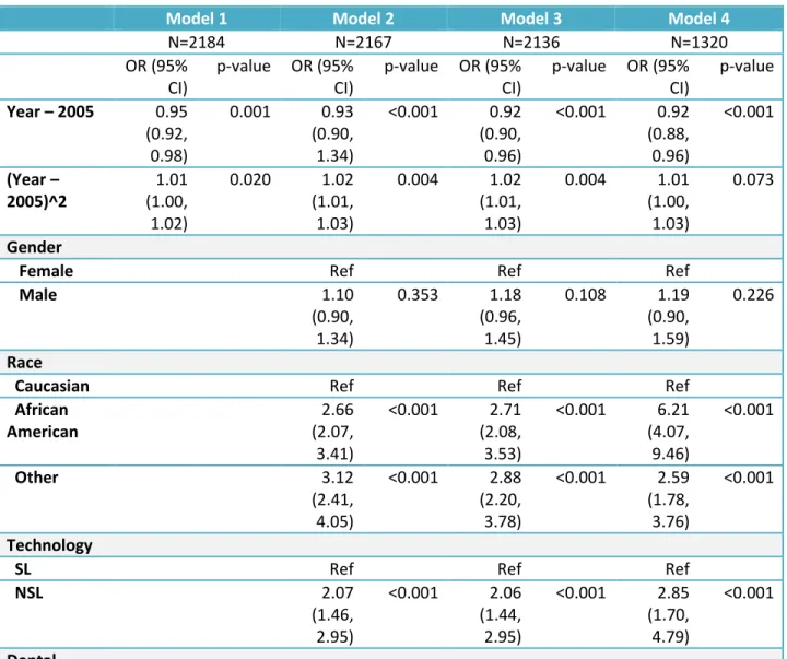
using (year – 2005) indicated that the extraction rate linearly decreased over year (p<0.05). The
models 2-4 proved that this decrease trend was not influenced by other confounding factors. The
odds ratio in the quadratic form (year-2005)2 confirmed that the extraction did increase after
2005, but it did not return to year 2000 because the linearly downward trend was significant.
Looking more closely at Model 4 which only included patients with crowding, we can observe in
Table 2 that in 2004, 2005 and 2006 patients had overall less crowding than the other years. This
could explain the lowest point in 2005.
The extraction rate decreased linearly over the years, but the trend is not affected by any of our
Discussion
We discovered a trend of decreasing numbers of tooth extractions in our sample of
patients treated at the UNC Graduate Orthodontic Clinic between 2000 and 2011 (n=2148). Our
patient pool was treated using an attending-resident model with twenty-five faculty members
having taught during this 11-year period. The patients were followed throughout treatment by
one faculty member and one (sometimes two) resident(s); thus, the patient sample is
representative of a variety of practitioners and their treatment philosophies. The total of 2184
subjects is a substantial sample size, which contributes to an epidemiological finding of the
extraction trend. Our data revealed that the percentage cases treated with tooth extractions varied
annually, ranging from 17% to 41%, with the greatest drop in 2005. Statistically, the extraction
rate was in linear decline from 40.9% in 2000 to 28.6% in 2011. A similar decreasing trend was
noted in previous studies. Janson20 conducted a retrospective study at the University of Sao
Paulo in Brazil to evaluate the frequency of different extraction patterns. His sample comprised
3,413 records since 1973. He divided the sample into 10 groups based on their extraction
protocol, the first group being the non-extraction patients. His results showed an overall increase
of the non-extraction group with a decrease of all the extraction protocols with the exception of
the two maxillary premolars extraction group, which remained stable. O’Connor found a similar
trend in his survey of 814 questionnaires returned by orthodontists8. There is a trend towards
non-extraction treatments20.
This linear decline over time was not influenced in our study by any explanatory
variables according to the logistic models (Table 3). Progressively adding the explanatory factors
provided a clear picture of the trend of the declined extraction rate in the past decade. No single
2005 was not statistically significant, which might be related to the low mean values of crowding
cases obtained in that year. The extraction rate, however, remained stable at 29% since 2007.
Although the explanatory variables could not explain the decreasing trend of the
extraction rate over time, eight major factors were found to be significantly related to the overall
extraction decision. The eight factors included year, ethnicity, Angle classification, skeletal
anterior-posterior classification, crowding, overjet, overbite, and technology. By averaging all
data from 2000 to 2011, a bivariance association analysis could detect a significant difference
between the extraction group versus the non-extraction group when each of the eight factors was
examined.
The ethnic diversity of our sample changed during these 11 years. The proportion of
Caucasian patients decreased from 82.3% in 2000 to 62.9% in 2011. This trend was also
observed by Proffit21 with an increase of African-Americans, Native-Americans, Hispanics, and
Asians patients seeking surgical treatment since 2000. Different ethnic groups present different
facial features and perceptions of esthetics, which ultimately dictate the decision to extract. As an
example, 42 Japanese dental students and 42 orthodontists were asked to rank their 3 favorite
profiles among 30 constructed profiles with different lip protrusion. The study showed that
Japanese preferred a retrusive profile even though natural Japanese facial features are somewhat
convex22. Proffit found in his study that more Class III patients and fewer Class II patients were
seeking surgery at UNC these days compared to ten years ago21. This change could be
attributable to the demographic changes but may also be due to the increased use of growth
modification appliances such as the Herbst appliance. These appliances offer a valid option for
Skeletal and dental anterior-posterior classifications were also significant factors for the
extraction decision. Extractions are frequently used in the correction of Class II and Class III
cases. They can be used as camouflage or as preparation for surgical treatment20-23. In our data,
patients presenting with a severe skeletal Class III underwent fewer extractions than the mild and
moderate Class III patients. This could be explained in that severe Class III patients seek surgical
treatment more so than their counterparts with milder discrepancies21, therefore not necessarily
needing extractions depending on the surgical plan.
Crowding, overjet, and overbite were also shown to influence the extraction decision.
Tooth extraction in orthodontics represents a major treatment option to relieve crowding3-12-24
and achieve acceptable overjet and overbite. For instance, we know that a non-extraction
treatment will likely procline the teeth and consequently decrease the overbite, and the opposite
is true with an increase of overbite in extraction cases25. Future analysis to find the threshold
values of crowding, overjet and overbite could provide information to guide orthodontists’
treatment decision.
Finally, our results showed that treatment technology could influence the practitioner’s
decision. 23.8% of the faculty members were using self-ligating brackets in 2011. This
proportion increased with 42% of them using self-ligation in 2015. 3 of the 14 attending faculty
members in the orthodontic clinic use passive self-ligating brackets (Damon or equivalent) and
rarely extract. Three other attending faculty members use active self-ligating brackets and their
extraction rate is also lower than the conventional twin bracket users group. The increased use
of these self-ligating brackets in clinical practice could be one of the variables explaining the
trend toward fewer extractions. Since the Russel Lock became the first self-ligating bracket on
the Damon SL in 1996, technological improvements have allowed them to gain greater
acceptability with clinicians. In the 11-year span of our study, self-ligating brackets have seen
increased use from 7% of cases in 2000 to 23.8% of cases in 2011.
Some patients may have concerns about orthodontic extractions and how their dental and
general health might be affected. Epidemiologic studies have linked the loss of teeth with an
increased predisposition for Alzheimer’s disease and early dementia. Tooth loss has been shown
to affect memory and learning in animal studies: the data suggests that tooth loss may inhibit
neurogenesis in the dentate gyrus of adult mice26. A recent 5 year prospective cohort study by
Okamoto27 finds a link between mild memory impairment and tooth loss in the elderly
population. One possible explanation would involve chemical components present during
periodontal disease, which should not concern our orthodontic patients. More studies are needed
to explore this detrimental effect.
Despite all the factors identified as having an effect on extraction rate, none were found
to be significant in explain the decreasing trend over time. This leads us to believe that the
decision to extract teeth is in the end highly irrational and seems to be a subconscious behavior
of the orthodontist. This decision is influenced by a multitude of variables and the same
orthodontist could probably opt for a different treatment plan for the same patient if asked at
different times.
Conclusions
The extraction rate fluctuated greatly since 2000 and has decreased linearly over the
decision: ethnicity, Angle Classification, skeletal anterior-posterior classification, presence of
root resorption, overjet, overbite, amount of crowding and technology used.
However, none of these variables were able to explain the decrease of the extraction rate
Tables
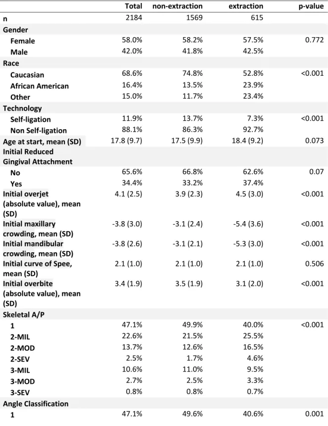
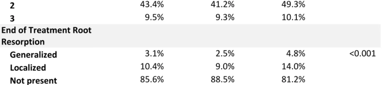
Table 1 – Influence of explanatory variables on the overall extraction rate.
Total non-extraction extraction p-value
n 2184 1569 615
Gender
Female 58.0% 58.2% 57.5% 0.772
Male 42.0% 41.8% 42.5%
Race
Caucasian 68.6% 74.8% 52.8% <0.001
African American 16.4% 13.5% 23.9%
Other 15.0% 11.7% 23.4%
Technology
Self-ligation 11.9% 13.7% 7.3% <0.001
Non Self-ligation 88.1% 86.3% 92.7%
Age at start, mean (SD) 17.8 (9.7) 17.5 (9.9) 18.4 (9.2) 0.073
Initial Reduced Gingival Attachment
No 65.6% 66.8% 62.6% 0.07
Yes 34.4% 33.2% 37.4%
Initial overjet
(absolute value), mean (SD)
4.1 (2.5) 3.9 (2.3) 4.5 (3.0) <0.001
Initial maxillary crowding, mean (SD)
-3.8 (3.0) -3.1 (2.4) -5.4 (3.6) <0.001
Initial mandibular crowding, mean (SD)
-3.8 (2.6) -3.1 (2.1) -5.3 (3.0) <0.001
Initial curve of Spee, mean (SD)
2.1 (1.0) 2.1 (1.0) 2.1 (1.0) 0.506
Initial overbite
(absolute value), mean (SD)
3.4 (1.9) 3.5 (1.9) 3.1 (2.0) <0.001
Skeletal A/P
1 47.1% 49.9% 40.0% <0.001
2-MIL 22.6% 21.5% 25.5%
2-MOD 13.7% 12.6% 16.5%
2-SEV 2.5% 1.7% 4.6%
3-MIL 10.6% 11.0% 9.5%
3-MOD 2.7% 2.5% 3.3%
3-SEV 0.8% 0.8% 0.7%
Angle Classification
2 43.4% 41.2% 49.3%
3 9.5% 9.3% 10.1%
End of Treatment Root Resorption
Generalized 3.1% 2.5% 4.8% <0.001
Localized 10.4% 9.0% 14.0%
Not present 85.6% 88.5% 81.2%
Table 2 – Extraction rate versus explanatory variables from 2000 to 2011 per year.
2000 2001 2002 2003 2004 2005 2006 2007 2008 2009 2010 2011 p-value
Extraction rate 40.9% 33.1% 31.6% 29.7% 26.5% 18.6% 24.7% 27.9% 27.2% 27.6% 24.8% 28.6% 0.048
Gender
Female 56.0% 57.5% 58.4% 63.9% 57.5% 52.9% 58.9% 56.3% 59.0% 57.2% 58.3% 59.0% 0.968
Male 44.0% 42.5% 41.6% 36.1% 42.5% 47.1% 41.1% 43.7% 41.0% 42.8% 41.7% 41.0%
Race
Caucasian 82.3% 81.7% 70.5% 78.1% 73.5% 68.6% 66.4% 64.5% 67.1% 62.0% 63.5% 62.9% <0.001
African American 11.5% 13.5% 21.7% 9.7% 16.8% 17.6% 18.5% 19.1% 13.5% 20.0% 15.0% 13.3%
Other 6.2% 4.8% 7.8% 12.3% 9.7% 13.7% 15.1% 16.4% 19.4% 18.0% 21.5% 23.8%
Technology
SL 7.0% 7.1% 7.8% 12.3% 7.1% 10.8% 8.9% 8.4% 2.9% 20.1% 20.9% 23.8% <0.001
NSL 93.0% 92.9% 92.2% 87.7% 92.9% 89.2% 91.1% 91.6% 97.1% 79.9% 79.1% 76.2%
Dental problem
1 49.1% 51.2% 48.4% 43.2% 55.9% 54.9% 56.8% 51.9% 41.2% 44.4% 42.3% 34.3% 0.008
2 42.0% 44.1% 39.8% 48.4% 38.7% 33.3% 37.0% 38.2% 48.7% 45.2% 46.3% 57.1%
3 8.9% 4.7% 11.9% 8.4% 5.4% 11.8% 6.2% 9.9% 10.1% 10.4% 11.3% 8.6%
Skeletal AP
1 44.7% 40.9% 43.4% 39.4% 50.4% 47.1% 47.9% 53.1% 45.6% 47.2% 50.9% 46.7% 0.370
2 36.8% 46.5% 39.3% 44.5% 34.5% 37.3% 39.0% 35.5% 43.9% 39.6% 34.0% 39.0%
3 18.4% 12.6% 17.2% 16.1% 15.0% 15.7% 13.0% 11.5% 10.5% 13.2% 15.0% 14.3%
Initial overjet
Mean 4.60 4.69 3.73 4.40 3.92 4.20 4.22 4.15 3.89 3.96 4.05 3.97 0.022
SD 2.79 3.00 2.43 3.03 2.49 2.83 2.26 2.53 2.48 2.41 2.27 2.21
Initial overbite
mean 3.72 3.80 3.41 3.33 3.34 3.59 3.40 3.26 3.25 3.29 3.35 3.26 0.223
SD 2.01 1.89 1.95 1.87 1.98 1.89 1.91 1.90 1.93 2.01 1.82 1.73
Initial max alignment
mean -3.75 -3.53 -4.15 -3.70 -3.33 -3.56 -3.37 -3.95 -3.87 -4.08 -4.13 -3.75 0.417
SD 2.56 2.73 3.51 2.79 2.80 2.36 2.67 3.18 3.38 3.16 3.06 2.67
Initial mand alignment
SD 2.59 2.75 2.99 2.72 2.51 2.21 2.07 2.94 2.88 2.43 2.54 2.15
EoTxRootResorp
G 2.7% 3.2% 7.1% 3.9% 2.7% 1.0% 1.4% 3.5% 2.1% 2.8% 3.1% 1.0% <0.001
L 23.9% 23.0% 14.5% 13.6% 9.7% 13.7% 8.9% 6.5% 4.2% 8.4% 6.2% 7.7%
N 73.5% 73.8% 78.4% 82.5% 87.6% 85.3% 89.7% 90.0% 93.7% 88.8% 90.7% 91.3%
Table 3 – Logistic regression model adjusted by potential risk factors for assessing extraction probability over time. Model 1 is the extraction per year. Model 2 is Model 1 plus gender, ethnicity and technology used. Model 3 is Model 2 plus Angle classification, skeletal anterior-posterior classification, overjet and overbite. Model 4 is Model 3 only including patients with crowding.
Model 1 Model 2 Model 3 Model 4
N=2184 N=2167 N=2136 N=1320
OR (95% CI)
p-value OR (95% CI)
p-value OR (95% CI)
p-value OR (95% CI)
p-value
Year – 2005 0.95
(0.92, 0.98)
0.001 0.93 (0.90, 1.34)
<0.001 0.92 (0.90, 0.96)
<0.001 0.92 (0.88, 0.96) <0.001 (Year – 2005)^2 1.01 (1.00, 1.02)
0.020 1.02 (1.01, 1.03)
0.004 1.02 (1.01, 1.03)
0.004 1.01 (1.00, 1.03)
0.073
Gender
Female Ref Ref Ref
Male 1.10
(0.90, 1.34)
0.353 1.18 (0.96, 1.45)
0.108 1.19 (0.90, 1.59)
0.226
Race
Caucasian Ref Ref Ref
African American
2.66 (2.07, 3.41)
<0.001 2.71 (2.08, 3.53)
<0.001 6.21 (4.07, 9.46)
<0.001
Other 3.12
(2.41, 4.05)
<0.001 2.88 (2.20, 3.78)
<0.001 2.59 (1.78, 3.76)
<0.001
Technology
SL Ref Ref Ref
NSL 2.07
(1.46, 2.95)
<0.001 2.06 (1.44, 2.95)
<0.001 2.85 (1.70, 4.79)
<0.001
problem
1 Ref Ref
2 1.56
(1.20, 2.03)
0.001 1.45 (1.00, 2.10)
0.050
3 1.25
(0.82, 1.89)
0.294 1.39 (0.77, 2.52)
0.280
Skeletal AP
1 Ref Ref
2 1.31
(1.01, 1.69)
0.042 1.40 (0.98, 1.99)
0.065
3 0.91
(0.63, 1.32)
0.908 0.82 (0.49, 1.38) 0.455 Initial overjet (absolute value) 1.08 (1.04, 1.13)
<0.001 1.16 (1.08, 1.25) <0.001 Initial overbite (absolute value) 0.83 (0.78, 0.88)
REFERENCES
1. Kelly JE, Sanchez M, Van Kirk LE. An assessment of the occlusion of the teeth of children 6-11 years. Washington (DC): U.S. Government Printing Office; Nov. PHS Publication. 1973; no. 74-1612.
2. Tweed CH. Clinical Orthodontics. 1966; Volume One pp. 1-54.
3. Boley J, Jeffrey M, Sachdeva R, Buschang P. Long-term stability of Class I premolar extraction treatment. Am J Orthod Dentofacial Orthop. 2003; 124:277-87.
4. Beattie JR, Paquette DE, Johnston LE. The functional impact of extraction and non-extraction treatments: A long-term comparison in patients with “borderline”, equally susceptible Class II malocclusions. Am J Orthod Dentofacial Orthop. 1994; 105:444-449.
5. Luppanapornlap S, Johnston LE. The effects of premolar extraction: a long-term comparison of outcomes in “clear-cut” extraction and non-extraction Class II patients. Angle Orthod. 1993; 63:257-272.
6. Janson G, Araki J, Estelita S, Camardella L. Stability of class II subdivision malocclusion treatment with 3 or 4 premolars extractions. Progress in Orthodontics. 2014; 15:67.
7. Takada K, Yagi M, Horiguchi E. Computational formulation of Orthodontic Tooth-Extraction Decision. Angle Orthod. 2009; 79:885-891.
8. O'Connor BMP. Contemporary trends in orthodontic practice: A national survey. Am J Orthod Dentofacial Orthop. 1993; 103(2):163-170.
9. Peck S, Peck H. Frequency of tooth extraction in orthodontic treatment. Am J Orthod. 1979; 76(5):491-496.
10. Weintraub JA, Vig PS, Brown C, Kowalski CJ. The prevalence of orthodontic extractions. Am J Orthod Dentofacial Orthop. 1989; 96(6):462-466
11. Proffit WR. Forty-year review of extraction frequencies at a university orthodontic clinic. Angle Orthod. 1994; 64(6):407-414.
12. Proffit WR, Fields HW, Sarver D. Contemporary orthodontics. 2007; 4th ed. St. Louis: CV Mosby Co.