THE EFFECT OF MODERATE INTENSITY INTERMITTENT EXERCISE ON METABOLIC BIOMARKERS IN BREAST CANCER SURVIVORS
Michelle M. Pebole
A thesis submitted to the faculty of the University of North Carolina at Chapel Hill in partial fulfillment of the requirements for the degree of Masters of Arts in the Department of Exercise
and Sport Science (Exercise Physiology).
Chapel Hill 2015
Approved by:
Anthony C. Hackney
Claudio L. Battaglini
Elizabeth S. Evans
ii © 2015 Michelle M. Pebole ALL RIGHTS RESERVED
iii
ACKNOWLEDGEMENTS
I would sincerely like to thank each of my board members for their help in completing
this project. First, to Dr. Battaglini for bringing his expertise in exercise oncology to this project;
his encouragement has pushed me forward with a smile on my face. Dr. Evans has been
instrumental to this project. Without her previous work, my analyses would not have been
possible. Dr. Evans has also been extremely patient with me throughout this whole process,
graciously responding to pestering e-mails from me throughout the process, as well as making
the trip over to Chapel Hill from Elon University numerous times. Ms. Amy Lane, a big thank
you for always keeping your door open to me when I came to ask questions. And lastly, I am
extremely thankful and grateful to Dr. Hackney. Without his unwavering guidance, patience,
and understanding, I would never have made it through this process with my sanity intact. I
would also like to express my gratitude to each subject, whose cooperation made it possible for
iv ABSTRACT
MICHELLE M. PEBOLE: The Effect of Moderate Intensity Intermittent Exercise on Metabolic Biomarkers in Breast Cancer Survivors
(Under the direction of Dr. Anthony C. Hackney)
PURPOSE: To examine the effect of one bout of exercise on the metabolic biomarkers
responses of breast cancer survivors (BCS) and healthy sedentary women controls (SW).
METHODS: 9 women who completed major treatments for Stage I-III invasive breast cancer
within 3-6 months and 9 SW without a history of cancer diagnosis completed a 30-minute bout
of intermittent cycle ergometry exercise at 60% of VO2peak. Blood was taken pre-exercise,
immediately post-exercise, and 2 hr post-exercise and analyzed for blood lactate, glucose and
free fatty acid (FFA) concentrations. RESULTS: Peak exercise lactate responses of BCS were
significantly lower than SW (p<0.01). No differences existed in glucose responses for BCS and
SW. BCS FFA response at 2 h post exercise was greater than SW (p<.05). CONCLUSIONS:
Findings support that BCS do not have the same metabolic responses to exercise as non-cancer
v
TABLE OF CONTENTS
I. INTRODUCTION... 9
Statement of Purpose………. 11
Research Questions………... 11
Hypotheses……… 11
Definition of Terms………... 12
Assumptions……….. 12
Limitations………. 12
Delimitations………. 12
Significance………... 13
II. REVIEW OF LITERATURE……….. 15
Breast Cancer Overview……… 16
Breast Cancer Pathophysiology………. 18
Breast Cancer Survivors and Exercise as a Supportive Intervention……… 20
Exercise Metabolism………. 22
Breast Cancer and Exercise Metabolism………... 25
Summary and Conclusions……… 26
III. METHODS……….. 28
Participants……… 28
vi
Statistical Analyses……… 33
IV. RESULTS………... 34
Subjects………. 34
Blood Lactate……… 35
Blood Glucose………... 36
Blood Free Fatty Acids………. 36
Plasma Volume Shifts………... 37
Percent Change Analysis………... 39
V. DISCUSSION………. 39
Introduction………... 39
Resting and Exercise Biomarker Responses………. 39
Exercise Energy Metabolism………. 41
Possible Mechanisms……… 43
Clinical Significance………. 44
Limitations and Strengths……… 45
Conclusion………. 46
vii
LIST OF TABLES Table
1. Subject physical characteristics (mean ± SD) …..………..…....35
viii
LIST OF FIGURES Figure
1. Blood lactate responses, Pre-Ex, 0hrPost, and 2hrPost exercise bout………....36
2. Blood free fatty acid responses, Pre-Ex, 0hrPost, and 2hrPost exercise bout ….…...38
3. The in vivo inner relationship between blood free fatty acid
and lactate concentrations in exercise situations and with
9
CHAPTER ONE Introduction
In 2014, an estimated 232,670 new breast cancer cases will occur in females, with 7,580
of those cases occurring here in North Carolina (American Cancer Society [ACS], 2014). Breast
cancer treatment typically involves breast-conserving surgery or mastectomy. With early
detection, long term survival is similar among women treated with breast-conserving surgery
plus radiation therapy and those treated with a mastectomy. Normally, with a diagnosis of breast
cancer comes an array of treatment options including surgery, radiation therapy, chemotherapy,
hormone therapy, and targeted therapy. Local therapies, such as surgery and radiation, are
intended to treat tumor without affecting the rest of the body (Abeloff, et al., 2008).
Contrastingly, systematic therapy such as chemotherapy, hormone therapy, and targeted therapy,
refers to administered drugs which can reach cancer cells throughout the body (ACS 2014).
With efficiency of current treatment options rising steadily, breast cancer survival is on the rise.
For all stage diagnoses of breast cancer, relative survival rates at 10 and 15 years after
diagnoses are 83% and 78%, respectively. Currently, approximately 61% of breast cancer
diagnoses are made at a localized stage, for which the 5 year survival rate is 99%. If the cancer
has spread to lymph nodes of the arm or other tissues, survival rate slightly decreases to 84%.
However, if the diagnosis is made at the late stage, where the cancer has spread to the collarbone
or distant lymph nodes and organs, survival rate drops dramatically to 24%, (ACS 2014).
Therefore, there is a large population of breast cancer survivors across the US; and with modern
10
healthy lifestyles in this population is physical activity. Exercise is important in this population
in that it helps with prevention of relapse as well as increasing quality of life. Many studies have
shown breast cancer survivors diagnosed in the early stage who are more physically active are
less likely to die from breast cancer, as well as lowering all-cause mortality (Avis et al., 2005).
Research has shown that exercise programs improve aerobic capacity, muscular strength,
flexibility, body composition, quality of life, physical functioning, and body image (Battaglini et
al., 2014; Schmitz, 2010). These improvements have been accompanied by decreases in body
size, fatigue, depression, anxiety and pain (Battaglini et al., 2009; Battaglini et al., 2014;
Schmitz, 2010). Furthermore, exercise before and after breast cancer diagnosis is associated
with a decreased risk of recurrence and/or death from breast cancer (Friedenreich et al., 2009;
Holmes et al., 2005; Irwin, 2009). The benefits of physical activity in this populations may come
from multiple interrelated biologic pathways involving decreasing adiposity, sex hormones
insulin resistance, adipokines, and chronic inflammation (Friedenreich et al., 2011). Despite
research showing that exercise elicits many positive outcomes for these patients, little is known
about exercise metabolism in breast cancer survivors. More studies on the acute metabolic
adaptations to exercise in this population may lead to a better understanding of their underlying
physiology. Currently, only a few studies have examined energy metabolism and substrate
utilization in breast cancer survivors. Those that have, found significantly lower lactate levels
following various intensity exercises when compared to age-matched healthy controls,
suggesting alterations in carbohydrate-lipid metabolism and substrate utilization in this
population (Evans et al., 2009; Tosti 2010). Considering the growth in this population, as well
as benefits of physical activity, additional research in this area is warranted. This research may
11 Statement of purpose
The purpose of this study was to examine the effect of a 30 minute acute bout of
moderate-intensity (60% VO2peak) intermittent exercise on metabolic biomarkers of energy
metabolism (blood lactate, glucose and free fatty acid concentrations) in female breast cancer
survivors versus sedentary controls.
Research Questions
RQ1: Will the acute exercise bout result in different lactate responses in the breast cancer group
as compared to the controls?
RQ2: Will the acute exercise bout result in different blood glucose responses post exercise when
compared to the control group?
RQ3: Will the acute exercise bout result in different blood free fatty acid responses post exercise
when compared to the control group?
Hypotheses
H1: The acute exercise bout will result in decreased lactate production post exercise in the breast
cancer group when compared to the control group.
H2: The acute exercise bout will result in decreased blood glucose concentrations post exercise
when compared to the control group.
H3: The acute exercise bout will result in increased blood free fatty acid concentrations post
12 Terms
Early Stage Breast Cancer: Breast cancer that has not spread beyond the breast or the axillary
lymph nodes.
Advanced breast cancer: Breast cancer that has spread beyond the breast and lymph nodes under
the arm or other parts of the body, also known as metastatic breast cancer.
Lobules: Gland of the breast that produces milk.
Ducts: Part of the breast that carry milk from the lobules to the nipple; the majority of breast
cancers start in the ducts of the breasts.
Stroma: Surround the ducts and lobules with fatty tissue, blood vessels, and lymphatic vessels.
Breast cancer survivor: For the purpose of this study, breast cancer survivors have diagnosis of
stage I, II, or III invasive breast cancer, have received chemotherapy, and completed all planned
surgery, chemotherapy, and radiation 3-6 months prior to enrollment.
Assumptions
1. All subjects enrolled in the study followed the pre-exercise guidelines prior to reporting
for laboratory for exercise.
2. VO2peak tests accurately approximate a patient’s VO2max.
Limitations
1. The control group was, on average, older than the experimental group.
2. Small sample size.
3. Occasional inability to obtain blood samples, leading to limited availability of samples.
13 Delimitations
1. Subjects were between 40-70 years old.
2. Subjects could not be regular users of anti-inflammatory medications.
3. Subjects must not have experienced a menstrual period for approximately 1 year prior to
enrollment.
4. The exercise bout was intermittent, which may be atypical for other exercise formats.
5. Subjects must have been diagnosed with stage I, II, or III invasive breast cancer, had
received chemotherapy, and completed all planned surgeries, chemotherapies, and
radiation therapy 3-6 months prior to enrollment.
6. The control group did not have a history of cancer diagnosis or treatment. Controls were
also sedentary and free from cardiovascular and musculoskeletal disease that would make
them unfit for exercise.
Significance
Breast cancer survival is on the rise. This population is one that stands to gain many
health advantages from physical activity and exercise. Exercise is an intervention that has shown
in other cancer populations to alleviate many negative effects of treatment, allowing patients to
lead a healthier, higher functioning, and more active lifestyle (Battaglini et al., 2014). However,
despite positive research findings, only 28% of breast cancer survivors exercise during treatment, with about 32% of survivors meeting ACSM’s exercise recommendations (Irwin, 2009).
Therefore, more knowledge about exercise energy metabolism in this population will be highly
beneficial in that it will give fitness professionals a more precise exercise prescription. More
14
Previous research has shown suppressed lactate levels in this population following
exercise. That being said, these findings suggest blood lactate responses post exercise are not a
good indicator of exercise intensity in breast cancer patients, contrary to findings in healthy
populations where lactate responses are frequently used as indicators of exercise intensity.
Furthermore, carbohydrate metabolism during exercise may be diminished in breast cancer
patients. Since carbohydrates are used during high intensity exercise, this may also have
implications for exercise prescriptions in this population in that higher intensity exercise may be
more challenging to perform. More research on blood lactate, blood glucose and free fatty acid
content will reveal more insight into substrate metabolism. This will be beneficial in considering
15
CHAPTER TWO: REVIEW OF LITERATURE
Cancer is the second leading cause of death in the United States, following heart disease
(Siegel et al., 2013). The lifetime probability of being diagnosed with cancer is slightly higher in
men (45%) than women (38%). While it is projected that 1,658,370 new cases of cancer will be
diagnosed in 2015, incidence rates are decreasing for all four major cancer sites (lung, breast,
colorectal, prostate), with the exception of female breast (American Cancer Society, 2015).
Furthermore, cancer death rates are steadily decreasing at slightly less than 2% per year
according to the recorded data from 2005-2009 (Siegel et al., 2013).
Modern medicine, coupled with the decreasing mortality rate, suggests more individuals
are successfully living longer with a prior diagnosis—either through remission or more effective
palliative care. Chances of survival increase when diagnosis is early, effective primary treatment
is prescribed, and proper secondary treatments and therapies are used to encourage better overall
quality of life (Smith et al., 2013). This holds true with breast cancer. With increasing survival
rates, it is important to comprehend the underlying physiology of cancer survivors as well as
promote healthy lifestyles in this population.
For the purpose of organization, this literature review is divided into the following
sections: 1) Breast Cancer Overview, 2) Breast Cancer Pathophysiology, 3) Breast Cancer
Survivors and Exercise as a Treatment 4) Breast Cancer and Exercise Metabolism 5) Summary
16 Section 1: Breast Cancer Overview:
As of January 1, 2012, approximately 14.5 million people in the United States are living
with a history of cancer, (American Cancer Society, 2015). While it is projected that 1.6 million
cases will be diagnosed in 2015, incidence rates are decreasing for all four major cancer sites,
with the exception of female breast cancer (American Cancer Society, 2015). Cancer is the
second leading cause of death in the United States, and accounts for about 1 of every 4 deaths
(Siegel et al., 2013). Fortunately, the 5-year survival rate for all cancer diagnoses made from
2003 until 2009 has increased to 68%; a significant increase from 49% in 1975-1977, (ACS,
2014). This improvement signifies both a progression in diagnosing and treating cancer, while also indicating increasing proportions of the population are living as cancer “survivors” (ACS,
2014). This study will focus on one of the most prevalent types of cancer in the United States,
breast cancer.
In 2014, an estimated 232,670 new breast cancer cases will occur in females, with 7,580
of those cases occurring here in North Carolina (ACS, 2014). Risk factors for breast cancer
include weight gain after the age of 18, being overweight or obese, use of hormone replacement
therapy, alcohol consumption, long term smoking, and physical inactivity, (ACS, 2014). Risk is
also increased with a family history of breast cancer diagnoses, although many researchers have
concluded most familial breast cancer results from a mixture of both genetics and lifestyle
choices. That is, inherited mutations in breast cancer susceptibility genes BRCA1 and BRCA2
occur much less than 1% in the population, but account for an estimated 5%-20% of all female
breast cancers (ACS, 2014). Factors associated with decreased risk of breast cancer include
17
weight, (ACS, 2014). Even with taking preventative measures, it is important to take necessary
breast cancer screening precautions.
Early detection is paramount in both treating and preventing breast cancer. For women at
average risk, breast cancer screening includes clinical breast cancer exams as well as
mammography (ACS, 2011). Early detection with mammography can catch breast cancer in an
early stage, when there are more treatment options. Declines in breast cancer mortality among
women since 1989 have been attributed to both improvements in early detections (like the
mammogram), as well as increases in treatment efficiency, (ACS, 2014). Annual screening for
breast cancer with both mammography and an MRI is recommended to start at the age of 40
(ACS, 2014).
Breast cancer treatment typically involves breast-conserving surgery or mastectomy.
With early detection, long term survival is similar among women treated with breast-conserving
surgery plus radiation therapy and those treated with a mastectomy. To determine if the tumor
has spread beyond the breast, underarm lymph nodes are usually removed and evaluated during
surgery (ACS, 2014). In early stage breast cancer patients, a sentinel lymph node biopsy may be
performed whereupon only the first lymph nodes to which cancer would have likely spread is
removed. This results in a lower change of long-term side effects such as lymphedema (ACS,
2014). Treatment involves several options.
Normally, with a diagnosis of breast cancer comes an array of treatment options
including surgery, radiation therapy, chemotherapy, hormone therapy, and targeted therapy.
Local therapies, such as surgery and radiation, are intended to treat the tumor without affecting
the rest of the body (Abeloff, et al., 2008). Contrastingly, systemic therapy such as
18
reach cancer cells throughout the body, (ACS 2014). With effectiveness of current treatment
options rising steadily, breast cancer survivorship is on the rise.
For all stages of breast cancer, relative survival rates at 10 and 15 years after diagnoses
are 83% and 78%, respectively. Currently, approximately 61% of breast cancer diagnoses are
made at a localized stage, for which the 5 year survival rate is 99%. If the cancer has spread to
lymph nodes of the arm or other tissues, survival rate slightly decreases to 84%. However, if the
diagnosis is made at the late stage, where the cancer has spread to the collarbone or distant
lymph nodes and organs, survival rate drops dramatically to 24%, (ACS 2014). Therefore, there
is a large population of breast cancer survivors across the US; and with modern advances in
diagnosis and treatment, that number will only increase. It is therefore very important to monitor
and consider the health of the population as measured by both prevention of relapse as well as
their quality of life. One way to promote healthy lifestyles in this population is physical activity.
Many studies have shown breast cancer survivors who are more physically active, especially
after diagnoses, are less likely to die from breast cancer, as well as lowering all-cause mortality
(Avis, Crawford, Manual, et al., 2005).
Section 2: Breast Cancer Pathophysiology
Cancer is born as a consequence of DNA damage intertwined with errors in proper cell cycle
behavior. All cancer stems from the rapid, uncontrolled growth of abnormally functioning cells
(Poste, 1980). The human body habitually produces these abnormally functioning cells; however,
safety and repair mechanisms of the genetic cell cycle serve to either correct cellular irregularity
or to ensure a discontinuation of cell specific growth. If both cellular missteps occur in concert,
19
in site-specific tumor growth (ACS, 2011). Breast cancer is made of a malignant tumor that
starts in the cells of the breast. These tumor cells can invade surrounding tissues and metastasize
to distant areas of the body. Breast cancer can be better understood by comprehending and
comparing normal versus infected breast structures.
Female breasts are made up of lobules, ducts, and stroma, each with a specific function.
Lobules produce milk, ducts carry breast milk from the lobules to the nipple, and stroma
surround the ducts and lobules with fatty tissue, connective tissue, blood vessels, and lymphatic
vessels (Abeldoff, Wolff, & Weber, 2008). Most breast cancers begin in cells that line the ducts
or lobules. Additionally, many lymph nodes are in close proximity to breast tissue, with most
lymph vessels in the breast connecting to axillary nodes under the arm (Avis et al., 2005). Other
significant lymphatic nodes in this region are located inside the chest and below/above the
collarbone. Different forms of breast cancer affect these structures.
The most common forms of breast cancers are ductal carcinoma in situ (DCIS), lobular
carcinoma in situ (LCIS), invasive ductal carcinoma (IDC), and invasive lobular carcinoma
(ILC). DCIS and LCIS are both considered pre-cancer states, whereas IDC and ILC are fully
cancerous (ACS, 2013). DCIS is a pre-invasive breast cancer signifying cells lining the ducts
have changed to look like cancer cells. However, these cells have not invaded through the walls
of the ducts to other breast tissue. Only some cases can go on to become invasive cancers, but
currently there are no diagnostic tools to determine which cases will do so (ACS, 2013). LCIS
cells look like cancer cell growth in the lobules of milk-producing glands of the breast, but do
not grow through the wall of the lobules. It differs from DCIS in that LCIS will not become
invasive if the cancer is not treated. However, women with this condition have 7 to 11 fold
20
IDC is the most common type of breast cancer, claiming about 80% of invasive breast cancer
cases; it starts in the milk duct of the breast, breaks through the wall of the duct, and grows in the
fatty tissue of the breast. This form of cancer is capable of metastasizing to other parts of the
body via the lymphatic system and bloodstream (ACS, 2013). Similarly, ILC starts in the
lobules and can metastasize to other parts of the body. About 1 in 10 invasive breast cancer
cases is an ILC. Less common types of breast cancer include inflammatory breast cancer, Paget
disease of the nipple, phyllodes tumor, and angiosarcoma.
Section 3: Breast Cancer Survivors and Exercise as a Supportive Intervention
Presently, with increased detection and treatment methods, breast cancer survivorship has
increased dramatically in the past few years. Today, there are roughly 12 million cancer
survivors alive in the United States alone, with that number steadily increasing (Hewitt,
Greenfield, Stovall, 2006). Historically, doctors advised patients primarily with rest and
avoidance of physical activity and exertion. However, recent research in the field of exercise
physiology and cancer rehabilitation reveals that exercise can improve physical functioning,
quality of life, and cancer-related fatigue in many cancer survivor groups, (Battaglini et al., 2004;
Battaglini et al., 2007; Schmitz et al., 2010). Furthermore, treatment for breast cancer is
expensive and comes with many deleterious side effects. Thus, non-pharmacologic methods,
such as physical activity and exercise, may offer an attractive supplement to traditional therapies
and aide to decrease morbidity and increase quality of life (Battaglini et al., 2009; Schmitz,
2010).
Current recommendations from the ACSM regarding cancer survivors focus on maintain
21
period. Present recommendations suggest increasing exercise levels to 3-5 days a week with
resistance training 2-3 days a week (American College of Sports Medicine [ACSM], 2014).
Research currently supports improvements in wellbeing even in patients undergoing systemic
cancer treatments with gradual increases in physical activity over one month. Exercise tolerance
in these individuals may vary greatly from day to day and person to person. Additionally, heart
rate may be a less reliable measure for monitoring exercise intensity in this population.
Therefore, using rate of perceived exertion (RPE) along with heart rate reserve (HRR) may be
helpful in evaluating intensity (ACSM, 2014). Aerobic exercise intensity should be moderate
(40% - 60% VO2peak or HRR; RPE moderate) to vigorous (65%-80% VO2peak or HRR; RPE
12-16). Flexibility exercises should be mindful of range of motion restrictions due to surgery and
chemotherapy/radiation therapy. Time recommendations do not differ from those of the healthy
population; 75 minutes a week of vigorous physical activity or 150 minutes of moderate intensity
activity, or a combinations of the two (ACSM, 2014). Resistance training should be at least 1 set
of 8-12 reps with exercises covering large muscle groups throughout the upper and lower body
(ACSM, 2014). In this population, slower progression may be needed, and awareness of
exercise on cancer-related symptoms in survivorship is needed (ACSM, 2014). Cancer survivors
who follow these recommendations will likely experience many health benefits.
There have been many studies examining the safety and efficacy of exercise training in
breast cancer survivors who have completed surgery, chemotherapy, and radiation therapy,
specifically. Research has shown that exercise programs improve aerobic capacity, muscular
strength, flexibility, body composition, quality of life, physical functioning, and body image
(Battaglini et al., 2009; Schmitz, 2010). When using estimated measurements derived from a
22
have been observed for breast cancer exercise groups, at improvements from baseline of 3.54
mL/kg/min, (Battaglini et al., 2014). These improvements have been accompanied by decreases
in body size, fatigue, depression and anxiety and pain (Battaglini et al., 2014; Spector et al.,
2014; Schmitz, 2010). Specific positive body composition changes with exercise training include
significant decreases in body fat percentage and increase in lean body mass (Battaglini et al.,
2014). Furthermore, exercise before and after breast cancer diagnosis is associated with a
decreased risk of recurrence and/or death from breast cancer, as well as dramatic increases in
quality of life (Friedenreich, et al., 2009; Holmes, et al., 2005; Irwin, 2009).
Yet, despite the numerous benefits of physical activity in cancer survivors, a large
proportion of cancer survivors do not perform regular physical activity, with many survivors
decreasing physical activity levels after diagnosis (Irwin, 2009). Only about 28% of breast cancer survivors exercise during treatment, with about 32% of survivors meeting ACSM’s
exercise recommendations (Irwin, 2009). Therefore, it is important to advocate physical activity
in cancer counselling as well as prescription by medical professionals.
Section 4: Exercise Metabolism
Throughout the day, and especially during exercise, the body meets energy needs via
substrate oxidation of primarily carbohydrates and lipids. During exercise, it is especially
important the body is efficient at utilizing these substrates for ATP production. However, recent
research suggests that there are several differences between exercise metabolism and substrate
oxidation in the healthy population as compared to breast cancer survivors.
Regulation of plasma glucose concentrations is one of the most tightly regulated
23
mg/dl (Brooks et al., 2005). This is required for central nervous function as well as other glucose
requiring systems, organs, and cells. During exercise, skeletal muscle greatly increases the
amount of glucose it uptakes in order to meet energy demands. Therefore, maintenance of blood
glucose homeostasis becomes a major physiological challenge during exercise (Brooks et al.,
2005). In healthy adults, glucose is stored as glycogen primarily in the liver, but also in the
skeletal muscle. To mobilize glucose, glycogenolysis occurs. Epinephrine and norepinephrine
secretion from the adrenal medulla at the start of exercise help stimulate muscle glycogenolysis
by activating the adenylate cyclase mechanism. Catecholamine secretion also stimulates hepatic
glycogenolysis in the liver. Therefore, increased catecholamine release is strongly associated
with increased glucose appearance in the blood (Brooks et al., 2005). The resulting glucose is
then used by working skeletal muscle. Normally, glucose rises at the start of exercise, and for
the remainder of the exercise bout, glucose may fall, but will remain within approximately 10%
of the normative value until exercise ends (Brooks et al., 2005). Additionally, it is well
established that with increases in exercise intensity comes greater reliance on carbohydrate as
fuel, and reductions in free fatty acid (FFA) utilization, in part due to rapid rises in
norepinephrine levels. Additionally, during hard exercise, inactive tissues become glucose
resistant, leaving glucose to be taken up by active muscle. (Brooks et al., 2005). Changes in FFA
oxidation result in changes in blood FFA concentrations.
Blood FFA concentrations change during exercise in healthy populations. During
prolonged moderate intensity exercise (above 50% VO2max), blood FFA levels rise continuously
(Brooks et al., 2005). Fasting increases the mobilization of triglyceride to FFA and glycerol
almost doubling the FFA concentrations in the blood. During more high intensity exercises,
24
catecholamines increase and pH declines, lipolysis is inhibited, and the esterification of FFA to
triglyceride is promoted (Brooks et al., 2005). Because of this, at higher intensity exercise (65%
VO2max or higher), blood FFA concentrations decline. This limits the amount of FFA for use as
fuel sources. Further, products of CHO catabolism inhibit mitochondrial enzymes that allow
FFA derivatives into mitochondria for oxidation (Brooks et al., 2005).
Lactate production also plays a key role in exercise metabolism. Lactate is produced as a
by-product of metabolism. During exercise metabolism, glucose is broken down into pyruvate,
which then quickly gets converted into lactate by the muscle lactate dehydrogenase isoform
(LDH-M) (Brooks et al., 2005). At rest and during light intensity exercise, lactic acid is
produced and removed at equal rates. In healthy adults, lactate turnover happens very rapidly
(Brooks et al., 2005). As exercise intensities increase, so do blood lactate levels; metabolic
clearance rate of lactate is also increased at these higher exercise intensities. However, at these
higher intensities lactate production outpaces lactate clearance, ultimately resulting in more
lactate accumulation (Brooks et al., 2005). Therefore, lactate removal is concentration
dependent; its levels must rise to force its removal. When exercise intensity increases, there is
greater reliance on carbohydrate as an energy substrate, thus increasing lactate production
through glycolytic mechanisms (Brooks, et al., 2005; Gladden 2004; van Loon, Greenhaff,
Constantin-Teodosiu 2001). Blood lactate levels increase non-linearly, and concentrations will
rise steadily beginning at about 60% VO2max. These elevated lactate levels may contribute to
altering several other physiological mechanisms in order to combat lactate build up and buffer a
decreased pH (Brooks et al., 2005). These mechanisms include increased ventilation,
25
supports that substrate oxidation and lactate metabolism differs in clinical populations such as
breast cancer survivors.
Section 5: Exercise Metabolism in Breast Cancer Survivors
Despite the small number of studies that have examined energy metabolism issues in
cancer patients, several differences have still been identified. For instance, research has shown
glucose intolerance is more frequent in cancer populations (Brooks et al., 2000; Edmonson,
1966; Glicksman & Rawson, 1956). Additionally, insulin resistance and greater levels of fasting
insulin are associated with breast cancer (Larsson et al., 2007). These findings suggest
alterations in carbohydrate metabolism in this population. Similarly, Evans and associates
indirectly examined carbohydrate metabolism during exercise by measuring blood lactate
responses (Evans et al., 2009). Results found post-treated breast cancer patients had significantly
lower lactate levels following high intensity exercise (70%VO2max) when compared to
age-matched healthy controls.
Additionally, Tosti et al. found significantly lower blood lactate responses to exercise at
various intensities including low (40% VO2max), moderate (60% VO2max), and high (70%
VO2max) (Tosti et al., 2010). Lower blood lactate concentrations during exercise suggest
alterations in carbohydrate metabolism in breast cancer survivors. This finding is new, and needs
more research to comprehend fully. In the same study, Tosti et al. found reduced carbohydrate
oxidation and greater fat oxidation in the cancer survivors as compared to healthy controls. These alterations in lactate metabolism are in line with Tosti’s findings on substrate utilization.
If less carbohydrate is being utilized, then less lactate should be produced. However,
26
reduction in carbohydrate metabolism and lactate responses in the cancer population are
unknown with speculations pointing towards altered endocrine responses, irregular glucose
metabolism, and treatment effects in this population.
Section 6: Summary and Conclusions
With efficiency of current treatment options rising steadily, breast cancer 10 and 15 year
survival rates are increasing steadily. Along with these increases come increasing breast cancer
survival populations. Therefore, it is important to monitor and comprehend the health of these
survivors as well as how this relates to their quality of life. One efficient way to promote healthy
lifestyles while improving quality of life is through physical activity and exercise. Many studies
have shown breast cancer survivors who are more physically active, especially after diagnoses,
are less likely to die from breast cancer, as well as lowering all-cause mortality (Avis et al.,
2005). Currently, breast cancer survivors are advised to perform physical activity 3-5 days a
week, with resistance training 2-3 days a week (ACSM, 2014). During these exercise bouts,
aerobic intensity is advised to be from moderate to vigorous intensities (40% - 80% VO2R; RPE
10-16) (ACSM, 2014). Time recommendations are the same as for healthy populations: 75
minutes of vigorous physical activity or 150 minutes of moderate intensity activity per week
(ACSM, 2014).
Following these recommendations leads to many health benefits including improved
aerobic capacity, muscular strength, flexibility, body composition, quality of life, physical
functioning, and body image (Schmitz, 2010). These improvements have been accompanied by
27
exercise before and after breast cancer diagnosis is associated with a decreased risk of recurrence
and/or death from breast cancer (Friedenreich et al., 2009; Holmes et al., 2005; Irwin, 2009).
Despite knowledge of the many benefits, little is known about exercise metabolism in
breast cancer survivors. Previous research has found that breast cancer survivors experience
reduced carbohydrate oxidation and greater fat oxidation as compared to healthy controls (Tosti
et al., 2010). Additionally, studies have found that in breast cancer populations, lactate
production is decreased at low, moderate, and high intensities (Evans et al., 2009; Tosti et al.,
2010). These findings suggest alterations in carbohydrate metabolism in this population.
However, mechanisms behind the reduction in carbohydrate metabolism and lactate responses in
the cancer population are unknown. This study aims to add to knowledge regarding acute
28
CHAPTER THREE: METHODOLOGY Participants
Research participants included a control and experimental group. The experimental
group included 9 women who completed major treatment for stage I-III invasive breast cancer
(BC) within 3-6 months of enrollment. The control group consisted of 9 sedentary women
without a history of cancer. All subjects were between the ages of 40-70 years, were not regular
users of anti-inflammatory medications, and had not experienced a menstrual cycle for
approximately 1 year prior to enrollment; they were recruited from the University of North
Carolina at Chapel Hill and the surrounding areas after obtaining approval from the Institutional
Review Board at UNC in the Department of Exercise and Sports Science and School of
Medicine, as well as the Protocol Review Committee of the UNC Lineberger Comprehensive
Cancer Center. Inclusion criteria for the breast cancer survivor group included:
Diagnosis of stage I, II, or III invasive breast cancer
Had received chemotherapy
Completed all planned surgery, chemotherapy, and radiation 3-6 months prior to
enrollment
Whereas, inclusion in the control group included:
29
Sedentary, defined by not meeting ACSM’s guidelines
Free from cardiovascular and musculoskeletal disease that would render aerobic exercise
participation unsafe
Research Design Overview
All blood analyses were done retrospectively as samples were obtained in an earlier study
conducted by Dr. Elizabeth Evans (2012). This paper will outline procedures subjects went
through prior to blood analysis, as consistent with Evans reporting. Throughout this study, each
subject completed three laboratory visits. During visit 1, subjects were given background
regarding the study, signed all IRB-approved written informed consent documents, received a
comprehensive medical and physical screening, and completed a peak oxygen uptake test
(VO2peak) on the cycle ergometer to assess maximal aerobic power. During visit 2, subjects
performed a 30-minute intermittent exercise bout on the cycle ergometer at 60% of VO2peak.
Immediately pre-exercise, immediately post-exercise, and 2 hours post-exercise blood samples
were obtained. Lastly, one resting blood sample was obtained at 24 hours post-exercise during
visit 3. However, these latter 24 hour post-exercise blood samples were not used in this
analyses. Pre-assessment guidelines included the following: fasting for at least 2 hours prior to
testing, no exercise for at least 12 hours prior to testing, maintaining adequate hydration, no
caffeine for at least 12 hours prior to testing, and no alcohol use for at least 48 hours prior to
testing.
30
To determine if subjects were healthy enough to proceed with the study protocol, each
completed a comprehensive medical history questionnaire, a 12-lead resting electrocardiogram
(ECG) and a physical examination (Whaley et al., 2006). After being cleared for participation, the subject’s height and body mass were taken using a portable stadiometer (Perspective
Enterprises, Portage, MI) and a calibrated balance-beam scale (Detecto, Webb City, MO). Body
mass index (BMI) was calculated from height (cm) and body mass (kg) measurements. A
Discovery Dual Energy X-ray Absorption (DEXA) scanner (Hologic, Inc., Bedford, MA)
reported body fat percentages for each subject. For the breast cancer survivor group, medical
treatment details were recorded. These treatments included surgery type, chemotherapy, whether
or not radiation therapy was received, along with other relevant cancer treatment and medication.
VO2peak was then measured via the Åstrand Cycle Ergometer Maximal Test protocol on a
Lode electronically-braked cycle ergometer (Lode, Groningen, The Netherlands) (Heyward,
2006). This protocol called for the subject to begin pedaling at a workload of 50 Watts for 3
minutes, and progressively added increments of 25 Watts every 3 minutes until volitional
fatigue. At the end of every minute of the test, heart rate and rating of perceived exertion (RPE)
were recorded (Borg, 1982). Expired gases were collected and analyzed using a TrueMax 2400
Metabolic System (Parvo Medics, Salt Lake City, UT). Minute-by-minute heart rates were
obtained from 12-lead ECG monitoring (GE CASE Cardiosoft V. 6.6 ECG diagnostic system,
General Electric, Palatine, IL). The highest VO2 measured by the metabolic system during the
last stage of the test was declared the VO2peak. The workload on the cycle ergometer that
corresponded to the subject’s VO2peak was peak work load. After test completion, subjects cooled
down by pedaling at a very light, self-selected workload. Research assistants monitored vitals
near-31
baseline levels (Whaley, Brubaker, Otto, 2006). Results obtained from the Åstrand test were
used to calculate the submaximal workload at which subjects exercised during visit 2.
Visit 2: Aerobic Exercise Session
The acute bout of moderate-intensity aerobic exercise (30 minutes) on the cycle
ergometer corresponded to 60% of the subject’s VO2peak. This intensity and duration
combination was comparable to protocols used in similar studies examining the effects of
aerobic exercise in breast cancer patients. Thus, this protocol is representative of exercise
prescriptions that are appropriate and used frequently in this population according to the
literature (Jones et al., 2011; Payne et al., 2008). However, previous testing with breast cancer
survivors at UNC-CH revealed cycling for 30 continuous minutes at 60% of VO2peak was
difficult for many breast cancer patients. To make sure all subjects could complete the exercise
session while maintaining the 60% of VO2peak workload, an intermittent protocol was utilized.
This protocol had subjects’ alternate 3-minute intervals of exercise (10x) with 1.5 minutes of
rest, for a total of 30 minutes of exercise in a 43.5-minute period. All exercise sessions started
between 7:00-10:00 am to control for daily physiologic variation in the research between
patients.
For the purpose of blood sampling, an angiocatheter was positioned in an antecubital vein
in the arm of the subject at the beginning of the laboratory visit. Subjects then laid supine for
approximately 20 minutes. A pre-exercise (Pre-Ex) blood sample was drawn into two 3-mL
K3EDTA Vacutainer® tubes. Following the rest, subjects sat calmly on the cycle ergometer for
3 minutes while pre-exercise metabolic data was collected. Subjects then warmed up for 4-5
minutes. After the warm up, subjects could stretch the lower body muscles in a self-selected
32
monitored during the first, third, seventh, and tenth exercise interval. To ensure subjects
maintained an exercise intensity as close as possible to 60% of VO2peak, workload was adjusted
throughout the exercise bout based off data from the metabolic system. Heart rate and RPE were
recorded every 3 minutes. The subject immediately dismounted the cycle ergometer when the
exercise bout was complete, and assumed supine position. An immediate post-exercise (0hPost)
blood sample was drawn into two 3-mL K3EDTA Vacutainer® tubes.
Subjects relaxed in the laboratory for the remainder of the visit. Ingestion of any food or
beverages was not permitted, with the exception of water. At 2 hours post-exercise (2hPost),
another blood sample was obtained in the same manner as previously described. Lastly, the
angiocatheter was removed and subject was bandaged.
Visit 3: 24-hour Follow-up Session
24 hours after completion of the aerobic exercise session the subjects returned to the
applied physiology lab (APL) in order to obtain the 24hourPost a resting blood sample. Standard
venipuncture procedures were used to transfer blood into two 3-mL K3EDTA Vacutainer® tubes.
Blood was taken from an antecubital vein in the arm. However, none of these bloods were used
in the analyses of the current study.
Determination of Plasma Blood Lactate, Glucose, and Free Fatty Acid
Following laboratory visits 2 and 3, whole blood samples were centrifuged at 4°C at 3000
rpm for 10 minutes using an IEC Centra-8R refrigerated centrifuge (International Equipment
Company, Needham Heights, MA). The plasma portion of the blood was removed and stored in
cyrofreeze vials at -80°C until the time of analysis. Blood glucose and free fatty acid
33
Lactate was analyzed using the DT60 Johnson & Johnson automated blood analyzer (Rochester,
NY).
Calculation of Plasma Volume Shifts
Plasma volume shifts during exercise were calculated utilizing the Dill and Costill
equation (Dill, Costill, 1974). Complete blood counts were analyzed at each study time point
using a COULTER® Ac•T diff ™ Hematology Analyzer (Beckman Coulter, Inc., Brea, CA).
This generated hematocrit and hemoglobin values used in plasma volume shift calculations.
Plasma volume shifts were calculated to evaluate the effect of exercise on fluid shifts in the
blood, which can affect concentrations of lactate, glucose, and free fatty acids.
Statistical Analyses
Statistical analyses were performed using SPSS version 19.0 with significance set a
priori at p ≤ 0.05. Blood glucose, free fatty acid, and lactate levels will be analyzed with a 2
(group) X 3 (measurement time) mixed model ANOVA to compare data sets. If significant
F-ratios are found with the ANOVA, a Bonferroni post-hoc test will be used to determine
individual mean differences.
Additionally, outcomes were converted to a percent change of pre exercise levels and
statistically analyzed with a 2 X 2 mixed model ANOVA. If significant F- ratios are found, a
34
CHAPTER FOUR: RESULTS
Subjects
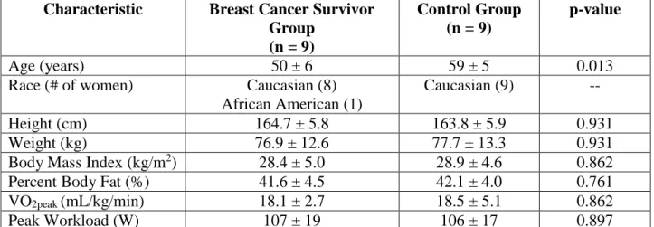
There were 18 subjects who participated in the study, 9 women in the breast cancer
survivor group, and 9 in the control group. Physical characteristics for all 18 subjects are
detailed in table 1. All the women in the breast cancer survivor group had a diagnosis of stage
I-II invasive breast cancer, had received chemotherapy, and completed all planned surgery,
chemotherapy, and radiation 3-6 months prior to enrollment. At the time of blood collection, 4
subjects in the survivor group were receiving additional adjuvant therapies, including Herceptin,
Femara, or Tamoxifen (Evans 2012). All women in the control group were sedentary, did not
possess any counter indications to exercise, and had never had a previous cancer diagnosis.
Subjects were matched on physical characteristics, with no significant differences between the
groups in physical characteristics, physical activity level, or menopausal status. However, it was
found that the control group was significantly older than the breast cancer survivor group
(p=0.013). Descriptive characteristics are presented in the form of mean ± standard deviation In
Table 1, which can be found on the next page.
35 Characteristic Breast Cancer Survivor
Group (n = 9)
Control Group (n = 9)
p-value
Age (years) 50 ± 6 59 ± 5 0.013
Race (# of women) Caucasian (8) African American (1)
Caucasian (9) --
Height (cm) 164.7 ± 5.8 163.8 ± 5.9 0.931 Weight (kg) 76.9 ± 12.6 77.7 ± 13.3 0.931 Body Mass Index (kg/m2) 28.4 ± 5.0 28.9 ± 4.6 0.862 Percent Body Fat (%) 41.6 ± 4.5 42.1 ± 4.0 0.761
VO2peak (mL/kg/min) 18.1 ± 2.7 18.5 ± 5.1 0.862
Peak Workload (W) 107 ± 19 106 ± 17 0.897
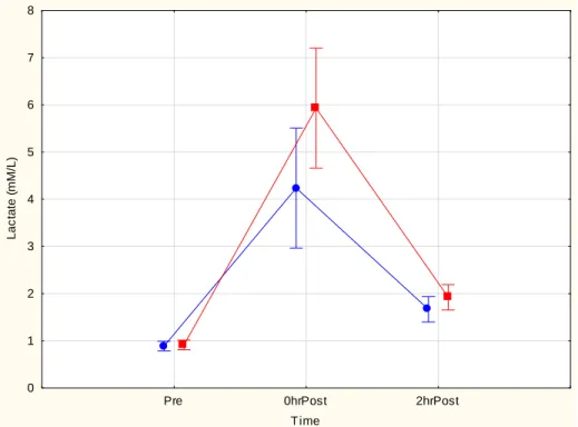
Blood lactate responses
Lactate responses are displayed in Figure 1. Resting Pre-Ex blood lactate levels did not
differ significantly between groups and exercise caused lactate to increase in both groups
(p<0.001). However, the 0hrPost blood lactate levels of the groups differed significantly, with
the level in the cancer patients being lower (p<0.01). At 2hrPost there were no group differences
observed and levels were not different from respective resting Pre-Ex values.
Figure 1. Blood lactate responses Pre-Ex, 0hrPost, and 2hrPost exercise bout. Mean ± 95% CI is
36
Pre 0hrPost 2hrPost
T ime 0
1 2 3 4 5 6 7 8
L
a
c
ta
te
(
m
M
/L
)
Blood glucose responses
Blood glucose levels Pre-Ex, 0hrPost, and 2hrPost are presented in Table 2. There were
no statistically significant differences in the response to exercise or between the control and
breast cancer group (p>0.05).
Table 2. Blood glucose responses Pre-Ex, 0hrPost and 2hrPost exercise bout. Mean ± 95% CI are
37
Group Time Mean
(mg/dl)
-95% (mg/dl)
+95% (mg/dl)
Cancer Pre-Ex 97.8 92.0 103.6
Cancer 0hrPost 93.5 87.0 100.0
Cancer 2hrpost 86.0 71.6 100.4
Control Pre-Ex 92.8 87.0 98.6
Control 0hrPost 83.3 76.8 89.7
Control 2hrpost 91.7 77.2 106.1
Blood free fatty acid responses
Blood free fatty acid responses immediately Pre-Ex, 0hPost, and 2hrPost are presented in
Figure 2. No group differences existed at Pre-Ex. Exercise caused free fatty acid concentrations
to increase in both groups significantly (p<0.01). At 2hrPost exercise, the cancer group’s free
fatty acid concentrations were still elevated from its respective Pre (p<0.01), while the control
was not significantly elevated and had returned to Pre-Ex levels. Furthermore, the cancer group
responses were significantly greater than the control responses at 2hrPost (p<0.01).
Figure 2. Blood free fatty acid responses Pre-Ex, 0hrPost, and 2hrPost exercise bout. Mean ±
38
Pre 0hrPost 2hPost
Time 0.1 0.2 0.3 0.4 0.5 0.6 0.7 0.8 0.9 F re e F a tt y A c id s ( m E q /L )
Plasma Volume Shifts
Plasma volume shifts during exercise and recovery were similar between groups and did
not differ significantly (p= 0.87 – 1.00).
Percent Change Analysis
All data were also statistically analyzed using percent change values, as noted in the
Methodology chapter. The results and the understanding of the data analysis did not change with
39
CHAPTER FIVE: DISCUSSION
The purpose of this study was to examine the effect of an acute, 30 minute bout of
moderate-intensity intermittent exercise on metabolic biomarkers of energy metabolism in
female breast cancer survivors. More specifically, this study examined blood lactate, glucose,
and free fatty acid concentrations in response to an intermediate 30 minute bout of cycling at
60% VO2peak. For the purposes of this document, the discussion section will be delimited to only
the variables discussed here regarding exercise energy metabolism (blood lactate, glucose, and
free fatty acids). Details and discussion of all other aspects of the study have been reported
elsewhere (Evans 2012).
Resting and Exercise Biomarker Responses
Resting values from both the control and experimental group for all biomarkers are
within normal resting reference values, suggesting this data is valid, and is comparable to those
in previous research studies. In general, the changes in the biomarkers in response to exercise
also agree with physiological responses in the healthy population (Brooks et al., 2005).
Specifically, resting blood glucose levels typically range from about 70 mg/dL to 110 mg/dL,
with the mean being approximately 100 mg/dL, (American Diabetes Association, 2000). Resting
blood glucose data from our subjects were within this range, with the means for the cancer group
40
lactate levels in healthy populations have been reported to range from 0.1 to 0.6 mEq/L and 0.5
to 2.0 mM/L, respectively (Brooks et al., 2005; Gollnick et al., 1986; McGee et al., 1992). In the
present study, mean resting free fatty acid concentrations were 0.28 mEq/L in the cancer group
and 0.23 mEq/L in the control group. Resting blood lactate values were also within reference
range, with the means being 0.8 mM/L in the cancer group and 0.9 mM/L in the control. Not
only do the resting values here align with previous research, but also the changes found in
response to exercise for each measurement do as well (Brooks et al., 2005).
During exercise, blood glucose rises at the start and remains within approximately 10%
of the normative value until exercise ends (Brooks et al., 2005). Data from this study yielded no
significant blood glucose findings. However, blood glucose did remain relatively constant,
staying within 10% of resting values from immediately Pre-Ex to 2 hours post exercise.
Additionally, in response to this moderate intensity exercise bout, blood FFA levels should rise
continuously (Brooks et al., 2005). In both the control and cancer group, exercise caused free
fatty acid concentrations to significantly increase. Furthermore, at 2 hours post exercise, the cancer group’s free fatty acid concentrations were still elevated from its respective Pre, while the
control was not significantly elevated (see discussion below). Blood lactate levels increase
non-linearly in response to exercise, with a steady rise beginning at about 60% VO2max (Brooks et
al., 2005). In both groups, blood lactate responses followed this model. Both groups yielded
significantly higher immediately post blood lactate concentrations when compared to resting
values, suggesting increased lactate accumulation. Although these findings did follow the
expected responses to exercise, several differences were found between the breast cancer and
41
Exercise Energy Metabolism: Control versus Cancer
Several differences are known between exercise energy metabolism in healthy
individuals and breast cancer survivors. Research has found significantly decreased blood
lactate responses to exercise following low, moderate, and high intensity exercise in post-treated
breast cancer survivors when compared to age-matched healthy controls (Evans et al., 2009;
Tosti et al., 2010). Additionally, research has identified reduced carbohydrate oxidation and
greater fat oxidation during exercise in the cancer survivors (Tosti et al., 2010). The present
findings support this previous research, while also expanding on it. Although there were no
significant differences between the two groups in blood glucose, there were differences in blood
FFA responses as well as blood lactate. Our data shows significantly higher FFA concentrations
2 hours post exercise in the post-treated cancer survivors when compared to the control group,
signifying a potentially greater fat oxidation in the cancer group. Although Tosti’s exercise was
9 continuous minutes, FFA concentrations were significantly increased immediately post
exercise. Both studies suggest increased fat oxidation in the breast cancer survivors (Tosti et al.,
2010). It is well recognized that when there are elevated FFA levels, this can inhibit glycolysis
and the metabolism of glucose hence, decreased lactate production (Brooks et al., 2005). The
strength of this relationship between blood lactate and FFA concentrations is illustrated in Figure
3.
Figure 3. The in vivo inter-relationship between blood FFA and lactate concentrations in exercise
42
The current analysis therefore supports previous research findings, while also expanding
on them. This study is the first to measure free fatty acid concentrations directly in the blood of
exercising breast cancer patients. Though our blood lactate findings are not novel, they do align
themselves with previous research, suggesting there is a difference in cancer energy metabolism
during exercise (Evans et al., 2009; Tosti et al., 2010). Together, these studies suggest decreased
blood lactate accumulation during exercise in breast cancer survivors when compared to healthy
controls – possibly due to compromised glycolytic processes. Additionally, our FFA findings
are new and novel. Although previous studies have found decreased carbohydrate oxidation and
increased fat oxidation in exercising cancer patients, this has been determined through expired
gas analysis. This is perhaps the first study to report these findings from direct FFA biomarker
measurements in the blood. These findings also suggest lower carbohydrate oxidation and
greater fat oxidation in the breast cancer group. Reasons for why the glucose did not change
significantly in response to exercise and support the lactate and FFA responses are unknown; but
in light of the comments of Brooks et al. (2005) this lack of significant change in glucose is not
43
Possible Mechanisms of Responses
Reduced blood lactate and carbohydrate oxidation, paired with increased fat oxidation
use in this cancer population has been documented several times. These findings could be
expected; decreased lactate production would signify decreased reliance on carbohydrate and
increased reliance on fat-lipid metabolism, (Brooks et al., 2005; Rasmussen & Winder, 1997).
However, the difference in the fat biomarker response between the study groups is a new
research finding, and nonetheless suggests that more research is needed to expand the body of
knowledge. Mechanisms behind the apparent increased fat oxidation in the breast cancer
patients are unknown, though it has been speculated differing hormonal status between the breast
cancer and healthy population groups may contribute (Evans 2012).
That is, Evans found in her doctoral dissertation research work different endocrine
hormones responses to a moderate intensity exercise bout, which may have implications for
regulations of exercise energy metabolism in breast cancer survivors. Normally, epinephrine
levels would increase during exercise; however, Evans found breast cancer survivors showed no
significant change in epinephrine in response to the exercise (Evans 2012). This irregular
epinephrine response may be a possible mechanism for the alteration in exercise metabolism in
breast cancer survivors. Epinephrine plays a role in stimulating muscle glycogenolysis during
exercise (Brooks et al., 2005; Hackney 2006). Thus decreased levels of epinephrine in the blood
may potentially delimit the availability of glucose in the muscle cell, therefore limiting the body’s ability to use glycolysis for energy (Brooks et al., 2005; Konatska et al., 1990). This
finding would suggest that an increased reliance on lipids to meet energy demands would be
44
exercise; this hormone plays a large role in increasing the lipolysis rate, further stimulating lipid
metabolism (Hackney 2006).
Additionally, cancer is often associated with hyperglycemia, glucose intolerance, and
insulin resistance, which may affect energy metabolism (Edmonson 1966; Glicksman & Rawson
1956; Marks & Bishop 1956; Tosti et al., 2010). These complications may reduce the body’s
capacity to use glucose as an energy substrate during exercise. Cancer treatments may also be
contributing to alterations in exercise metabolism (Lane & McKenzie, 2005; Kaur et al., 2010).
The ultimate goal of cancer treatment is termination of malignant cancer cells and destroying
their carbohydrate metabolic capacities to halt tumor growth. Therefore, most treatments target
diseased tissue with the ultimate goal of affecting only diseased tissues (ACS, 2014). However,
some healthy cells are inevitably affected (Kaur et al., 2010). Perhaps these treatments, by
unintentionally affecting healthy cells, significantly diminish the breast cancer patient’s ability to
perform carbohydrate metabolism. It is important to note, these explanations are only
speculation. More research on cancer exercise energy metabolism is needed to discern and
pinpoint these physiological mechanisms and explain these occurrences.
Clinical Significance
Regardless of the mechanism behind these responses, the findings have clinical
significance. These findings provide evidence that blood lactate responses to exercise are not a
valid indicator of exercise intensity in breast cancer survivors. In healthy populations, blood
lactate levels are a somewhat common parameter off which to base exercise prescriptions.
However, these results suggest exercise prescriptions written with lactate in this population may
45
carbohydrates during exercise, suggesting rapid production of energy via anaerobic pathways
may be decreased. Therefore, exercise at high intensities may not be appropriate for long
periods of time in breast cancer survivors. Increased research on exercise energy metabolism in
breast cancer survivors can provide insight as to what could be more appropriate intensity
measurements in the population, as well as uncover more information on breast cancer exercise
metabolism.
Limitations and Strengths
This study expanded on the previous body of knowledge, finding several significant
differences between healthy and cancer patient energy metabolism via direct metabolic
biomarkers in the blood. However, there are several limitations. To begin, the control group
was older than the breast cancer group by an average of 9 years. This may have affected
metabolism indirectly in the subjects, impacting the biomarkers indirectly. In addition, the
sample size was small. With additional subjects, perhaps statistical significance would have
been found in blood glucose. Despite a small sample size, statistical significance was still found
in blood lactate and free fatty acid concentration. Additionally, the sample was very
homogenous, comprised of only post treated breast cancer survivors. Therefore, it is unclear
whether these results can be generalized to other cancer diagnoses. Lastly, this study examined
only the response to the acute effects of exercise in this group. Thus, based on this limitation in
the findings, speculation is not valid on exercise training related effects.
Despite these limitations, this study does have several strengths. First, it supports
previous research as expected. Results here add to the increased evidence that breast cancer
46
acid findings. No previous study has examined FFA concentrations in response to exercise in
post treated breast cancer survivors. Finally, despite having a small sample size, there was
statistical significance in two of the three variables, suggesting the magnitude of the difference in
these variables is large. It has identified a clear metabolic difference between these two groups,
the mechanism for which is unknown. More research is needed to better understand and
advocate for these survivors.
Conclusions
This study found a reduced blood lactate response to moderate intensity exercise. More
research regarding the physiological mechanism behind this finding is needed to better
understand this altered metabolism. Reduced blood lactate responses to exercise also suggest
that lactate levels are not an appropriate method to prescribe exercise in this population.
Additionally, there were no significant findings with blood glucose. Free fatty acid metabolism
was found to be significantly increased in the breast cancer group 2 hours post exercise,
suggesting elevated levels of fat-lipid metabolism during exercise. Decreased carbohydrate and
increased fat oxidation rates during exercise in these survivors may alter and diminish their
ability to exercise at higher intensities. This research is important in educating clinicians and
exercise physiologists on the potential of different metabolic responses of cancer patients during
exercise, which should be taken into consideration when prescribing different exercise durations
and intensities. Therefore, more research is needed to confirm or refute the results of this
experiment so more precise and specific exercise prescriptions can be devised for breast cancer
47
REFERENCES
Abeloff MD, Wolff AC, Weber BL, et al. Cancer of the Breast. In: Abeloff MD, Armitage JO, Lichter AS, et al, eds. Clinical Oncology. 4th ed. Philadelphia, Pa: Elsevier; 2008: pp. 1875–1943.
American Cancer Society. Breast cancer facts and figures 2011-2012. Atlanta, GA: American Cancer Society; 2011.
American Cancer Society, 2013. Breast Cancer; 2013.
American Cancer Society 2014. Treatment and side effects. http://www.cancer.org/treatment/treatmentsandsideeffects/
American Cancer Society. Cancer facts and figures, 2014. Atlanta, GA: American Cancer Society.
American Cancer Society. Cancer Facts & Figures 2015. Atlanta: American Cancer Society; 2015.
American College of Sports Medicine. ACSM’s Guidelines for Exercise testing and
prescription. 9th edition. Exercise prescription for other clinical populations. 2014; pp. 263- 273.
American Diabetes Association. Screening for Type 2 Diabetes. Clinical Diabetes. 2000; 18(2): 69-74.
Avis N, Crawford S, Manuel J. Quality of life among younger women with breast cancer. J Clin Oncol. 2005; 23: 3322–3330.
Battaglini, CL, Bottaro M, Campbell JS, Novaes J, Simao R. Physical activity and levels of fatigue in cancer patients. Revista Brasileira de Medicina do Esporte. 2004; 2 (10): 98-104.
Battaglini CL, Boltaro M, Dennehy C, Rae L, Shields E, Kirk D, Hackney AC. The effects of an individualized exercise intervention on body composition in breast cancer patients
undergoing treatment. Sao Paulo Med. J. 2007; 125: 22-28.
Battaglini CL, Hackney AC, Garcia R, Evans E, Shea T. The effects of an exercise program in leukemia patients. Cancer Ther. 2009. 8(2): 130-138.
48
survivors: A systematic review of the literature. Wold J Clin Oncol. 2014. 5(2): 177-190.
Borg GA. Psychophysical bases of perceived exertion. Med Sci Sports Exerc. 1982; 14(5): 377-381.
Betof AS, Dewhirst MW, Jones LW. Effects and potential mechanisms of exercise training on cancer progression: A translational perspective. Brain Behav Immun. 2013; 30: S75-S87.
Brooks GA. The lactate shuttle during exercise and recovery. Med SciSports Exerc. 1986; 18(3): 360-368.
Brooks GA, Fahey, TD, Baldwin KM. Exercise physiology: human bioenergetics and its applications. 3rd Ed: 2000.
Costill DL, Coyle E, Dalsky G, Evans W, Fink W, Hoopes D. Effects of elevated plasma FFA and insulin on muscle glycogen usage during exercise. J Appl Physiol. 1997; 43: 695-699.
Costill DL, Dill DB. Calculation of percentage changes in volume of blood, plasma, and red cells in dehydration. J Appl Physiol. 1974 37(2): 247-248.
Edmonson JH. Fatty acid mobilization and glucose metabolism in patients with cancer. Cancer. 1966; 19: 277-280.
Evans ES. UNC-CH Doctoral Dissertation. The impact of acute aerobic exercise on natural killer cell, catecholamine, and cortisol responses in breast cancer survivorsUnder the direction of Dr. Claudio L. Battaglini, 2012.
Evans ES, Battaglini CL, Groff DG, Hackney AC. Aerobic exercise intensity in breast cancer patients: a preliminary investigation. Integr Cancer Ther. 2009; 8(2): 139-147.
Friedenreich CM, Gregory J, Kopciuk KA, Mackey JR, Courneya KS. Prospective cohort study of lifetime physical activity and breast cancer survival. Int J Cancer. 2009; 124: 1954–62.
Friedenreich CM. Physical activity and breast cancer: Review of the epidemiologic evidence and biologic mechanisms. Clinical Cancer Prevention. 2011; 188; 125-139.