ORIGINAL RESEARCH
Neonatal Pulmonary Macrophage Depletion Coupled to Defective
Mucus Clearance Increases Susceptibility to Pneumonia and
Alters Pulmonary Immune Responses
Yogesh Saini1,2, Kristen J. Wilkinson1, Kristy A. Terrell1, Kimberlie A. Burns1, Alessandra Livraghi-Butrico1, Claire M. Doerschuk1, Wanda K. O’Neal1, and Richard C. Boucher1
1
Marsico Lung Institute/University of North Carolina Cystic Fibrosis Center, School of Medicine, University of North Carolina at Chapel Hill, Chapel Hill, North Carolina; and2Department of Comparative Biomedical Sciences, School of Veterinary Medicine, Louisiana State University, Baton Rouge, Louisiana
Abstract
Resident immune cells (e.g., macrophages [MFs]) and airway mucus clearance both contribute to a healthy lung environment. To investigate interactions between pulmonary MFfunction and defective mucus clearance, a genetic model of lysozyme M (LysM) promoter–mediated MFdepletion was generated, characterized, and crossed with the sodium channelbsubunit transgenic (Scnn1b-Tg) mouse model of defective mucus clearance. Diphtheria toxin A–mediated depletion of LysM1pulmonary MFs in wild-type mice with normal mucus clearance resulted in lethal pneumonia in 24% of neonates. The pneumonias were dominated byPasteurella pneumotropicaand accompanied by emaciation, neutrophilic inflammation, and elevated Th1 cytokines. The incidence of emaciation and pneumonia reached 51% when LysM1MFdepletion was superimposed on the airway mucus clearance defect of
Scnn1b-Tg mice. In LysM1MF-depletedScnn1b-Tg mice, pneumonias were associated with a broader spectrum of bacterial species and a significant reduction in airway mucus plugging. Bacterial burden (CFUs) was comparable betweenScnn1b-Tg and nonpneumonic LysM1MF-depletedScnn1b-Tg mice. However, the nonpneumonic LysM1MF-depletedScnn1b-Tg mice exhibited increased airway inflammation, the presence of neutrophilic infiltration, and increased levels of inflammatory cytokines in bronchoalveolar lavagefluid compared withScnn1b-Tg mice. Collectively, these data identify key MF–mucus clearance interactions with respect to both infectious and inflammatory components of muco-obstructive lung disease.
Keywords:alveolar macrophages; macrophage depletion; sodium channelbsubunit transgenic mice; airway mucus obstruction; airway inflammation
Airway and alveolar host defense is mediated by multiple interconnected mechanisms, including airway mucociliary clearance and innate immune responses. Innate immune responses are performed by multiple cell populations (e.g., resident pulmonary macrophages [MFs]) and secretion of soluble antimicrobial factors in both airway and alveolar regions. Primary
defects in each of these layers of defense can produce disease and/or compensatory responses in other defenses, which may be beneficial to the host, or harmful, if they persist for protracted intervals. Elucidation of the interactions between defense mechanisms that maintain lung health is complex and requires direct experimental testing.
Mucus hyperconcentration, mucostasis, and mucus plugging are key pathophysiologic events in many airway diseases, including chronic obstructive pulmonary disease and cysticfibrosis. Although persistent microbial infection in static mucus can produce chronic inflammation, the potential proinflammatory effects of static, but sterile, mucus are poorly
( Received in original form March 17, 2014; accepted in final form June 18, 2015 )
This work was supported by a Flight Attendant Medical Research Institute grant (Y.S.), Cystic Fibrosis Research Development Program grant RDP R026 (W.K.O’N.), and National Institutes of Health grants P30 DK065988, P50 HL060280, P50HL107168, and P01HL108808 (R.C.B.).
Author Contributions: Conception and design—Y.S., W.K.O’N., and R.C.B.; acquisition, analysis and interpretation—Y.S., K.J.W., K.A.T., K.A.B., A.L.-B., C.M.D., W.K.O’N., and R.C.B.; drafting the manuscript for important intellectual content—Y.S., A.L.-B., C.M.D., W.K.O’N., and R.C.B.
Correspondence and requests for reprints should be addressed to Yogesh Saini, B.V.Sc. & A.H., Ph.D., Marsico Lung Institute/University of North Carolina Cystic Fibrosis Center, School of Medicine, University of North Carolina at Chapel Hill, 7011 Thurston Bowles Building, Chapel Hill, NC 27599-7248. E-mail: [email protected]
This article has an online supplement, which is accessible from this issue’s table of contents at www.atsjournals.org
Am J Respir Cell Mol Biol Vol 54, Iss 2, pp 210–221, Feb 2016 Copyright©2016 by the American Thoracic Society
understood. Resident MFs are key sentinel cells for both airway and alveolar surfaces, and their responses to failed mucus clearance are likely to contribute to disease pathogenesis. Upon an encounter with inhaled particles or organisms, pulmonary MFs assist in clearance and dampen or promote local inflammatory responses, depending upon their activation status and the nature of the stimulus. Like failed mucus clearance, loss of normal MFfunction has been reported to produce important pulmonary consequences, including pneumonia (1–3). We hypothesize that pulmonary MFs are key pulmonary defense cells that respond to mucus
obstruction, becoming activated in response to failed clearance by sensing and responding to hyperconcentrated mucus itself, and/or exogenous and endogenous inflammatory stimuli trapped and concentrated within adherent mucus.
To investigate the interplay between reduced mucociliary clearance and the MF components of host defense, we used the well characterized, epithelial sodium channelbsubunit (Scnn1b)transgenic mouse (Scnn1b-Tg). In this model, the overexpression ofScnn1btransgene is controlled by the rat club cell secretory protein (CCSP) promoter (2.25-Kb 59 flanking region of CCSP gene, also known asScgb1a1), which targets expression to airway club cells (previously referred to as Clara cells) with sporadic expression in alveolar type II cells (4, 5). Previously published studies have characterized the cell-specific expression ofScnn1btransgene and the resulting airway surface liquid dehydration (5, 6). The neonatal and adult muco-obstructive airways disease (5, 6) produced by airway surface liquid dehydration is characterized by
inflammation that includes increased MF size, persistent airway neutrophilia, and bronchial-associated lymphoid tissues (5–7). Morphologic (6, 7), functional (8), and recent gene array (9) studies indicate that pulmonary MFs are robustly activated soon after birth inScnn1b-Tg mice, suggesting that they contribute significantly to the development of lung inflammation.
To characterize the contribution of MFs to neonatalScnn1b-Tg mouse lung disease, mice with MFdepletion were generated using lysozyme M (LysM) cyclization recombinase (Cre)–mediated expression of gene encoding diphtheria toxin A (DTA) subunit. The LysM promoter was selected for these studies
because of its high level of expression in pulmonary MFs (10). The functional consequences of DTA expression on pulmonary MFpopulations were evaluated during the neonatal period in wild-type (WT) mice and withScnn1b-Tg expression. Putative“positive”compensatory
consequences of MFdepletion on the neonatalScnn1b-Tg lung phenotype (e.g., reduced inflammation) were sought in parallel with potential“negative”effects of compromising these key protective cells (e.g., increased on bacterial infection).
Materials and Methods
Transgenic Mice and Animal Husbandry
All animal protocols were approved by the Institutional Animal Care and Use Committee at the University of North Carolina (Chapel Hill, NC). The four distinct transgenic lines used are detailed subsequently here, and include: (1) LysM-Cre mice expressing Cre from the myeloid-specific LysM promoter; (2) membrane-targeted Tomato (mTOM)/ membrane-targeted enhanced green fluorescent protein (mEGFP)-floxed reporter mice where the Gt(ROSA)26Sor (a gene locus controlling constitutive expression of transgenes/constructs, abbreviated ROSA promoter for this article) locus expresses reporter gene mTOM (red) in the absence of Cre and mEGFP (green) upon Cre activation; (3) DTA-floxed mice with DTA expression induced by Cre activation; and (4)Scnn1b-Tg mice, which develop mucus obstruction. Studies were performed on neonates (5–7 days old), as indicated infigures and text.
Generation of Transgenic Mice
All mice used in the study were maintained in hot-washed, individually ventilated micro-isolator cages on a 12-hour dark/ light cycle, and were fed a regular diet and waterad libitum. LysM-Cre mice (B6.129P2-Lyz2tm1(cre)Ifo/J), mTOM/ mEGFP reporter mice (B6.129(Cg)Gt (ROSA) 26Sortm4(ACTB-tdTomato,-EGFP)Luo/J) and DTA-floxed
(B6.129P2Gt(ROSA) 26Sortm1(DTA)Lky/J) were procured from Jackson Laboratory (Bar Harbor, ME). At the time of procurement, LysM-Cre mice were on mixed C57Bl/6N and C57Bl/6J background, whereas mTOM/mEGFP
reporter mice and DTA-floxed mice were on C57Bl/6J background.Scnn1b-Tg mice used in the study were C57Bl/6N congenic.
Generation of mouse models of
fluorescent marker-labeled macrophages, macrophage depletion and crosses with Scnn1b-Tg mice. The myeloid cell–specific LysM promoter was used to drive expression of Cre in the myeloid lineage. The LysM-Cre mice used in this work have a targeted insertion of the Cre transgene in the exon 1 region of the LysM locus (11). To minimize the negative effect of Cre toxicity, we only generated and analyzed animals with a single copy of the Cre transgene, a strategy that also assured functionality of at least one allele of the LysM gene.
Generation and use of macrophage-reporter mice. LysM-Cre mice were crossed with the Gt(ROSA)26Sor-mTOM/mEGFP dual-reporter mouse strain. This strain contains an engineered transgene that is controlled by the ROSA promoter. In mice without Cre, the locus of crossover in phage 1 (LoxP)-flanked mTOM transgene/polyA stop cassette (redfluorescence at the membrane) is expressed from the ROSA promoter, and the mEGFP transgene (green fluorescence at the membrane) is silent due to the presence of LoxP-flanked poly(A) stop cassette (12). The presence of Cre results in recombination-mediated removal offlanked mTOM transgene/polyA stop cassette and induction of mEGFP transgene expression. Using this dual reporter transgene crossed to LysM-Cre mice, a mouse model was generated where LysM-Cre–positive cells would express the mEGFP.
Generation and use of LysM1
MF-depleted mice. Similar to the Gt(ROSA) 26Sor–mTOM/mEGFP dual-reporter strain, the Gt(ROSA)26Sor-DTA mouse strain contains an engineered transgene (i.e., a loxP-flanked neomycin/PolyA stop cassette) proximal to the DTA transgene that is controlled by the ROSA promoter (13). To achieve depletion of MFs, LysM-Cre mice were mated with the Gt(ROSA)26Sor-DTA mice. The presence of Cre in the Gt(ROSA) 26Sor-DTA mouse cells results in
LysM1MF-depleted Scnn1b-Tg mice. LysM-Cre1/1Scnn1b-Tg1/2mice were generated and mated with DTA1/2mice to generate LysM1MF-depletedScnn1b-Tg animals. LysM1MF-depletedScnn1b-Tg mice with a MF-specific reporter transgene were generated by mating LysM-Cre1/1 mTOM/mEGFP1/1mice with DTA1/2 Scnn1b-Tg1/2mice.
PCR Genotyping
Genomic DNA extraction from tail tissue was performed using the Direct-PCR extraction reagent (Viagen Biotech, Los Angeles, CA), according to the manufacturer’s instructions. Genotyping was performed by PCR for all the loci as previously published (5, 11–13). PCR conditions for all the alleles were: denaturation at 948C for 5 minutes, 38 cycles of amplification (948C for 45 seconds, 608C for 45 seconds, and 728C for 1 minute and 10 seconds, followed by a 7-minute extension at 728C). The amplified products were electrophoresed in 2% agarose in Tris borate EDTA for 30 minutes at 200 V. The ethidium bromide–stained gels were imaged in FluorChemQ imaging system (Alpha Innotech, San Jose, CA).
Bronchoalveolar Lavage Collection and Lung Tissue Processing
Bronchoalveolar lavagefluid (BALF) was harvested for differential cell counts and microbiological analyses as previously described (14).
Fluorescence Microscopy
To harvest lung tissues forfluorescent imaging, mice were anesthetized with Avertin (2,2,2-tribromoethanol), and, after exsanguination, lungs and tracheas were exposed through a midline thoracotomy. Lungs were inflatedin situwith 50% OCT in PBS. Whole lungs were embedded in OCT in tissue molds and frozen on dry ice. The lungs in the OCT frozen blocks were positioned to optimize longitudinal sectioning of primary bronchi. OCT-embedded frozen lung tissues were sectioned to a thickness of 5mm on a cryostat. Forfluorescent imaging, frozen sections werefixed in ice-cold acetone for 10 minutes. Fixed sections were washed twice in PBS and mounted with VECTASHIELD HardSet mounting medium with 49 ,6-diamidino-2-phenylindole (Vector Labs, Burlingame, CA). Sections were observed under Olympus FV1000 MPE SIM laser scanning confocal microscope (Olympus, Pittsburgh, PA) at the University of North Carolina Michael Hooker Microscopy Facility. For the determination of mTOM1and mEGFP1 cells, cytospin preparations were prepared andfixed in 10% neutral buffered formalin for 15 minutes. Thefixed Cytospin slides were washed with PBS and mounted with VECTASHIELD HardSet mounting medium with 49,6-diamidino-2-phenylindole (Vector Labs). Cytospin slides were observed under Olympus FV1000 MPE SIM laser scanning confocal microscope. mTOM1
(no mEGFP label) and mEGFP1(with or without mTOM label) MFs were counted based on morphology, as determined by differential interference contrast microscopy. A total of approximately 200 cells were counted to estimate the percentage of mTOM1and mEGFP1MFs.
Flow Cytometry
BAL cells were collected as described previously here. Cells werefixed in a one-step fix/lyse solution (eBiosciences, San Diego, CA), washed twice in PBS, and the pellets were suspended in staining buffer. BAL cells were analyzed for the mEGFP and mTOM fluorescence with Dako CyAn (Beckman Coulter, Inc., Pasadena, CA). Flow cytometric data were analyzed using Summit software Version 4.3 (Dako, Carpinteria, CA).
Cytokine Assay on BAL
Mouse TNF-a, keratinocyte chemoattractant (KC), macrophage inflammatory protein 2 (MIP-2), MIP-1a, MIP-1b, macrophage colony–stimulating factor (M-CSF), IL-10, IL-12, IL1a, IL-17, IL-4, IL-5, IL-6, IP-10, monocyte chemotactic protein-1 (MCP-1) and LPS-induced CXC chemokine (LIX) levels were measured in cell-free BAL using a Luminex-based assay (MCYTOMAG-70K; EMD Millipore Corp., Billerica, MA), according to the manufacturer instructions.
Histopathological Slide Preparation
The 10% neutral buffered formalin–fixed lungs were paraffin embedded, and 4- to
mTom
mEGFP
100
80
60
40
PERCENT
20
0
+
– – – –
– –
– –
+ + +
***
***
** *
+ +
– –
– –
+ +
+ +
+ +
100
80
60
40
PERCENT
20
0
100
80
60
40
PERCENT
20
0
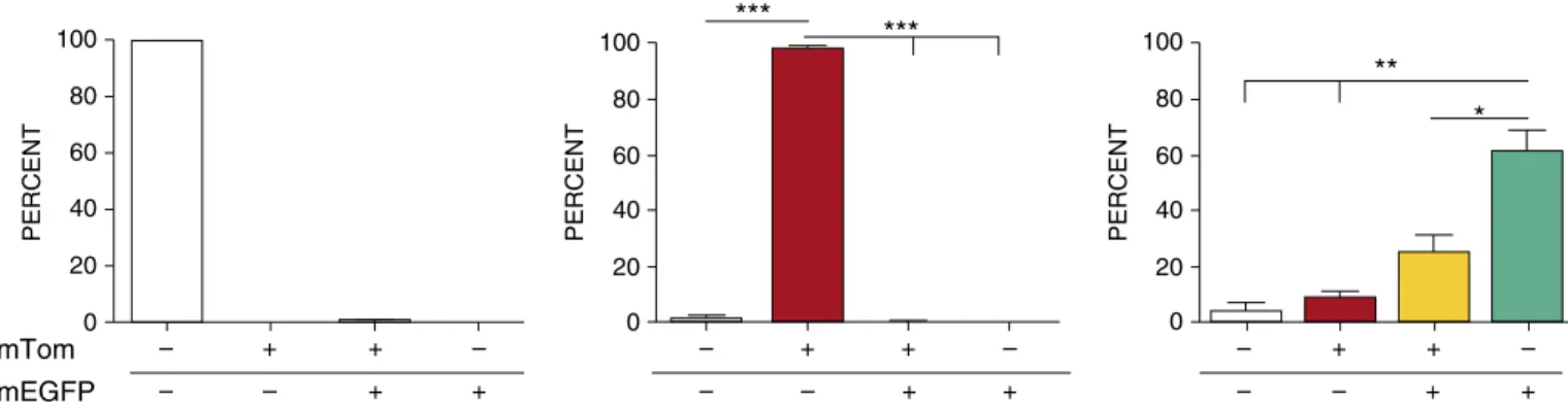
Figure 1.Lysozyme M (LysM) promoter expression activity in neonates. Flow cytometry analysis of neonatal bronchoalveolar lavage (BAL) cells harvested from LysM-cyclization recombinase (Cre)1single-transgenic mice (left panel), membrane-targeted Tomato (mTOM)/membrane-targeted enhanced green fluorescent protein (mEGFP)1single-transgenic mice (middle panel), and LysM-Cre1\mTOM/mEGFP1mice (right panel). The results reflect the distribution of BAL cells in the four quadrants from the flow analyses (Figure E2). Gt(ROSA)26Sor (a gene locus controlling constitutive expression of transgenes/constructs, abbreviated ROSA promoter for this article) locus drives mTOM (red) or mEGFP (green) expression in the absence or presence of Cre activity, respectively.White barsrepresent percentage of cells negative for mTOM and mEGFP signal;red barsrepresent percentage of cells positive for mTOM, but negative for mEGFP signal;yellow bars
6-mm-thick sections were cut. The lung tissues from 5- to 7-day-old mice were oriented to obtain longitudinal sections of primary bronchi. Sections were mounted on glass slides and stained with
hematoxylin and eosin for lung morphological assessments and Alcian blue/periodic acid–Schiff for
mucopolysaccharide assessment of intracellular and extracellular mucus.
Lung Histopathology
A previously reported semiquantitative grading system was used to score airway obstruction, mucus secretory cell abundance, airspace enlargement, and airway inflammation phenotypes (graded on a 0–3 scale) (14). The alveolar space consolidation phenotype was scored on the scale of 0–3: 0, no evidence of alveolar space consolidation; 1, less than 25% of left lung lobe with alveolar space consolidation; 2,>25–50% of the lobe with alveolar space consolidation; and 3,>50% of the lobe with alveolar space consolidation.
Statistical Analyses
Statistical analyses were performed using GraphPad Prism 5.0 (GraphPad Software, Inc., La Jolla, CA). One-way ANOVA followed by Tukey’spost hoctest for multiple comparisons was used to determine significant differences among groups.Pless than 0.05 was considered statistically significant. All data are expressed as means (6SEM).
Results
Characterization of LysM Promoter Activity in Pulmonary MFs
Before evaluating LysM-mediated DTA depletion of pulmonary MFs, a dual reporter LysM- Cre1\mTOM/mEGFP1 bitransgenic line was generated to
characterize the cell specificity of LysM-Cre in our mice. Flow cytometric evaluations of mTOM- and mEGFP-expressing cells were conducted on BAL cells collected from neonatal (5–7 days old) mice expressing LysM-Cre (to target Cre expression to myeloid cells under the LysM promoter) (11), ROSA-mTOM/mEGFP (reporter construct ROSA-mTOM/mEGFP containingfloxed mTOM) (12), or both transgenes (seeFigure E1 in the online supplement). In the absence of Cre activity, owing to the polyadenylation sequence
downstream of the loxP-flanked mTOM transgene, mEGFP expression did not occur in ROSA-mTOM/mEGFP mice. However, in the presence of Cre activity,
recombination-mediated excision of the mTOM region within thefloxed ROSA-reporter construct simultaneously eliminated mTOM expression and thus induced mEGFP expression (Figure E1).
In the absence of LysM-Cre, ROSA-mTOM/mEGFP reporter mice exhibited robust ROSA locus expression activity in BAL cells (96% of which were MFs [data not shown]), as indicated by mTOM1 expression in approximately 98% of harvested cells (Figure 1, middle panel, and Figure E2). In contrast, robust LysM promoter–driven Cre activity in MFs was evident by the presence of mEGFP1MFs
in bitransgenic, Cre-expressing reporter mice (Figure 1,right panel). In our mice, LysM-Cre activity–induced mEGFP expression was detected in approximately 87% of neonatal pulmonary MFs harvested by BAL.
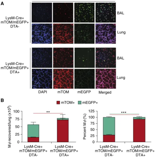
LysM-Cre–Driven DTA Expression Alters Pulmonary MFPopulations
The above studies indicated that
approximately 87% of BAL MFpopulations could be targeted for depletion using LysM-Cre–induced recombination in afloxed ROSA-DTA allele. Therefore, a bitransgenic (LysM-Cre1/1 ROSA-mTOM/mEGFP1/1) line was bred to an available transgenic line with a DTA-floxed allele targeted to the ROSA locus (13) to produce triple-transgenic mice (Figure E1).
**
0
LysM-Cre+ mTOM/mEGFP+
DTA-LysM-Cre+ mTOM/mEGFP+
DTA-LysM-Cre+ mTOM/mEGFP+
DTA+
DAPI mTOM mEGFP Merged
Lung Lung
BAL BAL
LysM-Cre+ mTOM/mEGFP+
DTA+ mTOM+
25 50 75 100
M recovered/lung (x10
3)
φ
***
0
LysM-Cre+ mTOM/mEGFP+
DTA-LysM-Cre+ mTOM/mEGFP+
DTA+
50 25 75 100 125
Percent M (%)
φ
A
B mEGFP+
Selective breeding (seeSUPPLEMENTAL MATERIALS ANDMETHODS) generated LysM-Cre–positive, ROSA-mTOM/mEGFP– positive, ROSA-floxed DTA–positive mice, in which both mTOM/mEGFP and DTA transcription were under the control of LysM promoter–driven Cre recombination. This feature facilitated tracking of DTA-expressing cells via the simultaneous expression of mEGFP1, providing the ability to monitor DTA-induced cell death using, as a readout, the proportion of mEGFP1cells in DTA1compared with DTA2lines. Triple-transgenic mice were obtained in the expected Mendelian ratios (data not shown).
Expression of the DTA transgene had clear effects on pulmonary MFpopulations. As predicted, DTA1mice exhibited a significant depletion of BAL mEGFP1 MFs compared with their DTA2 counterparts (Figure 2A). However, we observed an unexpected shift in BAL cell populations with DTA expression. Compared with DTA2mice (Figure 2B), the absolute numbers and the percentage of mTOM1MFs (defined as MFs based on their morphology) from the DTA1mice were increased (Figure 2B). Because of the increased numbers of mTOM1, the absolute MFnumbers were not reduced in DTA1neonates (Figure 2B). Of note, BAL neutrophils were consistently raised in neonatal DTA1mice (Figure E3).
Phenotype of WT andScnn1b-Tg Neonates in Presence or Absence of DTA Transgene Expression
Additional breeding was conducted to introduce theScnn1b-Tg into the LysM1 MF-depleted (DTA1) line. The mice obtained from these crosses were phenotyped in the neonatal period.
Altered MFphenotypes inScnn1b-Tg neonates with and without MFdepletion. Wefirst noted that the total MFnumber was increased inScnn1b-Tg compared with WT mice (Figure 3A). Interestingly, the increase in MFnumbers inScnn1b-Tg mice reflected an increase in mTOM1 MFs. The total numbers of MFs collected from LysM-Cre1/mTOM/mEGFP1/ Scnn1b-Tg neonates with and without DTA expression were comparable (Figure 3). However, there were major differences in the distribution of MFphenotypes. DTA expression reduced the absolute number and percentage of MFs with an active LysM promoter, as indicated by the
reduction in BAL mEGFP1MFs, and an increase mTOM1BAL MFs in DTA1/ Scnn1b-Tg neonates. Thus, in agreement with our data in DTA1WT mice (Figure 2B), DTA expression led to an absolute increase in LysM-inactive MFs (Figure 3).
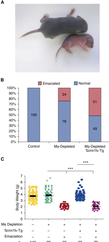
DTA1neonates were susceptible to emaciation, lung inflammation, bacterial pneumonia, and death. There were no visible phenotypic abnormalities at birth in any line. However, by 2–3 days after birth, 24% of DTA1/WT neonates exhibited an emaciation phenotype defined by reduced weight gain, flaky discoloration of skin, lethargy, and eventual death (Figure 4A). No DTA2/ Scnn1b-Tg neonates exhibited emaciation, but
the prevalence of emaciation increased to 51% in DTA1/Scnn1b-Tg neonates (Figure 4B). The severity of weight loss due to emaciation was similar between DTA1/Scnn1b-Tg and DTA1/WT neonates (Figure 4C).
DTA1neonates exhibited histologic evidence of pulmonary pathology. Histologically, there was no evidence of pathology in major nonrespiratory organs (liver, heart, intestine, spleen, and pancreas) in neonates (data not shown). In contrast, the lungs exhibited significant pathology.
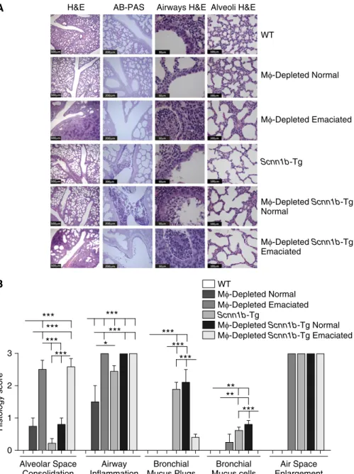
Alveolar space consolidation was the key histologicfinding associated with LysM1MFdepletion in neonates (Figures 5A and 5B). Nonemaciated DTA1/WT
0 0
25 50 75 100 125
LysM-CRE + + +
+ + +
– – +
– + +
mTom/mEGFP
DTA-floxed Scnn1b-Tg
Scnn1b -Tg
Scnn1b-Tg 100
200
M
φ
recovered/lung (x10
3) 300
WT
WT
**
** *** ***
***
Mφ-Depleted Scnn1b-Tg
Mφ-Depleted Scnn1b-Tg
+ + +
+ + +
– – +
– + +
Scnn1b -Tg
WT Mφ-Depleted
Scnn1b-Tg
Percent M (%)
φ
mTOM+ mEGFP+
A
B
neonates exhibited mild, heterogeneous alveolar space consolidation, whereas emaciated DTA1/WT neonates exhibited severe and more uniform alveolar
consolidation (Figures 5A and 5B). Consolidated alveoli were characterized by infiltrating neutrophils and enlarged, primarily apoptotic, MFs (as determined
byin situterminal deoxynucleotidyl transferase dUTP nick end labeling assay; Figure E4) that were more prominent in emaciated than in nonemaciated DTA1/WT neonates. No differences in the degree of alveolar space consolidation were noted in DTA1/Scnn1b-Tg compared with DTA1/WT neonates (Figures 5A and 5B).
Airway inflammation, as assessed histologically, was significant in all neonatal DTA1/WT mice compared with DTA2/WT (Figure 5B). The defective mucus
clearance that is a feature ofScnn1b-Tg mice was associated with more pronounced airway inflammation in nonemaciated and emaciated DTA1/Scnn1b-Tg neonates compared with nonemaciated DTA1/WT littermates (Figures 5A and 5B). The emaciation phenotype was strikingly associated with increased alveolar and airway inflammation in both WT and Scnn1b-Tg mice (Figures 5A and 5B).
Mucus obstructive phenotypes were not prominent in the airways of DTA1/WT neonatal mice. Mucus plugs and increased bronchial mucus cell numbers were observed inScnn1b-Tg neonates, consistent with the reported pathophysiology (5, 6). However, the extent of the mucus phenotype produced inScnn1b-Tg mice was dependent upon emaciation and MF depletion status. Interestingly, although the degree of mucus plugging was comparable in nonemaciated DTA1/Scnn1b-Tg and DTA2/Scnn1b-Tg neonates, the mucus retention phenotype was markedly reduced in emaciated DTA1/Scnn1b-Tg mice (Figures 5A and 5B).
An increase in the size of alveolar units was evident in allScnn1b-Tg lines, as previously described (Figures 5A and 5B). Previous studies have demonstrated that this histological feature reflects the failed development of alveolar septa and/or the loss of alveolar walls per acinar unit (i.e., an emphysema-like phenotype) (6, 15). Visually similar alveolar space enlargement was a consistent feature in the
nonconsolidated regions of emaciated DTA1/WT. In DTA1/Scnn1b-Tg mice, these lesions were observed irrespective of emaciation status (Figure 5B).
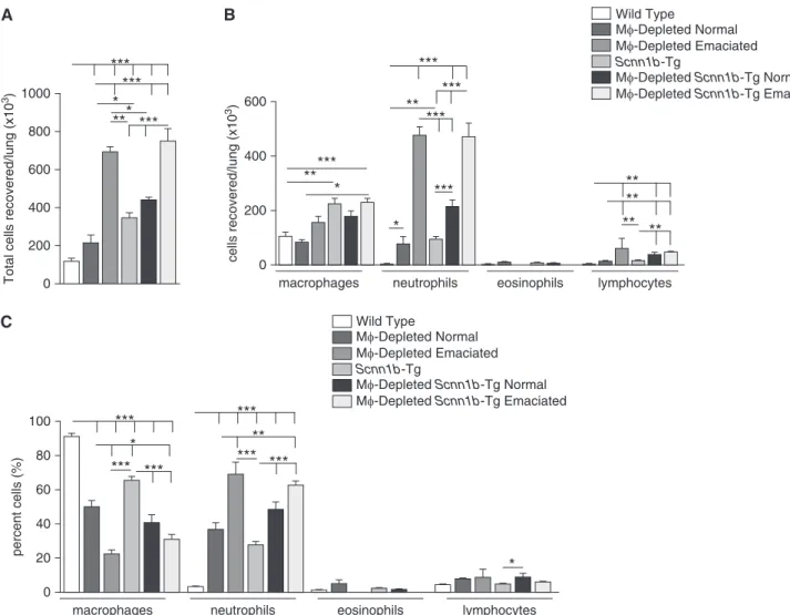
Total and differential BAL cell counts and BAL cytokine levels reflected
differential responses among the experimental groups to LysM1MF depletion, emaciation status, and the presence or absence ofScnn1b-Tg
expression (Figure 5; Table 1). In DTA1/WT
0%
0 1
Body Weight (g) 2
3 4 5 6 7
Control 10%
20% 30% 40% 50% 60% 70% 80% 90% 100%
Mφ-Depleted 100
24
51
76
49
Mφ-Depleted Scnn1b-Tg
Mφ Depletion – + + + +
– – – + +
–
142 99 32 66 70
– + – +
n= Scnn1b-Tg
Emaciation
*** ***
Emaciated Normal
B
C A
neonates, DTA expression produced no change in total macrophage numbers compared with DTA2/WT neonates (Figure 6, in agreement with Figure 2B). However, total BAL cells were significantly increased in DTA1/WT neonates
compared with DTA2/WT neonates due to increased neutrophil recruitment (Figures 6A–6C), consistent with cytological observations (Figure 2B and Figure E3).
Emaciated DTA1/WT neonates exhibited three to six times higher total cell numbers as compared with genotype-matched nonemaciated littermates, attributable not only to a large increase in neutrophil counts, but also to a small, but significant, increase in lymphocytes (Figures 6A–6C). Total BAL cells and macrophage numbers were increased in neonatalScnn1b-Tg mice compared with WT mice, but total counts
were not affected by DTA expression or as a function of emaciation status compared with their WT counterparts (Figures 6A–6C). Similar to nonemaciated DTA1/WT or DTA1/Scnn1b-Tg
neonates, BAL MFs recovered from the emaciated counterparts were
predominantly mTOM1(data not shown). The BAL inflammatory mediators tested were either below the detection limit or were present at minimal concentrations in DTA2/WT neonates (Table 1). LysM1 MFdepletion in WT neonates without emaciation resulted in a significant increase in the level of the neutrophil chemokine, KC (Table 1), consistent with the BAL neutrophilia (Figures 2B and 4C, Figure E3). Consistent with our previous reports (7, 14), BAL levels of KC, MIP2, MIP1a, MIP1b, and TNF-awere elevated in DTA2/Scnn1b-Tg neonates compared with DTA2/WT mice. Nonemaciated DTA1/ Scnn1b-Tg neonates exhibited a further elevation in the levels of these cytokines. Strikingly, the emaciation phenotype in DTA1/WT as well as DTA1/Scnn1b-Tg neonates was associated with a“cytokine storm,”as indicated by dramatic increases in 13 of 16 measured inflammatory mediators. Interestingly, only IL-5 was significantly reduced in emaciated compared with“normal”counterparts (Table 1).
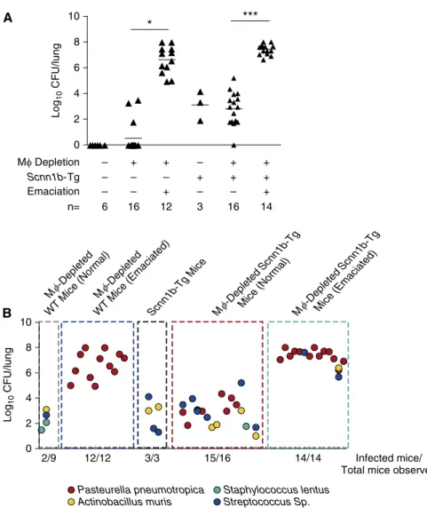
MFdepletion leads to neonatal bacterial pneumonia. The histological, cytokine, and BALfindings in emaciated neonates suggested bacterial infection, and studies were performed to test this hypothesis. Spleen cultures were negative for all genotypes, and no culturable bacteria were isolated from BAL of DTA2/WT neonates. However, a sporadic (3/16), small bacterial burden was observed in the BAL from nonemaciated DTA1/WT neonates (Figure 7A). In striking contrast, CFU counts were consistently approximately 5 logs higher (mean CFU = 2.33107) in BAL harvested from emaciated DTA1/WT neonates. Sequence analyses revealed that Pasturella pneumotropica,a common inhabitant of the mouse oropharynx (7), was the sole bacterial species culturable in emaciated DTA1/WT BAL (Figure 7B).
Total bacterial burden inScnn1b-Tg and nonemaciated DTA1/Scnn1b-Tg neonates were comparable and represented an admixture of bacterial pathogens previously reported inScnn1b-Tg mice (7). Streptococcusspp.,Actinobacillusspp., and
3
2
1
Histology score
0
Alveolar Space Consolidation
Airway Inflammation
Bronchial Mucus Plugs
Bronchial Mucus cells
Air Space Enlargement WT
Mφ-Depleted Normal Mφ-Depleted Emaciated
Mφ-Depleted Scnn1b-Tg Normal Mφ-Depleted Scnn1b-Tg Emaciated Scnn1b-Tg
WT AB-PAS
H&E Airways H&E Alveoli H&E
Mφ-Depleted Normal
Mφ-Depleted Emaciated
Mφ-Depleted Scnn1b-Tg Normal
Mφ-Depleted Scnn1b-Tg Emaciated
Scnn1b-Tg
*** *** ***
***
***
*** *
*** ***
***
**** *** B
A
Figure 5.MF-depleted neonates exhibit significant pulmonary pathology. (A) Representative
P. pneumotropica, routine colonizers in Scnn1b-Tg neonates, were the predominant organisms in nonemaciated DTA1/Scnn1b -Tg mice.P. pneumotropicapredominated, but was not the sole bacterial species in the emaciated DTA1/Scnn1b-Tg mice
(Figure 7B). The CFU counts in emaciated DTA1/WT and DTA1/Scnn1b-Tg neonates were similar.
Discussion
Mucus stasis has been associated with robust MFactivation inScnn1b-Tg mice (7–9) and human lung disease (16). MF activation is likely critical for both the protection of the lung as a compensatory mechanism and as a significant contributor to the development of the chronic inflammatory pathology in this model. Evaluation of muco-obstructive disease development in the presence and absence of MFs is one way to investigate the complex interrelationships between mucus stasis and MFfunction. This approach is challenging, however, as the pathophysiology associated with both MFdepletion and mucostasis are superimposed on normal lung developmental processes in both of our
models (9). Furthermore, MFactivation is known to be highly pleiotropic and sensitive to local environmental stimuli (17). The interpretation ofin vivofindings to address these interrelationships is critically dependent upon the specific features of the model.
For this study, due to the early onset of lung disease inScnn1b-Tg mice (5), it was necessary to develop a model that would exhibit chronic MFdepletion starting at birth. This requirement ruled out the use of chemical-/drug-induced approaches of MF depletion (18–20) and necessitated genetically induced depletion of MFs. One of the most widely used myeloid cell– restricted promoters, LysM, was selected to drive cell type–specific expression of Cre (11). The LysM promoter has been reported to be active in all myeloid cells (21), with one report indicating LysM promoter activity also in occasional alveolar epithelial type II cells (22).
In our study, LysM promoter activity in cells harvested by BAL, as indexed by Cre-mediated expression of mEGFP was age and disease dependent. Importantly, our studies identified a previously unappreciated age dependency in LysM promoter activity in BAL MFs. Whereas a vast majority of alveolar MFs harvested from normal adult
mice was positive for LysM promoter activity (data not shown), a significant percentage of MFs harvested from neonatal lavages exhibited a delay in the LysM-Cre–mediated recombination in the reporter transgene (Figure 2). Furthermore, in DTA1neonates, a significant LysM-Cre–negative alveolar MFpopulation was recruited into the pulmonary airspaces. Interestingly, these newly recruited MFs were not competent to routinely prevent the occurrence of bacterial infections in the emaciated subset of DTA1mice (Figure 4). The genetic model we have generated is best described as a model of LysM1MF depletion coupled to variable recruitment of LysM2, functionally defective MFs or monocytes, rather than a model of complete MFdepletion. Overall, the patterns observed with this model are consistent with a recent report that identified a large subset of MFresistant to LysM-Cre–mediated gene deletion in the peritoneum and liver after inflammatory stimulation (23), and complement other findings showing that mature tissue MFs and MFs derived from monocytes are phenotypically distinct (3, 24). Interestingly, in the study of Vannella and colleagues (23), the LysM-Cre–resistant
Table 1. Cytokine Responses in Neonatal Mice across Designated Groups
Cytokine WT
MF-Depleted WT“Normal”
MF-Depleted
WT“Emaciated” Scnn1b-Tg
MF-Depleted
Scnn1b-Tg
“Normal”
MF-Depleted
Scnn1b-Tg
“Emaciated”
Lower Limits of Detection
KC 2.360.9*† 45.8628.5†‡ 1,204.06303.5 85.4621.9* 154.8638.1‡ 1,522.06269.4 3.0 MIP2 14.761.4* 19.963.6‡ 7,463.061,365.0 273.5664.8* 540.16137.8‡ 5,898.061,436.0 16.0 Mip1a 1.460.0 1.460.0 9,180.06272.5* 32.468.8 123.5656.4 4,768.061,576.0* 18.0 LIX 18.760.0 18.760.0 799.06238.8 20.661.9* 200.5655.8* 1,236.06254.2 18.7 TNFa 0.660.0 1.761.1* 355.9668.3 11.164.2 21.268.4* 502.46138.3 3.1
IL-5 12.365.9 26.769.5 0.160.0 6.762.9 5.863.0 0.1260.0 2.9
IL10 1.760.2 1.160.2 466.96243.4 1.560.3 1.660.3 388.86168.2 3.1
M-CSF 0.060.0* 2.261.3 11.863.3 13.961.9 15.362.8 15.664.5* 2.8
IL-17 0.060.0 0.060.0 933.0631.4* 0.060.0 8.864.7 13.564.3* 3.1
IL-6 1.460.3* 13.967.2 1,620.06324.3 10.262.4* 17.465.4 1,355.06412.6 3.2 IP10 14.261.8 18.567.4 362.0637.0* 15.162.0 34.4614.6 189.5649.6* 3.1
MCP1 5.960.0 5.960.0 426.2664.6 5.960.0 29.4614.5 296.1668.9 17.0
IL-4 1.260.2 3.861.2 18.866.1* 1.660.3 3.4360.8 6.761.8* 3.2
MIP1b 15.460.0 18.863.3 9,189.06925.8 32.069.7 204.96108.3 6,195.061,507.0 18.9
IL-1a 24.969.0 118.9678.5 1,810.06881.3 32.969.6 135.9642.6 677.76354.0 18.8
IL-12 0.060.0 0.560.3 1.360.9 1.861.2 0.760.4 0.460.1 3.1
Definition of abbreviations: IP-10, interferon-g-inducible protein 10; KC, keratinocyte chemoattractant; LIX, LPS-induced CXC chemokine; MF, macrophage; MCP, monocyte chemotactic protein; M-CSF, macrophage colony–stimulating factor; MIP, macrophage inflammatory protein;Scnn1b-Tg, sodium channelbsubunit transgenic; WT, wild-type.
Cytokine levels (pg/ml) in bronchoalveolar lavage.Bold textindicates significantly higher values comparing “Normal” with “Emaciated” within the “MF-Depleted WT” and “MF-DepletedScnn1b-Tg” groups. The SEM values for some cytokines were 0.0 because these cytokine levels were below detection limits and were assigned the values equal to the lowest assay detection limit. Values less than the lower limit of detections were obtained by extrapolation.n= 5–6,P,0.05.
MFs were not characteristic of mature tissue MFs, and were thought to be more immature with reduced function, consistent with ourfindings. LysM-Cre–resistant MFs were also a prominent feature in the pulmonary macrophage population after allergic inflammatory challenge (25). The presence of these Cre-inactive cells is important with respect to the interpretation of results using LysM-Cre technology to delete cells of the myeloid lineage. The breeding strategy applied in our study, using Cre-dependent reporter gene expression coupled to DTA expression (or other lox-flanked alleles), was necessary to interpret the efficiency of LysM Cre– mediated MFdepletion.
Another feature of our model was the presence of BAL neutrophils associated with
LysM-mediated DTA1expression during the neonatal period. This feature is similar to the neutrophilia reported in MAFIA mice, a different model of macrophage depletion (18), and clodronate liposome–induced MFdepletion (19). Increased neutrophil counts in BAL were also found in a model specifically designed to reduce M2 MFs using
CD206-dependent Cre recombination (26). Kambara and colleagues (26) speculated that the neutrophilia reflected the
disruption of the normal antiinflammatory role of M2 macrophage populations. In parallel, proinflammatory mediators derived from DTA1, apoptotic MFs also could contribute to this phenomenon (27). MFs are also critical for clearing apoptotic cells generated during normal lung
development (28), and neutrophil recruitment may be a compensatory mechanism after MFdepletion in neonatal lungs with LysM-mediated DTA
expression. The functional consequences of LysM-mediated DTA expression in the neutrophil populations in our model will require further study.
Spontaneous bacterial infection is one of the consistent features ofScnn1b-Tg neonates, with infection due to both gram-positive and gram-negative species (7). These bacteria appear to originate from aspiration from the oral cavity, and are seemingly maintained in the neonatal Scnn1b-Tg mice at nonlethal densities due to the poor mucus clearance characteristic of this model. Interestingly, 24% of WT (non–Scnn1b-Tg) mice that expressed
0 200 400 600
Total cells recovered/lung (x10
3)
cells recovered/lung (x10
3)
800 1000
600
400
200
0
macrophages
macrophages
neutrophils eosinophils lymphocytes Wild Type
Mφ-Depleted Normal Mφ-Depleted Emaciated
Mφ-Depleted Scnn1b-Tg Normal Mφ-Depleted Scnn1b-Tg Emaciated
Scnn1b-Tg ***
***
**
* *** *** ***
***
** * ***
* *
0 20 40 60
percent cells (%)
80
100 ***
*
*** ***
** ** **
**
Wild Type
Mφ-Depleted Normal Mφ-Depleted Emaciated
Mφ-Depleted Scnn1b-Tg Normal Mφ-Depleted Scnn1b-Tg Emaciated
Scnn1b-Tg
*** **
*** ***
*
neutrophils eosinophils lymphocytes
A B
C
** ***
Figure 6.BAL analyses exhibit genotype-specific alteration in total and differential cell counts. (A) The BAL cell profile is dependent on MFdepletion and
LysM-mediated DTA expression (i.e., disrupted MFfunction) exhibited lethal emaciation by 2–3 days after birth, associated with alveolar airspace
consolidation, neutrophil-dominated inflammation, and a cytokine storm. The frequency of this occurrence increased to 51% in DTA1/Scnn1b-Tg mice. Strikingly, the only bacterial species identified in the DTA1/WT emaciated mice wasPasteurella pneumotropica, an opportunistic pathogen of the oropharynx of mice (29), whereas the Scnn1b-Tg line exhibited pneumonias with a broader species distribution typical of the species that normally colonize the Scnn1b-Tg line. These data support a model whereby mucus clearance (normal in WT mice) is sufficient for clearance of most
aspirated bacteria, but that specific bacteria (e.g.,P. pneumotropica) escape mucus clearance and require a second component of airways defense (i.e., MFfunction) for clearance. Although the mechanism by whichP. pneuomotropicaalone escapes mucus clearance in MF-deficient WT mice is not clear, the overall notion of the importance of MFs inP. pneuomotropica clearance is consistent with previous studies reporting that MF-specific Toll-like receptor 4 protects against intranasally administeredP. pneumotropica(30).
Although it is possible that altered neutrophil function due to LysM-Cre–mediated DTA1expression in these cells contributed to pneumonia, no evidence of LysM-Cre driven EGFP was
observed in BAL neutrophils. Furthermore, in previous studies, near-complete absence of airway neutrophils inScnn1b-Tg neonates, driven by Myd88 deletion, resulted in only approximately 1 log higher bacterial counts and no evidence of emaciation (7), compared with the approximately 5 log increment in bacterial load accompanied by emaciation in this study. Consequently, the bulk of evidence supports a direct effect of MFdepletion on the prevalence and severity of the bacterial pneumonias in DTA1/WT and DTA1/ Scnn1b-Tg mice, consistent with the known role of MFs during pulmonary bacterial infections (31, 32).
We had predicted that the reduced MF function in DTA1/Scnn1b-Tg mice would reduce the severity of airspace enlargement, given the reported role of MF-derived matrix metalloproteinase 12 to the development of this phenotype in theScnn1b-Tg model (33). We speculate that the persistent airspace enlargement may reflect at least two different scenarios. First, raised neutrophil elastase levels mediated by increased neutrophil numbers may have dominated emphysema pathophysiology, producing the observed worsening rather than amelioration of airspace enlargement (34, 35). Second, immature macrophage populations may exhibit protease:antiprotease imbalance with increased protease release (matrix
metalloproteinases) or/and decreased antiprotease release (tissue inhibitors of metalloproteinases [TIMPs]) (35, 36).
Mucus plugging typical of WTScnn1b -Tg neonates was almost completely absent in emaciated DTA1/Scnn1b-Tg mice. Because the mucus plugging in neonatal Scnn1b-Tg mice is at least partially related to Th2-type gene responses, the absence of mucus plugging likely reflects the observed shift in cytokine profile due to a high bacterial burden (14). Indeed, both eosinophil numbers and levels of BAL IL-5, a Th2 cytokine known to induce mucus cell metaplasia (37), were significantly reduced in emaciated DTA1/Scnn1b-Tg mice. The cytokine and cell quantitation data also suggest that emaciated neonates exhibited a neutrophil-dominated Th17 profile, with increased levels of Th17 cytokines (e.g., TNF-a, IL-6, and IL-17). Cross-talk between neutrophils, MFapoptosis–induced neutrophilia (27), and Th17 responses (38) enhanced in the presence of bacterial infection likely produced the Th17-dominated inflammation in this scenario.
* ***
0
Mφ Depletion
Mφ -Depleted
WT Mice (Normal)
Mφ -Depleted
WT Mice (Emaciated) Mφ
-Depleted
Scnn1b-Tg
Mice (Normal)
Mφ
-Depleted
Scnn1b-Tg
Mice (Emaciated) Scnn1b-Tg Mice
– + + – + +
– – – + + +
– – + – – +
6 16 12 3 16 14
Scnn1b-Tg Emaciation n= 2 4
Log
10
CFU/lung
6 8 10
0
2/9 12/12 3/3 15/16 14/14
Pasteurella pneumotropica Actinobacillus muris
Staphylococcus lentus Streptococcus Sp.
Infected mice/ Total mice observed 2
4 6
Log
10
CFU/lung
8 10
A
B
Figure 7.Microbiological analyses of BAL fluid from MF-depleted neonates. (A) BAL CFU counts by genotype and emaciation status, as indicated. The CFU values were log10 transformed with an offset of
Ourfindings that the nonemaciated DTA1/Scnn1b-Tg mice exhibit increased inflammation and equivalent mucus plugging as compared with DTA2/Scnn1b-Tg mice contrasts with recentfindings in models of Th2-type allergic lung inflammation. In the allergic models, MFdepletion attenuated, rather than enhanced, goblet cell metaplasia and other Th2 inflammatory responses in the absence of bacterial pneumonia (25, 39, 40).
The contrast in results highlights the importance of considering the role of MFs within the context of the specific disease model being studied. These data also predict that development of therapies directed at MFfunction with indication for multiple pulmonary diseases will not be straightforward.
In conclusion, our genetic approaches to explore the interactions between defects in mucus clearance and macrophage function on pulmonary host defense in mice produced several important observations. First, MFdepletion led to reduced pulmonary bacterial clearance and alveolar infection in a fraction of WT mice, which became more prevalent in the context of the impaired mucus clearance. Second, the model identified populations of LysM-Cre–inactive alveolar MFs in multiple contexts, including WT neonatal mice and mucus-obstructedScnn1b-Tg mice, which provides an opportunity to explore the mechanisms leading to monocyte/
macrophage recruitment from the blood to the lung across developmental ages and
during disease. Third, the failure of macrophage depletion to ameliorate the phenotype inScnn1b-Tg mice demonstrates the complexity of the interplay between defective mucus clearance and macrophage function in muco-obstructive lung diseases.n
Author disclosuresare available with the text of this article at www.atsjournals.org.
Acknowledgments: The authors thank the University of North Carolina (UNC) Michael Hooker Microscopy Facility for microscopic studies, the Immunotechnology Core at the UNC Center for Gastrointestinal Biology and Disease for multiplex cytokine measurement, and the UNC Flow Cytometric Core Facility for flow studies. They also thank Ms. Syanne D. Olson and Alexandra C. Infanzon for editorial assistance.
References
1. Cheung DO, Halsey K, Speert DP. Role of pulmonary alveolar macrophages in defense of the lung againstPseudomonas aeruginosa.Infect Immun2000;68:4585–4592.
2. Kooguchi K, Hashimoto S, Kobayashi A, Kitamura Y, Kudoh I, Wiener-Kronish J, Sawa T. Role of alveolar macrophages in initiation and regulation of inflammation inPseudomonas aeruginosapneumonia. Infect Immun1998;66:3164–3169.
3. Hashimoto S, Pittet JF, Hong K, Folkesson H, Bagby G, Kobzik L, Frevert C, Watanabe K, Tsurufuji S, Wiener-Kronish J. Depletion of alveolar macrophages decreases neutrophil chemotaxis to Pseudomonasairspace infections.Am J Physiol1996;270: L819–L828.
4. Hackett BP, Gitlin JD. Cell-specific expression of a Clara cell secretory protein–human growth hormone gene in the bronchiolar epithelium of transgenic mice.Proc Natl Acad Sci USA1992;89: 9079–9083.
5. Mall M, Grubb BR, Harkema JR, O’Neal WK, Boucher RC. Increased airway epithelial Na1absorption produces cysticfibrosis–like lung disease in mice.Nat Med2004;10:487–493.
6. Mall MA, Harkema JR, Trojanek JB, Treis D, Livraghi A, Schubert S, Zhou Z, Kreda SM, Tilley SL, Hudson EJ,et al. Development of chronic bronchitis and emphysema inb-epithelial Na1channel–overexpressing mice.Am J Respir Crit Care Med2008;177:730–742.
7. Livraghi-Butrico A, Kelly EJ, Klem ER, Dang H, Wolfgang MC, Boucher RC, Randell SH, O’Neal WK. Mucus clearance, MyD88-dependent and MyD88-independent immunity modulate lung susceptibility to spontaneous bacterial infection and inflammation.Mucosal Immunol 2012;5:397–408.
8. Trojanek JB, Cobos-Correa A, Diemer S, Kormann M, Schubert SC, Zhou-Suckow Z, Agrawal R, Duerr J, Wagner CJ, Schatterny J,et al. Airway mucus obstruction triggers macrophage activation and matrix metalloproteinase 12–dependent emphysema.Am J Respir Cell Mol Biol2014;51:709–720.
9. Saini Y, Dang H, Livraghi-Butrico A, Kelly EJ, Jones LC, O’Neal WK, Boucher RC. Gene expression in whole lung and pulmonary macrophages reflects the dynamic pathology associated with airway surface dehydration.BMC Genomics2014;15:726.
10. Kreisel D, Nava RG, Li W, Zinselmeyer BH, Wang B, Lai J, Pless R, Gelman AE, Krupnick AS, Miller MJ.In vivotwo-photon imaging reveals monocyte-dependent neutrophil extravasation during pulmonary inflammation.Proc Natl Acad Sci USA2010;107: 18073–18078.
11. Clausen BE, Burkhardt C, Reith W, Renkawitz R, F ¨orster I. Conditional gene targeting in macrophages and granulocytes using LysMCre mice.Transgenic Res1999;8:265–277.
12. Muzumdar MD, Tasic B, Miyamichi K, Li L, Luo L. A global double-fluorescent Cre reporter mouse.Genesis2007;45:593–605. 13. Voehringer D, Liang HE, Locksley RM. Voehringer D, Liang HE,
Locksley RM. Homeostasis and effector function of lymphopenia-induced“memory-like”T cells in constitutively T cell–depleted mice. J Immunol2008;180:4742–4753.
14. Livraghi A, Grubb BR, Hudson EJ, Wilkinson KJ, Sheehan JK, Mall MA, O’Neal WK, Boucher RC, Randell SH. Airway and lung pathology due to mucosal surface dehydration in beta-epithelial Na1channel– overexpressing mice: role of TNF-alpha and IL-4Ralpha signaling, influence of neonatal development, and limited efficacy of glucocorticoid treatment.J Immunol2009;182:4357–4367. 15. Livraghi-Butrico A, Grubb BR, Kelly EJ, Wilkinson KJ, Yang H, Geiser
M, Randell SH, Boucher RC, O’Neal WK. Genetically determined heterogeneity of lung disease in a mouse model of airway mucus obstruction.Physiol Genomics2012;44:470–484.
16. Byers DE, Holtzman MJ. Alternatively activated macrophages and airway disease.Chest2011;140:768–774.
17. Hussell T, Bell TJ. Alveolar macrophages: plasticity in a tissue-specific context.Nat Rev Immunol2014;14:81–93.
18. Burnett SH, Kershen EJ, Zhang J, Zeng L, Straley SC, Kaplan AM, Cohen DA. Conditional macrophage ablation in transgenic mice expressing a Fas-based suicide gene.J Leukoc Biol2004;75: 612–623.
19. Nakamura T, Abu-Dahab R, Menger MD, Sch ¨afer U, Vollmar B, Wada H, Lehr CM, Sch ¨afers HJ. Depletion of alveolar macrophages by clodronate-liposomes aggravates ischemia–reperfusion injury of the lung.J Heart Lung Transplant2005;24:38–45.
20. Miyake Y, Kaise H, Isono K, Koseki H, Kohno K, Tanaka M. Protective role of macrophages in noninflammatory lung injury caused by selective ablation of alveolar epithelial type II Cells.J Immunol2007; 178:5001–5009.
21. Jakubzick C, Bogunovic M, Bonito AJ, Kuan EL, Merad M, Randolph GJ. Lymph-migrating, tissue-derived dendritic cells are minor constituents within steady-state lymph nodes.J Exp Med2008;205: 2839–2850.
22. Markart P, Faust N, Graf T, Na CL, Weaver TE, Akinbi HT. Comparison of the microbicidal and muramidase activities of mouse lysozyme M and P.Biochem J2004;380:385–392.
23. Vannella KM, Barron L, Borthwick LA, Kindrachuk KN, Narasimhan PB, Hart KM, Thompson RW, White S, Cheever AW, Ramalingam TR, et al. Incomplete deletion of IL-4Raby LysM(Cre) reveals distinct subsets of M2 macrophages controlling inflammation andfibrosis in chronic schistosomiasis.PLoS Pathog2014;10:e1004372. 24. Gundra UM, Girgis NM, Ruckerl D, Jenkins S, Ward LN, Kurtz ZD,
macrophages are phenotypically and functionally distinct.Blood 2014;123:e110–e122.
25. Lee YG, Jeong JJ, Nyenhuis S, Berdyshev E, Chung S, Ranjan R, Karpurapu M, Deng J, Qian F, Kelly EA,et al. Recruited alveolar macrophages, in response to airway epithelial–derived monocyte chemoattractant protein 1/CCL2, regulate airway inflammation and remodeling in allergic asthma. Am J Respir Cell Mol Biol2014;52:772–784.
26. Kambara K, Ohashi W, Tomita K, Takashina M, Fujisaka S, Hayashi R, Mori H, Tobe K, Hattori Y.In vivodepletion of CD206 M2 macrophages exaggerates lung injury in endotoxemic mice.Am J Pathol2015;185: 162–171.
27. Hohlbaum AM, Gregory MS, Ju ST, Marshak-Rothstein A. Fas ligand engagement of resident peritoneal macrophagesin vivoinduces apoptosis and the production of neutrophil chemotactic factors. J Immunol2001;167:6217–6224.
28. Li MO, Sarkisian MR, Mehal WZ, Rakic P, Flavell RA.
Phosphatidylserine receptor is required for clearance of apoptotic cells.Science2003;302:1560–1563.
29. Mikazuki K, Hirasawa T, Chiba H, Takahashi K, Sakai Y, Ohhara S, Nenui H. Colonization pattern ofPasteurella pneumotropicain mice with latent pasteurellosis.Jikken Dobutsu1994;43:375–379. 30. Hart ML, Mosier DA, Chapes SK. Toll-like receptor 4–positive
macrophages protect mice fromPasteurella pneumotropica–induced pneumonia.Infect Immun2003;71:663–670.
31. Ghoneim HE, Thomas PG, McCullers JA. Depletion of alveolar macrophages during influenza infection facilitates bacterial superinfections.J Immunol2013;191:1250–1259.
32. Broug-Holub E, Toews GB, van Iwaarden JF, Strieter RM, Kunkel SL, Paine III R, Standiford TJ. Alveolar macrophages are required for protective pulmonary defenses in murineKlebsiella pneumonia: elimination of alveolar macrophages increases neutrophil recruitment but decreases bacterial clearance and survival.Infect Immun1997;65:1139–1146.
33. Hautamaki RD, Kobayashi DK, Senior RM, Shapiro SD. Requirement for macrophage elastase for cigarette smoke–induced emphysema in mice.Science1997;277:2002–2004.
34. Gehrig S, Duerr J, Weitnauer M, Wagner CJ, Graeber SY, Schatterny J, Hirtz S, Belaaouaj A, Dalpke AH, Schultz C,et al. Lack of neutrophil elastase reduces inflammation, mucus hypersecretion, and emphysema, but not mucus obstruction, in mice with cystic fibrosis–like lung disease.Am J Respir Crit Care Med2014;189: 1082–1092.
35. Stockley RA. Neutrophils and the pathogenesis of COPD.Chest2002; 121(5 suppl)151S–155S.
36. Tetley TD. Macrophages and the pathogenesis of COPD.Chest2002; 121(5 suppl)156S–159S.
37. Lee JJ, McGarry MP, Farmer SC, Denzler KL, Larson KA, Carrigan PE, Brenneise IE, Horton MA, Haczku A, Gelfand EW,et al. Interleukin-5 expression in the lung epithelium of transgenic mice leads to pulmonary changes pathognomonic of asthma.J Exp Med1997; 185:2143–2156.
38. Pelletier M, Maggi L, Micheletti A, Lazzeri E, Tamassia N, Costantini C, Cosmi L, Lunardi C, Annunziato F, Romagnani S,et al. Evidence for a cross-talk between human neutrophils and Th17 cells.Blood 2010;115:335–343.
39. Mathie SA, Dixon KL, Walker SA, Tyrrell V, Mondhe M, O’Donnell VB, Gregory LG, Lloyd CM. Alveolar macrophages are sentinels of murine pulmonary homeostasis following inhaled antigen challenge. Allergy2015;70:80–89.
40. Zasłona Z, Przybranowski S, Wilke C, van Rooijen N, Teitz-Tennenbaum S, Osterholzer JJ, Wilkinson JE, Moore BB, Peters-Golden M. Resident alveolar macrophages suppress, whereas recruited monocytes promote, allergic lung