R E S E A R C H A R T I C L E
Open Access
Antimutagenic and anticancer activity of
Darjeeling tea in multiple test systems
Udayan Bhattacharya
1, Shanta Adak
2, Niladri Shekhar Majumder
1, Biswajit Bera
3and Ashok K Giri
1*Abstract
Background:Darjeeling tea, a most popular variety of black tea, though consumed by the people in different parts of world but its beneficial health effects have not been investigated in details. In this study, the antimutagenic and anticancer effect of Darjeeling tea extract (DTE) has been evaluated.
Methods:Antimutagenic activity of the DTE was carried out in two different strains ofSalmonella typhimuriumby AMES test against a known mutagen benzo[a]pyrene (B[a]P) with S9 activation. Moreover, anticlastogenic property of DTE was also measured by micronuclei formation (MN) against B[a]P with S9 activation in human lymphocytes. The anticancer activity of the same was studied on U937 cell line. Here, Human PBMCs were used as the normal cell control to identify selective anticancer activity of the extract against U937 cells.
Results:The results showed significant antimutagenic activity on bacterial strains. A significant decrease in MN was also observed in the DTE treated human lymphocyte cultures pretreated with B[a]P when compared with B[a]P treated cultures alone. The study clearly exhibited anticancer activity of the extract on U937 cell line. Further studies also revealed that apoptosis induction is an important mechanism behind the anticancer effect of DTE.
Conclusion:Overall, this study indicates that DTE has significant antimutagenic and anticancer activities on bacterial and mammalian cells respectively.
Keywords:Darjeeling tea, Antimutagenic, Anticancer, Apoptosis
Background
Tea, made from the tender shoots (two leaves and a bud) of the plantCamellia sinensisL. is the second most popular beverage of the world after water. According to its processing, tea can be classified into few categories of which two types are most common viz. green tea and black tea. Since last few decades tea has received a great deal of attention due to their beneficial health effects [1]. It has been found that tea is rich in substances, such as polyphenols, which are capable of reducing the risk of a variety of human illnesses, including cancer or uncon-trolled cell proliferation [2]. It is known that transition of a normal cell to a cancer cell is a multi-step process which is triggered by genetic mutations.
Epidemiological and laboratory studies have indicated that regular consumption of tea has been associated with reduced risk of several forms of cancer in human and
also in mouse experimental model [3,4]. Scientific stud-ies have also revealed that tea is able to reduce the DNA mutation rate. It is known that tea extracts have antimu-tagenic [5-8] and anticancer activities [9-17]. Though Black tea is more common than green tea, until recently, tea research related to human health, has mostly been focused on green tea. However, recent studies indicate that black tea can also provide health benefits similar to that of green tea [18].
Darjeeling teas are the highest grown teas in the world in terms of altitude and preferred for its flavour, aroma and quality throughout the world from centuries [19]. It is also considered to be one of the most delicate kinds of tea worldwide. But, there is hardly any report on its anti-mutagenic and anticancer activities to date. Therefore, this study was aimed to investigate the antimutagenic and anticancer potential of Darjeeling tea extract (DTE) by different standard laboratory methods.
* Correspondence:[email protected]
1
Molecular and Human Genetics Division, Indian Institute of Chemical Biology, 4, Raja S. C. Mullick Road, Jadavpur, 700 032 Kolkata, India Full list of author information is available at the end of the article
Methods
Materials
Darjeeling tea (cultivated at 2800 m of elevation) sample was kindly provided by Tea Board, Kolkata, India. Agar and nutrient broth were purchased from HiMedia la-boratories Ltd (India). RPMI 1640 medium liquid from Hyclone (Utah, USA), penicillin, and streptomycin were obtained from Pan Biotech (Aidenbach, Germany), gen-tamycin from Cambrex Bio Science Walkersville Inc (MD, USA), cell proliferation reagent WST-1
[2-(4-Iodophenyl)-3-(4-nitrophenyl)-5-(2,4-disulfophenyl)-2H-tetrazolium] was obtained from Roche Diagnostics (Indianapolis, Indi-ana, USA), Alexa Fluor 488 annexin V/Dead Cell Apop-tosis Kit with Alexa Fluor 488 annexin V and PI for Flow Cytometry, fetal bovine serum (FBS), phytohemagglutinin (M form) and amphotericin (B from) were purchased from Invitrogen (Carlsbad, CA, USA), caspase protease assay kit was purchased from Chemicon International Corporation (Temecula, CA, USA), Mitochondria/Cytosol fractionation kit from BioVision (Mountain View, CA,
USA), Primary antibody of cytochrome c andβ-actin and
polyclonal secondary antibody were obtained from Cell Signaling Technology (Danvers, MA, USA). The Bio-Rad Protein Assay Kit was from Bio-Rad Laboratories (Hercules, CA, USA). Biotin, histidine, nicotinamide adenine dinucleo-tide phosphate (NADP), glucose-6- phosphate, ampicillin trihydrate, tetracycline, cytochalasin B, benzo[a]pyrene (B[a]P), dimethyl sulphoxide (DMSO), Dithiothreitol (DTT), colchi-cines, Tween-20, NBT/BCIP, absolute ethanol, isopropanol, agarose, and tris-acetate buffer were purchased from from Sigma-Aldrich Co. (St. Louis, MO, USA).
Bacterial strains
For antimutagenicity assays, Salmonella strains TA98
and TA100 were used. These strains were kindly pro-vided by Dr. Bruce N. Ames, Biochemistry Division, University of California, Berkeley, USA.
Animal
Charles River male rats of 150–175 g. were used for the preparation of liver homogenate (S9) for bacterial anti-mutagenicity assays. Four animals were kept per cage with husk bedding which were received from the animal house of our institute and were fed balanced rodent pel-let diet (Gold Mohar, Lipton Ltd., Chandigarh, India) and water. The animals were kept in an environment with controlled 12 h light and 12 h dark cycle. Ambient temperature and relative humidity were kept 22° ± 2°C and 55% ± 5% respectively. All experimental procedures on animals were approved by the Institutional Animal Ethics Committee of CSIR-Indian Institute of Chemical Biology.
Preparation of S9 fraction
The procedure of Ames et al. [20] and Garner et al. [21] was used for the preparation of rat liver homogenate (S9). Charles river male rats of 150–175 g were fed 0.1% phenobarbital in their drinking water for seven days. On day 6, no foods were provided for these rats. The next day, they were killed for the rat liver homogenate. S9 mix was prepared following the method of Maron and Ames [22]. All steps were performed at 0° to 4°C with cold and sterile solutions and glassware. S9 fractions were distributed in 2 ml aliquots in small sterile plastic tubes, quickly frozen and stored at−80°C.
Preparation of Darjeeling tea extract (DTE)
Darjeeling tea (100 g) was brewed in boiling water (1000 ml) for 2 minutes and filtered. Then the extract was evaporated to dryness by rotatory evaporator to yield around 8 g of whole tea extract denoted as DTE.
Mutagenicity and antimutagenicity assays in preincubation tests
Standard mutagenicity assays in preincubation tests were performed for both mutagenicity and antimutagenicity assays [22]. DTE was dissolved in DMSO and different
concentrations of DTE (1, 10, 100, 500, 1000 μg/plate)
were used in both mutagenicity assay and antimutageni-city assay against known mutagen B[a]P. A similar ex-periment was carried out with B[a]P (50 μg/plate) alone, with metabolic activation (S9 mix), which served as posi-tive control. For preincubation tests bacteria, different con-centrations of DTE, positive compound B[a]P (50μg/plate) and phosphate buffer were co-incubated in the S9 mix for 20 min at 37°C and then top agar was added to it. Within 1 h the plates were inverted and placed in a dark, vented incubator at 37°C for 48 h. Four plates were used for each concentrations tested and for both positive and negative controls.
Finally, the revertant colonies on the test plates were counted. The experiment was repeated twice with simi-lar set up. All these experiments were carried out using liver homogenate (S9) fractions for both the strains. Presence of background lawn on all the plates was con-firmed. The spontaneous reversion rates of these two
different Salmonella strains were checked and those
were similar as reported by other authors.
Micronucleus assay (MN) in vitro in human lymphocytes
approved protocol. Consent was obtained from each hu-man subjects for the use of their blood in research. After collection, whole blood (0.7 ml) was added to 7 ml of RPMI 1640 supplemented with L-glutamine, 15% FCS, penicillin (100 IU/ml), streptomycin (100μg/ml) and 2% phytohemaglutinin M-form. From each blood samples ten cultures were carried out. After the 24 h of initial in-cubation B[a]P (100μM) plus S9 mix were added to four cultures from each samples and then incubated for 3 h [24]. The cultures were then washed twice with RPMI medium containing 1% FCS. Then four concentrations
(25, 50, 100, 200μg/ml) of DTE were dissolved in DMSO
and were added to the above mentioned cultures. After initial 24 h of incubation, one culture from each sample was treated with only B[a]P and S9 mix which served as positive control. One culture from each sample was also treated with DMSO plus S9 mix only which will serve as negative control at the same time point. Other remaining cultures were treated with different concentrations (25, 50, 100, 200μg/ml) of DTE only which also serve as DTE treated control at the same time in parallel. Then all the ten cultures from each sample were further incubated for another 20 h and then cytochalasin B was added to each culture at the final concentration 6μg/ml and the cultures were incubated for an additional 28 h. After a total of 72 h of incubation, the cells were centrifuged at approximately 1000 rpm for 5 min. Supernatant was discarded and cell pellets were treated with a hypotonic solution. Cells were mixed with freshly prepared methanol: acetic acid (3:1) for 10 min and then centrifuged at approximately 1000 rpm for 5 min. After air drying the slides were stained with 4% Giemsa in phosphate buffer, pH 7.3, for 10 min. At least 2000 cytokinesis-blocked binucleated human lymphocytes with preserved cytoplasm were scored whenever possible from each of the six donors for all the concentrations tested. MNs were identified in accordance with the criteria followed by Fenech et al. [25].
Cell culture
U937 (Human Histiocytic Lymphoma) cell was obtained from National Centre for Cell Science (NCCS), Pune, India. The cells were cultured in RPMI 1640 medium, supplemented with 10% heat inactivated and sterile fetal bovine serum (FBS), penicillin (100 units/ml),
strepto-mycin (100μg/ml), gentamycin (100μg/ml) and
ampho-tericin B (1.2 mg/ml) and incubated at 37°C in a
humidified atmosphere containing 5% CO2inside a CO2
incubator.
Isolation of PBMCs (Peripheral blood mononuclear cells)
PBMCs were isolated from heparinized whole blood of normal human by density gradient centrifugation on Histopaque. Briefly, fresh heparinized blood was mixed with equal volume of PBS. 10 ml of diluted blood was
gently layered on 5 ml of Histopaque in a 15 ml conical tube and centrifuged at 2000 rpm at 4°C for 40 minutes with no brake. Cells were collected from the white inter-phase between the plasma and transparent fraction and resuspended in RPMI-1640 with 10% heat-inactivated fetal bovine serum and maintained at 37°C in a
humidi-fied atmosphere containing 5% CO2inside an incubator
for 2 hours on cell culture dishes. After that non-adherent cells were washed out gently and the rest of the PBMCs were used in cell viability assay as normal cells.
Cell viability assay using WST-1
PBMCs and U937 cells were obtained from culture medium and loaded in a haemocytometer for cell count before plating of the cells. 105cells were plated per well in 96 well plates and treated with DTE. Four
concentra-tions (25, 50, 100, 200 μg/ml) of DTE including a
con-trol (0.05% DMSO) were administered in triplicate.
Campothecin B (5μM, final concentration) was used as
a positive control to check the assay was functioning correctly. The plates were incubated for 24 h, 48 h and 72 h respectively at 37°C in a humidified atmosphere
containing 5% CO2. Cell viability was measured using
WST-1 following manufacturer’s protocol. Briefly, 10 μl of WST-1 reagent was added to each well and incubated for 3 h. Absorbance was measured at 450 nm using an ELISA reader (Beckman Coulter, CA, USA) with a refer-ence serving as blank. The viability of control cells were taken as 100% and were measured from the absorbance value. The viability of treated cells were calculated simi-larly from the absorbance values and expressed as per-centage of control viability.
Detection of apoptosis by flow cytometry
U937 cells (1X10⁶in each case) were treated with
differ-ent concdiffer-entrations (25, 50, 100 μg/ml) of DTE for
48 hours along with positive and negative controls as mentioned in the previous section. Apoptosis was mea-sured using AnnexinV-PI Apoptosis Detection Kit. U937 cells were harvested and PI and Annexin V conjugated fluos were added to the medium and then analyzed on flow-cytometer (Becton Dickinson, San Diego, CA; with 488 nm argon laser light source; 530 nm band pass filter for Alexa fluor 488-fluorescence and 623 nm band pass filter for PI- fluorescence) using CellQuest software. A total of 10,000 events were acquired and the cells were properly gated for analysis.
Detection of apoptosis by confocal microscopy
U937 cells (106cells/ml) were cultured in a 60-mm cul-ture dishes initially for 24 h and then treated with 0, 25,
50, and 100μg/ml doses of DTE for 48 h along with the
labeling the nuclei, followed by incubation with FITC-AnnexinV to detect phosphatidylserine at the outer surface of cell membrane. Stained nuclei and phophati-dylserine were visualized and photographed using a Nikon A1R laser scanning confocal microscope (Nikon Inc, Melville, New York, USA). DAPI (excitation at 350 nm and emission at 470 nm) and Fluorescein Isothiocyanate (FITC) (excitation at 490 nm and emission at 525 nm) were excited by the argon–krypton laser. Apoptosis was charac-terized by detecting externalization of phosphatidylserine.
Measuring activity of caspase-3
Activity of Caspase-3 was assayed using caspase-3 fluore-metric assay kit. U937 cells (2×10⁶) were treated with three different concentrations of DTE (0, 25, 50, 100μg/ml) for 48 h and the activity was determined fluorimetrically (exci-tation, 400 nm and emission 505 nm).
Preparation of cytosolic fraction and western blot analysis of cytochrome c
U937 cells were harvested after treatment with 0, 25, 50 and 100μg/ml concentrations of DTE for 48 h. Isolation of a highly enriched mitochondrial fraction and cytosolic fraction of cells was performed using a Mitochondria/ Cytosol fractionation kit. Briefly, U937 cells (5 × 10⁷) were centrifuged at 600 g for 5 min at 4°C, resuspended in ice-cold PBS and centrifuged at 600 g for 5 min at 4°C. Then the cells were resuspended in 1.0 ml of cytosol ex-traction buffer mix containing DTT and protease inhibi-tors and incubated on ice for 10 min. The cells were homogenized on ice. The homogenate was centrifuged at 700 g for 10 min at 4°C and the supernatant was collected and centrifuged at 10,000 g for 30 min at 4°C. Then, the supernatant was collected as the cytosolic fraction. This isolated cytosolic fraction was subjected to western blot analysis for cytochrome c release assay. Cytochrome c ex-pression was analyzed with anti-cytochrome c polyclonal antibody. An alkaline phosphatase-conjugated goat anti-rabbit secondary antibody was used for this experiment. For western blot analysis the protein concentration of the clear supernatant was evaluated using Bio-Rad Protein Assay Kit. Aliquots of equal amounts of proteins were subjected to SDS–PAGE. Proteins were then electrophor-etically transferred to nitrocellulose membrane and non-specific sites were blocked with 5% skimmed milk in 1% Tween-20 (Sigma-Aldrich) in 20 mM TBS (pH 7.5) and reacted with the primary polyclonal antibody against cyto-chrome c and b-actin for 4 h at room temperature. After washing the tris-buffered saline containing 0.1% Tween-20, the membrane was then incubated with al-kaline phosphatase-conjugated goat anti-rabbit secondary antibody. The protein bands were visualized using
NBT–BCIP.
Statistical analysis
GraphPadInstat software was used for statistical ana-lysis. All data are expressed as the mean ± S.D. of three independent observations. The differences between the control and the treatment groups were determined by one-way analysis of variance (ANOVA). Post test was done using Dunnette Multiple Comparison Test to de-termine the significant levels.
Results
Antimutagenicity study by Ames test
Table 1 shows the results of antimutagenicity assays of the DTE (concentrations 1, 10, 100, 500, 1000 μg/plate) inSalmonellastrains TA98 and TA100 by preincubation tests with metabolic activation. The antimutagenicity as-says in preincubation tests showed significant antimuta-genic effects of DTE, specially at higher concentrations, against the known positive mutagen B[a]P with S9 acti-vation. In these assays the protective effects of DTE was found to be upto 58.6% and 44% on TA98 and TA100 respectively at the highest dose against B[a]P.
Anticlastogenecity study by MN assay
[image:4.595.306.539.462.700.2]Table 2 shows the results of anticlastogenic effects of four different concentrations (25, 50, 100, 200μg/ml) of DTE against B[a]P as measured by micronuclei (MN) formation in human lymphocyte cultures. A statistically
Table 1 Antimutagenic effect of fractions Darjeeling tea extract (DTE) in Salmonella strains TA98 and TA100 in preincubation tests
DTE (μg/plate) Mean ± SD plus S9 mix
TA 98
Control (100μl of DMSO) 52 ± 12
B[a]P (50 ug/plate) 256 ± 23
B[a]P + 1 247 ± 21
B[a]P + 10 240 ± 18
B[a]P + 100 212 ± 20 **
B[a]P + 500 167 ± 16 **
B[a]P + 1000 106 ± 17 **
TA 100
Control (100μl of DMSO) 138 ± 12
B[a]P (50μg/plate) 786 ± 34
B[a]P + 1 770 ± 25
B[a]P + 10 737 ± 21
B[a]P + 100 695 ± 36 *
B[a]P + 500 512 ± 24 **
B[a]P + 1000 440 ± 37 **
significant decrease in MN was observed in all the cul-tures of B[a]P post-treated with DTE at different concen-trations when compared with the cultures that received B [a]P only. In MN assay the protective effect of DTE was found within a range of 16.4–56.8% in human lymphocyte (in vitro). The effect was maximum at 100μg/ml concen-tration against the known positive mutagen B[a]P.
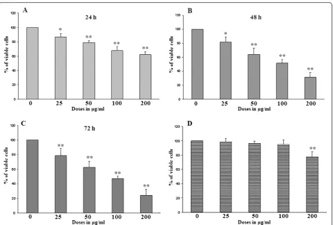
Cell viability assay using WST-1
The effects of DTE on the viability of U937 cells were determined by obtaining the percentage of viable cells at
varying concentrations (0, 25, 50, 100, 200 μg/ml) of
[image:5.595.57.291.134.290.2]DTE at different time points such as 24 h (Figure 1A), 48 h (Figure 1B) and 72 h (Figure 1C) of treatment re-spectively. The results obtained in this assay clearly shows that in each time points DTE have significant growth inhibitory effect on the selected cell line. The study also showed inhibition of U937 cell viability upon treatment with DTE which is both dose and time dependent. A similar experiment was performed on Table 2 Anticlastogenic effect of Darjeeling tea extract
(DTE) against benzo[a]pyrene (B[a]P) in human lymphocyte culture as measured by micronuclei formation
Clastogen (μM) DTE (μg/ml) Number of micronucleated cells/1000 binucleated
cells (mean + _S.D.)
- 25 1.33 ± 1.22
- 50 1.27 ± 0.98
- 100 1.35 ± 1.08
- 200 2.63 ± 1.46
B[a]P 100 - 18.69 ± 3.39
B[a]P 100 25 15.62 ± 2.37 *
B[a]P 100 50 12.56 ± 2.77 **
B[a]P 100 100 8.07 ± 1.68 **
B[a]P 100 200 10.49 ± 3.12 **
Each data represented as Mean ± SD of six samples per concentration. Results at of each concentration of DTE plus positive mutagen benzo[a]pyrene (B[a]P) were compared with the positive mutagen treated group alone (all in presence of S9 mix) by Dunnett’s multiple comparison. *P < 0.05, **P < 0.01.
Figure 1Effects of Darjeeling tea extract (DTE) on the viability of U937 cells (A, B,C) and on peripheral blood mononuclear cells (PBMCs) (D) in the cell viability assay using WST-1 at different time points.U937 cells (1 × 10⁶per treatment) were treated with different
[image:5.595.59.540.340.664.2]PBMCs (Data not shown for 24 h and 72 h of
treat-ment). At above 100 μg/ml dose DTE showed toxicity
towards PBMCs after 48 h of treatment (Figure 1D).
Further experiments were conducted taking 100μg/ml
as highest concentration of DTE and the treatment time was optimized as 48 h.
Induction of apoptosis in U937 cells upon treatment with DTE
Induction of apoptosis by DTE on U937 cells was stud-ied by means of Annexin V/propidium iodide double staining method. The cells were treated with different
concentrations of DTE (0, 25, 50, 100 μg/ml) for 48 h.
and analysed by flow cytometry. The result indicates that
the different doses of DTE (25, 50, 100 μg/ml) can
[image:6.595.56.540.343.655.2]in-duce apoptosis in U937 cells significantly and it ranged from 7.4% to 35.9% from lower to higher concentrations (Figure 2).
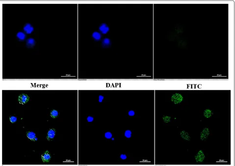
The induction of apoptosis by DTE was also evident from the confocal microscopic study. The DTE treated
cells showed typical apoptotic feature i.e. externalization of phosphatidylserine on the outer surface of the treated cells. Only a representative picture has been incorpo-rated here (Figure 3).
Effect of DTE on caspase-3 activity
Activation of caspases is a hallmark sign of apoptotic cell death. Among the caspase group caspase-3 is one of the most common and important caspase protease that pro-motes the execution phase of apoptosis. We examined whether this specific caspase was involved in DTE induced death of U937 cells. The activity of caspase-3 was exam-ined after 48 h of treatment at different concentrations (0, 25, 50, 100μg/ml). All fractions induced statistically signifi-cant (p < 0.05 to p < 0.01) increase of the activity of caspase-3 in U937 cells upon DTE treatment (Figure 4A).
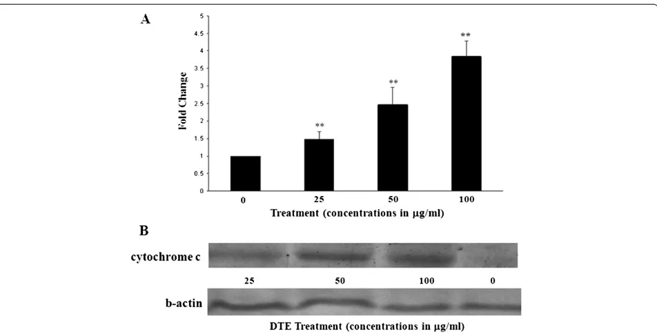
Cytochrome c release in the cytosol
To confirm the result further, we investigated the release of cytochrome c in cytosolic fraction of U937 cells to
Figure 2Flow cytometric detection of apoptosis induction by three different doses of DTE at 48 h of treatment.U937 cells (1 × 10⁶per
detect apoptosis. Western blot analysis showed that cytochrome c appeared in the cytosolic fractions after 48 h of incubation with DTE in a dose dependent man-ner (Figure 4B).
Discussions
Ongoing research for the last few decades have shown that tea and its polyphenolic constituents can bring about many protective biological activities including anti-mutagenic and anticancer activities and consequently, tea appears to be a good natural agent for chemoprevention. Sufficient data have been generated asserting that prevent-ive effects of green tea consumption on occurrence of various types of cancer. Black tea and its polyphenols have potentials to protect human beings from different detri-mental health effect. Earlier reports are available on the antimutagenic potential of black tea or its extracts on bac-terial system [5].
The main objective of this study was to investigate the antimutagenic and/or anticancer activities of Darjeeling tea extract (DTE). In this study, at first the mutagenic ef-fects of DTE on bacterial system was monitored and no significant mutagenic effect was observed in the concen-trations tested for antimutagenicity assay (data not shown). All the solvent control data and the mutagenicity assay data were with the normal range as mentioned by Maron and Ames [22]. Our experimental data indicates that DTE exhibits protective effects in vitro on bacterial system espe-cially at the higher concentrations against the known mutagen B[a]P in presence of S9 mix. Result of this experi-ment was in agreeexperi-ment with a previous study where black tea polyphenols theaflavins and thearubigins showed sig-nificant antimutagenic activity against the same mutagen on bacterial systems [26].
[image:7.595.60.540.89.427.2]To validate the data further the antimutagenic poten-tial of DTE was tested in mammalian cell culture system
using the same mutagen in presence of S9 mix. This assay result corroborates with the previous finding by showing less number of micronuclei formed in presence of DTE at different concentrations when compared with the mutagen B[a]P treated group only.
Development of cancer is often related with multiple mutations and hence it can be hypothesised that DTE could have anticancer activity as it was found to be a po-tent antimutagenic substance. Earlier it has been reported that tea polyphenols having antimutagenic potential also have anticancer effects [27]. Therefore, we subsequently tried to evaluate the anticancer effect of DTE on cancer-ous U937 cells. The objective of cell viability assay was to assess the direct growth inhibitory effect of the DTE on the proliferation and survival of the U937 cells [28]. Our results depicted inhibitory effects of DTE on the U937 cells at different time point. Decrease in the percentage of viable cells over a range of concentrations (0, 25, 50, 100,
200 μg/ml) of DTE at 24, 48 and 72 hours of treatment
was observed. The decrease in the percentage of vi-able cells upon DTE treatment was found to be dose dependent.
“Apoptosis”is a type of programmed cell death which plays a very significant role in eliminating the hyperpro-liferating cells from our body. Induction of apoptosis in
cancer cells, thus, can be considered as a protective mechanism against development and progression of this disease. In this study, we hypothesised that the loss of cell viability of U937 cells upon treatment with DTE could be due to the induction of apoptosis by the ex-tract. Hence, we extended our study to find out the apoptosis induction by the extract. It has been observed that treatment with DTE resulted apoptosis in U937 cells as evident from Annexin V/propidium iodide double staining method. This result suggests that the inhibition of cell viability could be due to induction of apoptotic death of U937 cells upon DTE treatment. The confocal micros-copy result also showed externalization of phosphotydle serine on the surface of DTE treated U937 cells which support the earlier evident of apoptosis induction.
[image:8.595.61.537.87.329.2]Activity of caspase-3 is in many cases related with the apoptosis induction in cancer cells and an increase in the activity of this proteolytic enzyme is considered as a signature of the executionery phase of this process [29]. Here we found that, DTE, at all selected concentrations, was able to upregulate the activity of caspase-3 upon treatment in U937 cells. Another classical feature of apoptotic death is release of mitochondrial cytochrome c in cytosol [30]. We also investigated this phenomenon on U937 cells after DTE treatment. It was found that in
almost all concentrations DTE was able to release cyto-chrome c in cytosol of U937 cells at 48 hours of treat-ment further proving the fact that DTE was able to induce apoptosis in the selected cancer cell line.
Conclusion
Hence, this study demonstrated the antimutagenic and anticancer activities of the Darjeeling tea extract. DTE
showed antimutagenic property onSalmonellastrains in
bacterial mutagenicity assay and on human lymphocytes in vitro with metabolic activation. DTE also showed in-hibitory effect on the viability of the U937 cells. We fur-ther found that DTE exerts its antiproliferative effect by inducing apoptotic death in U937 cells. Therefore, it could be concluded that DTE have anticancer effects and could have chemopreventive properties.
Abbreviations
WST-1:2-(4-Iodophenyl)-3-(4-nitrophenyl)-5-(2,4-disulfophenyl)-2H-tetrazolium; B[a]P: Benzo[a]pyrene; DTE: Darjeeling Tea Extract; DTT: Dithiothreitol; DMSO: dimethyl sulfoxide; MN: Micronuclei; NADP: Nicotinamide adenine dinucleotide phosphate.
Competing interests
This is to certify that there is no actual or potential conflict of interest including any financial, personal or other relationships among the authors or with any other people or organizations related to this article.
Authors’contributions
UB carried out the antimutagenic and anticancer activity study and drafted the manuscript. He played vital role in acquisition, analysis and interpretation of data, SA carried out the anticlastogenic study and also helped in analysis and interpretation of data, NSM participated in antimutagenic and anticancer activity study. BB participated in the design of the study and provided critical intellectual input. AKG designed and coordinate the study and approved the final version of the manuscript. All authors read and approved the final manuscript.
Acknowledgements
The authors are grateful to Dr. Bruce N. Ames, Professor of Molecular Biology and Biochemistry, Department of Molecular Biology and Cell Biology, University of California, Berkeley, USA for providing Salmonella strains TA98 and TA100. Authors would also like to thank Tea Board of India for providing samples of Darjeeling tea for the research work. The authors are also grateful to Dr. Anupam Banerjee and Dr. Samit Adhya of our institute for providing us the support to obtain confocal pictures.
Author details
1
Molecular and Human Genetics Division, Indian Institute of Chemical Biology, 4, Raja S. C. Mullick Road, Jadavpur, 700 032 Kolkata, India. 2
Department of Zoology, Seth Anandram Jaipuria College, Kolkata, India. 3Tea Board, Kolkata, India.
Received: 12 March 2014 Accepted: 11 August 2014 Published: 2 September 2014
References
1. Stagg GV, Millin DJ:The nutritional and therapeutic value of tea-a review. J Sci Food Agric1975,26:1439–1499.
2. Stoner GD, Mukhtar H:Polyphenols as cancer chemopreventive agents. J Cellular Biochem Suppl1995,22:169–180.
3. Kada T, Kaneko K, Matsuzaki S, Matsuzaki T, Hara Y:Detection and chemical identification of natural bioantimutagens: a case of the green tea factor. Mutat Res1985,150:127–132.
4. Weishburger JH, Chung FL:Mechanism of chronic disease causation by nutritional factors and tobacco products and their prevention by tea polyphenols.Food Chem Toxicol2002,40:1145–1154.
5. Khan JA, Jalal JA, Ioanndes C, Moselhy SS:Assessment of the antimutagenic effect of Doash tea extract fractions.Toxicol Ind Health2012,28:867–875.
6. Horn RC, Vargas VM:Mutagenicity and antimutagenicity of teas used in popular medicine in the salmonella/microsome assay.Toxicol In Vitro 2008,22:1043–1049.
7. Catterall F, Copeland E, Clifford MN, Ioannides C:Contribution of theafulvins to the antimutagenicity of black tea: their mechanism of action.Mutagenesis1998,13:631–636.
8. Weisburger JH, Hara Y, Dolan L, Luo FQ, Pittman B, Zang E:Tea polyphenols as inhibitors of mutagenicity of major classes of carcinogens.Mutat Res1996,371:57–63.
9. Liu X, Zhang DY, Zhang W, Zhao X, Yuan C, Ye F:The effect of green tea extract and EGCG on the signaling network in squamous cell carcinoma. Nutr Cancer2011,63:466–475.
10. Mao JT, Nie WX, Tsu IH, Jin YS, Rao JY, Lu QY, Zhang ZF, Go VL, Serio KJ: White tea extract induces apoptosis in non-small cell lung cancer cells: the role of peroxisome proliferator-activated receptor-{gamma} and 15-lipoxygenases.Cancer Prev Res (Phila)2010,3:1132–1140.
11. Zhang D, Al-Hendy M, Richard-Davis G, Montgomery-Rice V, Sharan C, Rajaratnam V, Khurana A, Al-Hendy A:Green tea extract inhibits proliferation of uterine leiomyoma cells in vitro and in nude mice.Am J Obstet Gynecol 2010,202: 289, e1-9(289):e1–e9.
12. Fukuda I, Tsutsui M, Sakane I, Ashida H:Suppression of cytochrome P450 1A1 expression induced by 2,3,7,8-tetrachlorodibenzo-p-dioxin in mouse hepatoma hepa-1c1c7 cells treated with serum of (− )-epigallocatechin-3-gallate- and green tea extract-administered rats.Biosci Biotechnol Biochem 2009,73:1206–1208.
13. Angelo LS, Kurzrock R:Turmeric and green tea: a recipe for the treatment of B-chronic lymphocytic leukemia.Clin Cancer Res2009,15:1123–1125. 14. Wang Y, Zhang H, Holmgren A, Tian W, Zhong L:Inhibitory effect of green
tea extract and (−)-epigallocatechin-3-gallate on mammalian
thioredoxinreductase and HeLa cell viability.Oncol Rep2008,20:1479–1487. 15. Newell AM, Yousef GG, Lila MA, Ramírez-Mares MV, de Mejia EG:Comparative
in vitro bioactivities of tea extracts from six species of Ardisia and their effect on growth inhibition of HepG2 cells.J Ethnopharmacol2010,130:536–544. 16. Roomi MW, Ivanov V, Kalinovsky T, Niedzwiecki A, Rath M:Anticancer effect
of lysine, proline, arginine, ascorbic acid and green tea extract on human renal adenocarcinoma line 786–0.Oncol Rep2006,16:943–947. 17. Yang CS, Maliakal P, Meng X:Inhibition of carcinogenesis by tea.Annu Rev
Pharmacol Toxicol2002,42:25–54.
18. Du Y, Wu Y, Cao X, Cui W, Zhang H, Tian W, Ji M, Holmgren A, Zhong L: Inhibition of mammalian thioredoxinreductase by black tea and its constituents: implications for anticancer actions.Biochimie2009, 91:434–444.
19. Gohain B, Borchetia S, Bhorali P, Agarwal N, Bhuyan LP, Rahman A, Sakata K, Mizutani M, Shimizu B, Gurusubramaniam G, Ravindranath R, Kalita MC, Hazarika M, Das S:Understanding Darjeeling tea flavour on a molecular basis.Plant Mol Biol2012,78:577–597.
20. Ames BN, Durston WE, Yamasaki E, Lee FD:Carcinogens are mutagens: a simple test system combining liver homogenates for activation and bacteria for detection.Proc Natl Acad Sci1973,70:2081–2285.
21. Garner RC, Miller EC, Miller JA:Liver microsomal metabolism of aflatoxin B 1 to a reactive derivative toxic to Salmonella typhimurium TA 1530. Cancer Res1972,32:2058–2066.
22. Maron DM, Ames BN:Revised methods for the Salmonella mutagenicity test.Mutat Res1983,113:173–215.
23. Fenech M:The in vitro micronucleus technique.Mutat Res2000,20:81–95. 24. Sasaki YF, Yamada H, Shimoi K, Kator K, Kinae N:The clastogen-suppressing effects of green tea, Po-lei tea and Rooibos tea in CHO cells and mice. Mutat Res1993,286:221–232.
25. Fenech M, Chang WP, Kirsch-Volders M, Holland N, Bonassi S, Zeiger E:Human MicronNucleus project. HUMN project: detailed description of the scoring criterian for the cytokinesis—block micronucleus assay using isolated human lymphocyte cultures.Mutat Res2003,10:65–75.
27. Bhattacharya U, Mukhopadhyay S, Giri AK:Comparative antimutagenic and anticancer activity of three fractions of black tea polyphenols
thearubigins.Nutr Cancer2011,63:1122–1132.
28. Zhang Y, Yasumoto Y, Mei C, Arima T:Vitamin E inhibits proliferation of primary cultured human mesangial and endothelial cells.Nephron2001, 89:291–296.
29. Cohen GM:Caspases: the executioners of apoptosis.Biochem J1997, 15(326):1–16.
30. Skulachev VP:Cytochrome c in the apoptotic and antioxidant cascades. FEBS Lett1998,27:275–280.
doi:10.1186/1472-6882-14-327
Cite this article as:Bhattacharyaet al.:Antimutagenic and anticancer activity of Darjeeling tea in multiple test systems.BMC Complementary and Alternative Medicine201414:327.
Submit your next manuscript to BioMed Central and take full advantage of:
• Convenient online submission
• Thorough peer review
• No space constraints or color figure charges
• Immediate publication on acceptance
• Inclusion in PubMed, CAS, Scopus and Google Scholar
• Research which is freely available for redistribution