REVIEW
Signaling in the stem cell niche: regulating cell fate, function and
plasticity
Carlos Andrés Chacón-Martıneź 1,*, Janis Koester1,* and Sara A. Wickström1,2,3,‡
ABSTRACT
Stem cells have the ability to self-renew and differentiate along multiple lineages, driving tissue homeostasis and regeneration. Paradigms of unidirectional, hierarchical differentiation trajectories observed in embryonic and hematopoietic stem cells have traditionally been applied to tissue-resident stem cells. However, accumulating evidence implicates stemness as a bidirectional, dynamic state that is largely governed by the niche, which facilitates plasticity and adaptability to changing conditions. In this Review, we discuss mechanisms of cell fate regulation through niche-derived cues, with a particular focus on epithelial stem cells of the mammalian skin, intestine and lung. We discuss a spectrum of niche-derived biochemical, mechanical and architectural inputs that define stem cell states during morphogenesis, homeostasis and regeneration, and highlight how these diverse inputs influence stem cell plasticity.
KEY WORDS: Epithelium, Intestine, Lung, Niche, Skin, Stem cells
Introduction
Epithelial tissues such as the lung, gastrointestinal tract and skin undergo continuous cell replacement in a process termed homeostasis (Blanpain and Fuchs, 2014; Leeman et al., 2014). This process depends on the dynamic activity of tissue-resident stem cells and the surrounding environment–the niche–in which they reside. These same stem cells are also called into action to regenerate damaged tissue following injury, to facilitate rapid repair and to prevent tissue overgrowth, in a process that is again regulated by the niche. Such stem cells receive and respond to various feedback signals from their immediate environment to be able to react to the changing needs of tissues. Secreted signals originate from the stem cells themselves (autocrine), from the neighboring niche cells ( paracrine) or from other tissues (systemic), and generally form part of central stem cell regulatory pathways. Other forms of signaling, such as contact-dependent Notch signaling, signaling from the extracellular matrix (ECM) through adhesion receptors, as well as mechanical signals from physical and topological cues, can also provide key signals to stem cells.
The original definition of a stem cell is that it possesses the capacity for both long-term self-renewal and multi-lineage differentiation (Becker et al., 1963; Till and McCulloch, 1961). Although this still holds true, we now know that stem cells are a heterogeneous population of cells with varying transcriptional
profiles and self-renewing capacities, yet are functionally equivalent in their ability to maintain tissue homeostasis and restore tissue integrity upon injury (Goodell et al., 2015; Krieger and Simons, 2015; Wabik and Jones, 2015). The term ‘niche’, which was originally conceptualized by Schofield (1978), refers to the specific microenvironment in which stem cells reside. The niche consists of stem cells themselves as well as their progeny, but also multiple heterologous cell types and a niche-specific ECM. The niche thus provides soluble, adhesive and physical signals to stem cells, which are crucial for maintaining stem cell functions. Owing to the complex composition of epithelial stem cell niches, they function by integrating a plethora of signals, both local and systemic, to ensure appropriate and coordinated responses of stem cells to the changing needs of tissues (Scadden, 2014).
The traditional paradigm of a unidirectional, hierarchal differen-tiation trajectory–beginning with a multipotent self-renewing stem cell and proceeding through transit-amplifying cell stages before transitioning into the terminal differentiated state–was uncovered in the embryonic and hematopoietic stem cell fields (Weissman, 2000). These concepts have further been applied to epithelial tissue-resident stem cells. However, accumulating evidence suggests that stemness, especially in the context of epithelial tissues, is a bidirectional, dynamic state that is largely governed by the stem cell niche, allowing plasticity and adaptability to changing conditions (Chacón-Martínez et al., 2017; Ritsma et al., 2014; Rompolas et al., 2013; Sun et al., 2014; Takeda et al., 2011). These discoveries highlight the importance of the biochemical composition and biophysical architecture of the niche, which influence stem cell state and fate (Morrison and Spradling, 2008; Scadden, 2014). Given this central role of the niche in regulating epithelial stem cell behavior, the identification of niche factors and signaling pathways that can promote stem cell function or enrich for cells with stem cell properties could have significant implications in regenerative medicine and tissue engineering. In this Review, we discuss how niche inputs regulate stem cell functions during morphogenesis, homeostasis and regeneration, focusing mainly on three well-studied mammalian systems: the lung, gastrointestinal tract and skin epithelia. We first provide an overview of the stem cell dynamics within these tissues. We then describe recent research that has identified key biochemical and biomechanical signals within the niche that control stem cell fate, and highlight how these signals facilitate stem cell heterogeneity and plasticity to ensure robust tissue function and repair.
Stem cells drive dynamic tissue turnover in epithelia
Epithelial tissues are continuous sheets of tightly adherent cells that line most body surfaces, organs, tracts and cavities. They regulate water and nutrient absorption, and physically protect tissues from the external environment. Owing to this front-line defensive function, epithelial tissues such as the skin epidermis, intestine and lung must self-renew in order to rapidly replace lost or damaged cells. Indeed, the intestinal epithelium is one of the fastest
self-1Paul Gerson Unna Group‘Skin Homeostasis and Ageing’, Max Planck Institute for
Biology of Ageing, D-50931 Cologne, Germany.2Helsinki Institute of Life Science,
Biomedicum Helsinki, University of Helsinki, FI-00014 Helsinki, Finland.3Wihuri
Research Institute, Biomedicum Helsinki, University of Helsinki, FI-00014 Helsinki, Finland.
*These authors contributed equally to this work
‡Author for correspondence ([email protected])
DEVEL
O
renewing tissues and completely regenerates within 3-5 days (reviewed in van der Flier and Clevers, 2009). This single-layered simple epithelium extends invaginations, termed crypts, into the underlying connective tissue (Fig. 1A). Rapidly cycling intestinal stem cells (ISCs), which are positive for the marker LGR5, are located at the crypt base interspaced between Paneth cells (Barker et al., 2007; Hertzog, 1937), whereas slow-cycling, label-retaining HOPX+, LRIG+, BMI1+, TERT+, DLL1+ISCs are located at the +4 position relative to the crypt base (Barker et al., 2007; Montgomery et al., 2011; Takeda et al., 2011). Once these stem cells migrate out of their niche, they differentiate either into the absorptive or secretory lineages and finally into one of four differentiated cell types: enterocytes, mucin-secreting goblet cells, peptide hormone-secreting neuroendocrine cells and microbicide-hormone-secreting Paneth cells (reviewed by Crosnier et al., 2006).
The turnover dynamics of the multilayered, stratified skin epithelium, the epidermis, are very similar to those of the intestinal epithelium, fully renewing every 7-10 days (Potten et al., 1987). Epidermal stem cells reside in the basal layer of this stratified epithelium (Fig. 1B), where they initiate a transcriptional program of terminal differentiation while moving upwards to give rise to the spinous layer, the granular layer and, finally, the cornified layer of dead cells. In addition to the epidermis, the skin harbors multiple specialized appendices such as the hair follicle, which is maintained by its own stem cell population: the hair follicle stem cells (HFSCs, Fig. 1B) (Oshima et al., 2001; Reynolds and Jahoda, 1991). Hair follicles self-renew through cyclical bouts of growth (anagen), degeneration (catagen) and rest (telogen). At the start of the hair cycle, quiescent HFSCs residing in the so-called bulge niche are triggered to proliferate through complex signaling crosstalk with neighboring niche cells, and migrate to supply the cells needed for hair follicle downgrowth (Gonzales and Fuchs, 2017; Müller-Röver et al., 2001).
In contrast to the rapidly renewing intestine and epidermis, the airways of the lungs are lined by a pseudo-stratified epithelium that can take as long as 6 months to be replaced, but has a remarkable A Small intestine
EGF WNT3 DLL4 Lactate
WNT
Globet cell
Paneth cell Transit-amplifying
cell
LGR5+ ISC
Enteroendocrine cell Enterocyte
+4 stem cell Mesenchymal cell Key
Basement membrane
Extracellular matrix
C Airway
Neuroendocrine cell Secretory cell
Basal stem cell Ciliated cell
Mesenchymal cell Basement membrane
Extracellular matrix Key
JAG2 FGF10 WNT
B Skin
Bulge Isthmus
Infundibulum
Dermal papilla IFE
Sebaceous gland
BMPs JAG1
WNTs
SHH
WNT
ECM signals
Progenitor
Secondary hair
germ Sebocyte
Treg IFE stem cell HFSC
Mesenchymal cell
Basement membrane Key
[image:2.612.48.302.48.722.2]Extracellular matrix
Fig. 1. Niche signals controling lineage hierarchies and dynamics during homeostasis.(A) In the small intestine, actively cycling LGR5+intestinal stem
cells (ISCs) are located at the crypt base and are interspaced between Paneth cells. Slow-cycling, label-retaining (HOPX+, LRIG+, TERT+, BMI+and DLL1+)
stem cells located at the +4 position relative to the crypt base function as a stem cell reserve and can replenish LGR5+ISCs upon their loss. ISCs give rise to
four different lineages: enterocytes, goblet cells, neuroendocrine cells and Paneth cells. Stromal cells and Paneth cells provide ISCs with essential niche signals such as WNTs to support intestinal homeostasis. (B) The stratified epidermis (interfollicular epidermis; IFE) of the skin is continuously renewed by epidermal stem cells (also termed epidermal progenitor cells). Epidermal stemness is maintained by autocrine WNT signals and by contact with the basement membrane. Once these cells initiate differentiation, they move upwards and give rise to various differentiated layers. The hair follicle is compartmentalized into multiple micro-niches that are maintained by distinct stem cell populations. CD34+hair follicle stem cells (HFSCs) reside in the
bulge niche, where they are activated through signaling with their immediate progeny (hair germ cells) and mesenchymal dermal papilla cells to initiate differentiation and hair follicle growth. Further instructive signals are provided by the surrounding niche basement membrane, proximal dermal fibroblasts and T cells. (C) The lung airway epithelium consists of basal cells, secretory cells, ciliated cells and neuroendocrine cells. Basal cells can act as stem cells, self-renewing and differentiating into secretory and ciliated cells. Interactions between niche mesenchymal cells and basal stem cells are important for maintaining basal stem cells in an undifferentiated state. On the other hand, continuous Notch signaling from basal stem cells to their secretory progeny supports progeny maintenance and prevents their further differentiation into ciliated cells.
DEVEL
O
ability to regenerate after injury (reviewed by Rock and Hogan, 2011). The airway epithelium consists of basal cells, secretory cells, ciliated cells and neuroendocrine cells (Fig. 1C), while the alveolar epithelium, which facilitates gas exchange, contains alveolar type 1 (AT1) and alveolar type 2 (AT2) cells. In mice, basal cells have been shown to act as the main stem cell population in the proximal airway epithelium, with the ability to both self-renew and give rise to multiple cell types, and there is evidence that the same may be true in humans (Tata and Rajagopal, 2017). AT2 cells can both self-renew and give rise to AT1 cells, and thus are considered the stem cell of the alveolar epithelium. Recently, a newly identified subset of AXIN2+AT2 cells was shown to constitute a major progenitor pool in the distal lung and to effectively regenerate the alveolar epithelium upon injury (Zacharias et al., 2018). It should be noted, however, that these lineage hierarchies can vary under different conditions and in different locations of the lung, and care must thus be taken when discussing and interpreting data relating to these different cell populations (Chen and Fine, 2016).
Niche signals that originate from stem cell progeny
A major theme that has developed in recent years is the crucial role of stem cell-to-daughter cell crosstalk in regulating homeostasis and the appropriate response to injury across multiple tissues. This is well illustrated by recent research on the regulation of ISC function. As mentioned above, LGR5+ISCs reside at the bottom of intestinal crypts interspersed between their own specialized progeny, the Paneth cells (Barker et al., 2007; Hertzog, 1937). Paneth cells play a role in immunity and host-defense, but also secrete important signaling molecules such as WNT3, EGF and Notch ligand DLL4 (Ganz, 2000; Sato et al., 2011), suggesting that they might signal to ISCs. In line with this, it has been shown that co-culturing LGR5+ ISCs with a Paneth cell-enriched population or adding exogenous WNT3A, enhances the efficiency of LGR5+ ISCs in forming differentiated intestinal organoids in vitro (Sato et al., 2011). Consistently, depletion of Paneth cellsin vivousing three different genetic mouse models leads to reduced stem cell numbers (Sato et al., 2011), indicating that daughter cells of LGR5+ISCs provide essential niche signals for these stem cells. However, there is controversy about the role of Paneth cells as two subsequent studies showed that complete ablation of Paneth cells does not affect LGR5+ ISC maintenance and proliferation (Durand et al., 2012; Kim et al., 2012), challenging the initial findings. However, in these later studies, alternative pathways that upregulate WNT/β-Catenin signaling are observed. Consistently, later reports showed that deletion of epithelial Wnt3, although necessary for organoid cultures, has no effect on stem cell function in adult mice, as stromal secretion of WNTs could fully support intestinal homeostasis (Farin et al., 2012; Kabiri et al., 2014; San Roman et al., 2014). Interestingly, WNT alone is not sufficient to promote LGR5+ ISC self-renewal, but additional signals from R-spondins are required. WNT stabilizes R-spondin receptor expression (LGR4, LGR5, LGR6), enabling R-spondin to drive stem cell expansion (Yan et al., 2017). Collectively this indicates that Paneth cells are a dispensable source of WNTin vivo, and thus the outcome of Paneth cell depletion might be dependent on the approach used and its indirect impact on the WNT signaling status of the niche. A recent paper has provided alternative mechanisms to explain the function of Paneth cells in the ISC niche, albeit only in anin vitroorganoid system. Comparative metabolomics of the two cell types revealed that LGR5+ISCs display higher mitochondrial activity compared to Paneth cells (Rodríguez-Colman et al., 2017). It has previously been suggested that efficient oxidative metabolism and low ROS
levels are crucial processes for stem cell self-renewal and quiescence, whereas mitochondrial status, aerobic glycolysis and ROS production are associated with differentiation (Ho et al., 2017; Khacho et al., 2016). Following on from this, it was shown that Paneth cells provide lactate to LGR5+ISCs, which fuels oxidative phosphorylation leading to production of ROS and subsequent enhanced differentiation (Rodríguez-Colman et al., 2017). Together, these findings suggest that stem cell progeny within the niche support stem cell functions through multiple mechanisms, but it seems likely that several niche-resident cells, acting in a redundant fashion, provide the most crucial signals such as WNT. This would facilitate robust niche function, ensuring that no particular niche cell population is indispensable for proper stem cell activities.
A similar feedback mechanism–from progeny back to stem cells
–is seen in the hair follicle, where early HFSC progenitors signal back to HFSCs to promote their activity during hair regeneration. In this context, progenitor cell-derived sonic hedgehog (SHH) sustains HFSC activation during the hair follicle growth phase for as long as the progeny and the dermal papilla are in close proximity to the bulge HFSC niche (Hsu et al., 2014). This provides a self-organizing feedback loop to precisely scale HFSC activation to the degree of hair follicle growth.
Signals that originate from stem cells
Besides daughter cells sending feedback signals to their parent stem cells, stem cells themselves signal to their progeny, as exemplified by a recent study in the lung epithelium (Pardo-Saganta et al., 2015b). Basal stem cells in the lung continuously signal through the Notch ligand JAG2 to secretory daughter cells, thereby supporting their maintenance. Without this signal, a large proportion of secretory cells terminally differentiate into ciliated cells. Therefore, basal stem cells regulate homeostasis of their daughter cells, providing an elegant feedback loop to control the balance between the number of stem cells and their progeny (Pardo-Saganta et al., 2015b).
Besides paracrine signaling with niche cells, stem cells participate in self-signaling loops. Stem cells in the epidermis, for example, express several Wnt genes, and inhibition of WNT secretion leads to their premature differentiation, indicating that autocrine WNT signaling maintains the undifferentiated stem cell state during homeostasis (Lim et al., 2013). During lung injury, upon the loss of differentiated luminal cells, the epithelium is restored by basal stem cells (Rock et al., 2009). Subsequently, two distinct basal cell subpopulations emerge – one defined by expression of the intracellular domain of Notch2 (N2ICD) and another by expression of MYB, a transcription factor acting downstream of Notch signaling (Tan et al., 2013). N2ICD+cells differentiate into secretory cells, while MYB+cells differentiate into ciliated cells. Consequently, blocking Notch signaling leads to increased numbers of MYB+basal stem cells (Pardo-Saganta et al., 2015a). Similarly, in the intestine, Notch signaling promotes LGR5+ stem cell proliferation, while preventing differentiation into to the secretory cell lineage. Accordingly, deletion ofNotch leads to secretory cell hyperplasia (Fre et al., 2005; Stanger et al., 2005; van Es et al., 2005; VanDussen et al., 2012). Interestingly, blocking WNT signaling in the intestine rescues this secretory cell hyperplasia (Tian et al., 2015), indicating that Notch signaling tunes local WNT activity, thereby coordinating balanced self-renewal and differentiation within the niche. Collectively, this intricate complexity of the sources, factors and contexts of niche signals is beginning to reveal how stem cell behavior is adjusted to ensure precise lineage output responses to maintain or restore
tissue integrity.
DEVEL
O
Other cell types that signal in the niche
Stromal cells within the niche are also active players in maintaining adult stem cells, acting via the secretion of key signaling factors such as WNT, Notch and BMP (Roberts et al., 2017). In the hair follicle, both the mesenchymal cells of the dermal papilla and dermal adipocyte precursor cells secrete factors that activate stem cells (Sennett and Rendl, 2012). More globally, waves of BMPs secreted by dermal fibroblasts are involved in maintaining the synchronized quiescent state of HFSCs (Plikus et al., 2008). The interaction between fibroblasts and stem cells has also been shown to be important for establishing a functional basal stem cell niche in the lung airway (Ruiz et al., 2014). Using kidney capsule engraftments and culture of lung explants, airway epithelium basal stem cells were found to act via SDF1 to recruit and activate fibroblasts, which subsequently secrete TNFα and reciprocally activate the basal stem cells, generating a self-sustained feedback loop. Activated basal stem cells showed increased SDF1 expression, which in turn induces fibroblasts to express IL8 and VEGF, factors that promote angiogenesis and recruitment of endothelial cells. Interestingly, the basal stem cells in close proximity to fibroblasts remain in an undifferentiated state to ensure self-renewal capacity (Ruiz et al., 2014). Similar self-organizing feedback loops have been observed during lung regeneration, where niche cells play key roles in lung stem cell regulation. For example, endothelial cell signaling plays a key role in lung bronchioalveolar stem cell differentiation (Lee et al., 2014). Bronchioalveolar stem cells can differentiate into multiple lineages, but BMP4 signaling in endothelial cells induces thrombospondin 1 expression, which subsequently acts on bronchioalveolar stem cells to trigger their specific differentiation into the alveolar lineage. Interestingly, BMP is upregulated in bronchioalveolar stem cells and AT2 cells after alveolar injury, and expression of thrombospondin 1 is required for efficient injury repair (Lee et al., 2014), demonstrating how stem cells rely on feedback signals from the niche to regulate their regenerative functions.
Roles for distinct stromal sub-types have also been uncovered. The mesenchymal niche of the lung epithelium, for example, appears highly heterogeneous and specialized: each mesenchymal lineage has a distinct localization and transcriptional profile, leading to niche-specific regulatory functions (Zepp et al., 2017). The mesenchymal niche of alveolar stem cells is PDGFRα positive, responds to WNT signals and promotes AT2 cell self-renewal (Zepp et al., 2017). Similarly, a distinct region-specific population of LGR6+ mesenchymal niche cells regulates airway progenitor differentiation and self-renewal via FGF10 signaling. The epithelial progenitors, in turn, signal to mesenchymal cells via WNT signaling, promoting their proliferation (Lee et al., 2017). A self-organizing niche feedback loop between the mesenchyme and stem cells has also been observed in the mucociliary epithelium of the trachea. In this context, BMP signaling through SMAD1/5/8 is transiently decreased upon injury by reduced expression of BMP ligands in the mesenchyme, as well as decreased expression of BMP receptors and increased expression of BMP antagonists in both the epithelium and the mesenchyme (Tadokoro et al., 2016). This leads to accumulation and multilayering of basal stem cells. Interestingly, this expansion is followed by active extrusion of apoptotic cells from the crowded epithelium through constriction of their neighbors, restoring homeostatic cell density. These findings suggest that BMP signaling normally reduces proliferation and might be important for maintaining the steady state; counteracting this signaling after injury restores the damaged tissue (Tadokoro et al., 2016). Collectively, these findings suggest that the presence of
self-organizing feedback circuits between stem cells and their surrounding niche cells could function as checkpoints to prevent stem cell differentiation in the absence of properly maintained niche homeostasis or during regenerative growth, which can then be switched off to allow differentiation once homeostasis is re-established.
Emerging data implicate immune cells as another important component of stem cell niches. Specifically, regulatory T cells (Tregs) have an important immunosuppressive function (Bettelli et al., 2006) and play a role in injury repair (Arpaia et al., 2015; Nosbaum et al., 2016). A subset of highly activated Tregs accumulates around hair follicles during late telogen and colocalizes with HFSCs, whereas they are less abundant during anagen (Ali et al., 2017). Hair depilation in mice triggers onset of the anagen growth phase, but mice depleted of Tregs show a markedly reduced anagen induction and hair regrowth, accompanied with shortened hair follicles. HFSCs isolated after depilation were shown to have differential expression of Notch target genes in Treg-depleted mice. Furthermore, the deletion of
Jag1in Tregs results in a reduction of key differentiation genes in HFSCs. These results suggest that Tregs support HFSC function through JAG1-Notch signaling (Ali et al., 2017). Interestingly, colocalization of Tregs with adult stem cells has also been observed in the hematopoietic stem cell niche, where they generate an immunoprivileged microenvironment that promotes stem cell persistence (Fujisaki et al., 2011). Whether the trophic function of Tregs represents a general feature of adult stem cell niches, and whether other immune cells contribute to stem cell-niche signals, remains an interesting avenue for future research.
Niche signals modulate cellular plasticity following injury In the classical view of strict cell hierarchy within tissues, stem cells reside on top of a lineage hierarchy, and once they make the decision to commit to differentiation the process is irreversible (Sánchez Alvarado and Yamanaka, 2014). In recent years, this concept has been challenged as numerous studies demonstrate that progenitor cells and even more differentiated cell types exhibit enormous plasticity (Fig. 2), endowing them with the ability to transdifferentiate and dedifferentiate under certain homeostatic and regenerative conditions in processes orchestrated by the niche (Chacón-Martínez et al., 2017; Ritsma et al., 2014; Rompolas et al., 2013; Sun et al., 2014; Takeda et al., 2011).
Dedifferentiation refers to the process in which differentiated cells revert to a less differentiated state within their lineage. In mammalian epithelia, the advent of lineage tracing has begun to unearth the widespread nature of dedifferentiation of stem cell progeny back to stem cell states. Such studies have revealed that cell fate plasticity is particularly apparent during injury and subsequent regeneration, when tissue and niche architecture are disrupted and remodeled. For example, the replenishment of LRG5+ISCs after their genetic ablation suggests that these stem cells are dispensable for tissue homeostasis, as slow-cycling BMI1+cells have the ability to mobilize into the empty niche and dedifferentiate into LGR5+ ISCs (Muñoz et al., 2012; Tian et al., 2011). In a similar fashion, LGR5+progeny such as DLL1+ secretory progenitors and label-retaining cells, located at crypt position +3/+4, were shown to have the ability to regenerate LGR5+ISCsin vivoupon acute stem cell loss (Buczacki et al., 2013; van Es et al., 2012). Of note, whilein
vitrocultures of FACS-purified LRG5+cells give rise to organoids, also known as mini-guts (Sato et al., 2009), purified DLL1+cells do not generate organoids using standard culture conditions (in the presence of EGF, Noggin and R-spondin1). However, the addition
DEVEL
O
of WNT3 to the culture medium allows full organoid formation by DLL1+ cells, suggesting a crucial role of niche-derived WNT signaling in secretory progenitor dedifferentiation (van Es et al., 2012). Interestingly, the loss of LGR5+ISCs (induced by diphtheria toxin administration in a genetic mouse model to lineage trace enterocyte precursors, which are characterized by the expression of the alkaline phosphatase intestinal ALPI), revealed the ability of enterocytes to dedifferentiate into LGR5+ ISCs. Of note, ALPI+ cells lose this plasticity as they differentiate and move out of the crypt niche. Single-cell gene expression analyses revealed that regeneration of LGR5+cells by ALPI+enterocytes does not involve transition through a DLL1-expressing progenitor cell state (Tetteh et al., 2016), indicating that several routes of dedifferentiation exist. Together, these studies have revealed a high degree of plasticity within the intestine in response to injury, which is guided by the positioning of cells in the crypt niche and the WNT-enriched signaling microenvironment of the crypt (Fig. 2A).
A similar dedifferentiation phenomenon has been observed in the gastric epithelium, where Troy+ (TNFRSF19+) and LGR5+ differentiated chief cells populate the base of gastric glands. These cells do not have stem cell functions during homeostasis. However, upon damage-induced loss of stem cells in the isthmus region adjacent to the opening of the gland, chief cells become active and are able to replenish isthmus stem cells as well as parietal, mucous and neuroendocrine cells in a WNT-dependent manner (Leushacke et al., 2017; Stange et al., 2013). Taken together, these findings show that, upon stem cell loss after injury, exposure to defined stem cell-niche signals such as WNT instructs multiple classes of early progeny to re-acquire a stem cell state.
Similar roles of differentiated cells as stem cell reservoirs have been identified in other tissues such as the skin interfollicular
epidermis (IFE) and the hair follicle during regeneration following injury (Fig. 2B). Unlike HFSCs, IFE stem cells constantly proliferate to renew the epidermis. Although initially thought to be maintained by quiescent stem cells and rapidly dividing transit-amplifying cells (Barrandon and Green, 1987; Potten and Morris, 1988), a single progenitor population with equal probability to self-renew and differentiate has been suggested to be responsible for maintaining the IFE (Clayton et al., 2007; Mascré et al., 2012; Rompolas et al., 2016). However, the existence of functionally heterogeneous stem cell populations in specific locations of the mouse tail epidermis has been reported (Gomez et al., 2013; Mascré et al., 2012; Sada et al., 2016). In addition, a degree of transcriptional heterogeneity and the existence of an LGR6+subpopulation within the mouse back skin IFE has been observed (Füllgrabe et al., 2015; Joost et al., 2016; Snippert et al., 2010a), but the functional significance of this heterogeneity is unclear. Thus, although the precise molecular identity of the IFE stem cell remains elusive, transcriptional and functional heterogeneity is most likely controled by anatomical location and therefore region-specific, currently unknown, niche signals.
HFSCs represent further evidence of niche-induced plasticity. These stem cells do not normally contribute to IFE maintenance but migrate to sites of epidermal wounds, occupy the IFE niche and, through currently unknown signals from this new niche, subsequently adopt IFE fate and contribute to tissue repair after injury (Blanpain et al., 2004; Claudinot et al., 2005; Ito et al., 2005). Interestingly,in vivotwo-photon imaging coupled to lineage tracing and laser ablation revealed that HFSCs are in fact dispensable for hair regeneration; their committed, immediate progeny, as well as other K14+ cells located in the IFE, infundibulum and sebaceous glands, can repopulate the empty stem cell niche, adopt stem cell
Differentiation
Key
De-/trans-differentiation ? Unknown signaling pathways
C Proximal airway
Basal cell
Ciliated cell Secretory
cell YAP cell-cell contact B Skin
HFSC IFE SC
Infundibulum cell or sebocyte Progeny
SHH
?
?
Differentiated IFE cell
?
? A Intestine
LGR5+ ISC +4 SC
Paneth cell Transit-amplifying cell
Enterocyte Globet
cell
Enteroendocyte cell ?
[image:5.612.65.551.52.300.2]WNTs WNTs
Fig. 2. Niche-controled differentiation trajectories and plasticity in epithelia.(A) In the intestine, progenitors and also enterocytes can dedifferentiate to LGR5+intestinal stem cells (ISCs) through WNT-mediated niche signals. (B) In the skin, the immediate progeny of hair follicle stem cells (HFSCs) as well as more
distant populations located in the interfollicular epidermis (IFE), infundibulum and sebaceous glands can repopulate the bulge stem cell niche upon HFSC loss. The precise signals that control this plasticity are unclear butin vitrostudies implicate sonic hedgehog (SHH) signaling in this process. In response to wounding, HFSCs are able to migrate into the IFE to regenerate the epidermis and, vice versa, IFE stem cells can generate hair follicles upon transplantation. Although experimental evidence for many dedifferentiation events is compelling (solid arrows) for others it is preliminary (broken arrows). (C) In the lung proximal airway epithelium, secretory cells can dedifferentiate into basal cells through signals that involve direct cell-cell contact and the transcription factor YAP.
DEVEL
O
features and actively contribute to subsequent hair regeneration cycles (Rompolas et al., 2013). Similarly, diphtheria toxin-mediated ablation of the LGR5+subpopulation of HFSCs initially results in abrogation of hair follicle regeneration, but eventually the LGR5+ stem cells as well as normal hair growth is restored by the CD34+ HFSC population (Hoeck et al., 2017). Niche-derived signals contributing to this phenomenon could emanate from the surrounding ECM (Morgner et al., 2015), from contiguous stem cell progeny (Hsu et al., 2011; van Es et al., 2012) and/or from neighboring mesenchymal cells (Chi et al., 2013). For example, it has been shown that the hair follicle bulge ECM niche has a specific composition, with very low levels of the basement membrane protein laminin 511 compared with the adjacent hair germ that harbors the activated progeny, and that contact of stem cells with laminin 511 induces WNT signaling (Morgner et al., 2015). This establishes a mechanism by which a niche-specific ECM could control stem cell differentiation. Intriguingly, disrupting the physical contiguity between the epidermal and mesenchymal niche components impairs the dedifferentiation of committed epidermal cells into HFSCs and subsequent hair regeneration (Rompolas et al., 2013). This indicates that direct epidermal-dermal proximity within the niche is indispensable for specifying the HFSC fate during dedifferentiation. On the other hand, exogenous manipulation of key niche factors such as SHH is sufficient to trigger dedifferentiation of progenitors to the HFSC statein vitro (Chacón-Martínez et al., 2017).
Cellular plasticity within the lung has also been reported (Fig. 2C). In the proximal airway, committed secretory cells can dedifferentiate into basal stem cells upon acute loss of these stem cells (Tata et al., 2013). Using diphtheria toxin to specifically ablate CK5+ basal stem cells, secretory cells were found to proliferate rapidly to compensate for basal stem cell loss. Around 8% of these cells lost markers of secretory cells, while gaining expression of basal stem cell markers (Tata et al., 2013). Interestingly, the dedifferentiation process of secretory cells is modulated by direct cell-cell contact with basal stem cells (Tata et al., 2013) and by the mechanosensitive transcription factor YAP (Zhao et al., 2014), which points to a reciprocal mechanism that relies on single-cell level interactions and sensing the density of the stem cell layer to ensure tissue integrity.
Collectively, these findings imply that localized signals from the mesenchyme, proximal progenitors and the ECM most likely provide the necessary signals for progenitors entering the niche to take over a stem cell (or stem cell-like) state. However, the role of inflammatory and danger signals produced from ablated cell populations has not been sufficiently addressed so far, and it is possible that such signals may also contribute. It is also conceivable that environmental insults trigger unexpected cellular responses to ensure tissue level function, but whether dedifferentiation has a role in tissue homeostasis has not been extensively studied. In vivo lineage tracing andin vitroorganoid cultures of HOPX+cells, slow-cycling label-retaining cells located at the +4 position have demonstrated that, under homeostatic conditions, the progeny of these cells can populate the entire crypt base, including regions where LGR5+ ISCs reside. Likewise, LGR5+ ISCs give rise to HOPX+cells, indicating that intestinal adult stem cells located in distinct niches display plasticity under homeostatic conditions (Takeda et al., 2011). A potential caveat here is that HOPX and other +4 markers have been shown to exhibit broader expression patterns, even overlapping with LGR5+cells (Muñoz et al., 2012), and thus the presence of HOPX transcript cannot be used as a proxy for stem cell identity (Li et al., 2014). However, a marker-free tracing study
of these quiescent +4 label-retaining cells later showed that these cells can indeed, in response to injury, give rise to LGR5+ ISCs (Buczacki et al., 2013). Further studies are thus required to fully establish the role of +4 cells in homeostasis. One way to interpret all these findings is to postulate that positioning of a cell within the anatomic tissue structure, and thereby its exposure to local niche factors, determines cell identity, preventing stem cell loss and ensuring robust tissue function. Future studies addressing these aspects in homeostatic and regenerative dedifferentiation will hopefully provide a better understanding of this process.
Niche architecture regulates stem cell fate
Cell division has been regarded as a crucial step in cell fate commitment. Developmental studies have established that asymmetric cell divisions generate two daughter cells with intrinsically distinct fates: a stem cell and a committed progenitor that goes on to differentiate. In contrast, symmetric cell divisions generate either two identical stem cells, which enables self-renewal, or two committed progenitors to ensure differentiation (Williams and Fuchs, 2013). The type of cell division that occurs, and thereby the decision to either differentiate or self-renew, was thought to be cell-autonomous in epithelia, similar to stem cells in various other model organisms such as C. elegans (reviewed by Cowan and Hyman, 2004). However, single-cell lineage tracing studies of LGR5+ISCs as well as LGR5+gastric and pyloric stem cells in their
in vivoniche have uncovered that differentiation follows a stochastic drift model wherein spatial niche constraints orchestrate non-cell autonomous fate decisions (Leushacke et al., 2016, 2013; Lopez-Garcia et al., 2010; Snippert et al., 2010b). In this model, tissue homeostasis is accomplished at the population level by neutral competition of stem cells, rather than by predefined cell-autonomous fate decisions of single cells. Furthermore, and in line with the decisive role of niche constraints, imaging studies in the skin and the intestine have shown that the functional heterogeneity of stem cells can be traced back to their anatomical position within the niche (Ritsma et al., 2014; Rompolas et al., 2013). In the hair follicle, the main determinant of whether or not a stem cell is likely to participate in the regeneration process is based on its proximity to the niche borders. Within the bulge niche, stem cells in the lower half are more likely to proliferate and generate differentiated progeny, whereas cells located in the upper half are either quiescent or generate only low numbers of spatially restricted progeny (Rompolas et al., 2013). In a similar fashion, intestinal transit-amplifying cells are generated upon stem cell division by their physical displacement from the stem cell niche (Ritsma et al., 2014; Snippert et al., 2010b). This fate change can also be triggered by the cell division of neighboring cells, uncoupling fate determination from cell division (Ritsma et al., 2014). Moreover, in lung alveoli, the physical proximity of AXIN2+ AT2 alveolar stem cells to single WNT-producing fibroblasts controls stem cell differentiation and transdifferentiation through WNT signaling (Nabhan et al., 2018). Daughter cells of these alveolar stem cells move away from the WNT source and thus differentiate due to lack of constant WNT signaling. Altogether, these findings indicate that close physical proximity to niche signals determines stemness and that stem cells stochastically differentiate through competition for niche space. Interestingly, similar principles have been observed in the well-characterized stem cell niche of the Drosophila gonad, where niche cells secrete crucial stem cell factors such as BMP ligands but also additional factors that limit their diffusion, thereby restricting the signal to cells in direct proximity of the niche
(reviewed by Lehmann, 2012).
DEVEL
O
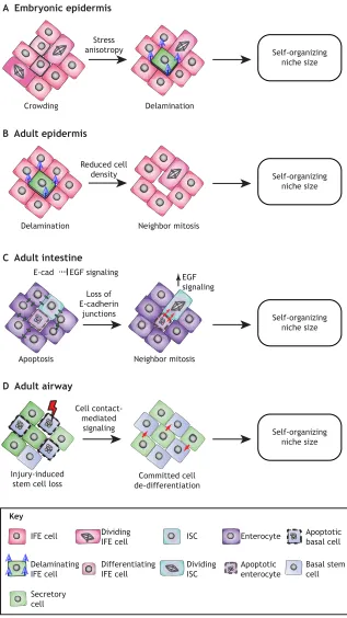
Mechanical cues modulate stem cell dynamics and organization
Consistent with the idea of niche space being the rate-limiting step for stem cell differentiation, it was recently shown that cell density and lateral crowding play crucial roles in maintaining tissue homeostasis by regulating stem cell fate decisions at the single-cell level in the embryonic IFE (Miroshnikova et al., 2018). The basal layer of the actively stratifying embryonic IFE exists in a mechanically jammed, solid-like state. Proliferation in this jammed
cell layer causes crowding and local cell stress anisotropy, which trigger differentiation and, at the same time, induce mechanical changes in these differentiating cells. Specifically, it has been shown that a differentiation-associated reduction in cell cortical tension and increased cell-cell adhesion subsequently trigger delamination of the differentiating cells from the basal cell layer (Miroshnikova et al., 2018), providing a robust self-organizing mechanism for embryonic epidermal stratification (Fig. 3A). Live imaging of the adult homeostatic IFE has also shown that the space
Crowding Delamination
Stress anisotropy A Embryonic epidermis
Self-organizing niche size
Delamination Neighbor mitosis
Reduced cell density B Adult epidermis
Self-organizing niche size
Apoptosis Neighbor mitosis
Loss of E-cadherin
junctions C Adult intestine
E-cad EGF signaling
EGF signaling
Self-organizing niche size
Injury-induced stem cell loss
Committed cell de-differentiation Cell
contact-mediated signaling D Adult airway
Self-organizing niche size
Key
IFE cell
Delaminating IFE cell
Dividing ISC
ISC Enterocyte
Apoptotic enterocyte
Basal stem cell Apoptotic basal cell
Secretory cell
Dividing IFE cell
[image:7.612.48.365.176.740.2]Differentiating IFE cell
Fig. 3. Coordination of cell fate controls niche size.
Epithelial stem cell niches sense changes in cell density that trigger fate changes in neighbors, generating self-organizing feedback loops to control niche size. (A) In the actively stratifying embryonic epidermis, cell divisions in the expanding epidermis lead to crowding and subsequent stress anisotropy within the cell layer. This triggers differentiation and subsequent delamination. (B) In the homeostatic adult epidermis, cell loss in the terminally differentiated cell layers triggers differentiation and delamination in the basal layer, liberating space. This alters basal cell size of the neighbors of the delaminating cell, triggering cell division of the largest cell to restore homeostatic cell density. (C) In theD. melanogaster
intestine, apoptosis signals neighboring stem cells to divide (red arrows) through loss of cell-cell contacts mediated by E-cadherin. This increases EGF signaling in stem cells to restore cell density. (D) In the lung proximal airway epithelium, basal stem cell loss through injury (red lightning) triggers secretory cells to dedifferentiate (red arrows) into functional basal stem cells through a cell contact-dependent signal. EGF, epidermal growth factor; IFE, interfollicular epidermis; ISC, intestinal stem cell.
DEVEL
O
liberated by delamination of a epidermal cell triggers the division of its neighboring cell (Mesa et al., 2017 preprint), which is consistent with the idea that basal cell density affects stem cell fate decisions in the epidermal layer. Interestingly, whereas in the actively stratifying embryonic epidermis cell divisions trigger crowding, in the homeostatic adult tissue cell divisions do not impact delamination but divisions occur in response to delamination (Fig. 3B). This could indicate that delamination and stem cell divisions are mechanically coupled in both the embryonic and adult epidermis, but that stem cells in the embryo are constantly cycling to provide sufficient material for the growing tissue, whereas adult stem cells divide only upon demand to replace terminally differentiated dying cells. A similar mechanism of cell density-driven homeostasis seems to operate in the Drosophila midgut (Fig. 3C), where stem cell division is triggered by removal of apoptotic cells in a process dependent on cell-cell contacts and their ability to coordinate EGF signaling (Liang et al., 2017). Another recent study in the adult Drosophila midgut further highlights the importance of mechanical signals, in particular mechanical stress in epithelial stem cell homeostasis. Here, a specific population of enteroendocrine precursor cells senses mechanical stimuli that regulate their differentiation in a process mediated by intracellular calcium increase through the stretch-activated calcium channel Piezo, allowing these cells to respond to gut compression or extension (He et al., 2018). Similarly, mechanical tension in response to injury has been shown to promote alveolar regeneration in the lung (Liu et al., 2016. Fig. 3D).
Mechanical signals are involved not only in determining the fate of individual cells, but also in establishing and positioning stem cell niches themselves. In the developing chick gut, buckling forces fold the epithelium, causing local tissue folds with high SHH concentrations. These high SHH microenvironments alter adjacent mesenchymal cell fate to form local signaling centers that signal back to the epithelium to establish intestinal crypts as stem cell niches (Shyer et al., 2015). Similarly, force-driven structural rearrangements are not only crucial for creating the hair follicle shape but also trigger lineage commitment in the developing avian skin. Here, spontaneous dermal cell aggregation and contractility compress the epidermis focally, leading to the mechanosensitive activation ofβ-catenin and subsequent initiation of the hair follicle-specific transcriptional program (Shyer et al., 2017). Early studies of follicle patterning hypothesized that a molecular pre-pattern emerges first and changes cellular behaviors, which then cause changes in tissue structure (Noramly and Morgan, 1998; Widelitz and Chuong, 1999). However, the study by Shyer and co-workers now indicates that initial follicle fate markers, such as β-catenin, are turned on simultaneously with architectural changes, instead of preceding them, and that the developing skin is able to form spaced aggregates withoutβ-catenin activation, suggesting that pre-patterning does not occur (Shyer et al., 2017). Collectively, these studies highlight that the mechanical regulation of stem cell positioning, density, contractility and compression within the niche provide key instructive signals that contribute to providing robust, self-organizing principles that control stem cell functions (Fig. 3).
Conclusions and perspectives
Recent developments in the fields of single cell sequencing, high resolution imaging and bioengineering have made it possible to make significant leaps in our understanding of adult stem cell biology. It has become evident that the stem cell niche provides a spectrum of cues that ensure plasticity of the stem cell compartment
and prevent stem cell loss. Furthermore, the precise location of stem cells within their niche provides additional fine-tuning of transcriptional programs, thereby facilitating stem cell heterogeneity, which is important for regulating the appropriate responses to the rapidly changing needs of the tissue. A key emerging paradigm is the presence of self-organizing feedback loops that are based on direct cell-cell contact and density sensing. These self-organizing circuits allow coordinated behaviors of stem cells within a niche. Coupling stem cell loss to neighbor division, or vice versa, provides simple and efficient control of niche size and tissue architecture, and could represent a universal feature of stem cell regulation, at least in epithelia.
Despite these recent advances, several key questions remain unanswered regarding the precise molecular mechanisms of stem cell heterogeneity and plasticity. In particular, it will be important to understand which regulatory mechanisms and phenomena are relevant for homeostasis and which of them are used primarily or even exclusively in response to stress or damage. In addition, more comprehensive characterization of stem cell niches is still required, as the roles of inflammatory cells, neuronal cells and other niche-proximal cell types are only beginning to emerge. Moreover, many tissues show a decline in regenerative potential during aging coupled with a loss of stem cell homeostasis and function (López-Otín et al., 2013; Signer and Morrison, 2013); in fact, a direct mechanistic link between organismal lifespan and stem cell activity has been demonstrated, pioneered by work in Drosophila ISCs (Biteau et al., 2010). Thus, understanding how aging affects the composition and function of stem cell niches and whether niche-targeted therapies could be used to enhance tissue homeostasis and regeneration potential during aging will be exciting avenues of future research.
Our increasing understanding of the regulatory networks and environmental factors dictating stem cell functions has already enabled the design ofin vitrostem cell models, such as organoids (Latil et al., 2017; Nichane et al., 2017; Sato et al., 2009; Yin et al., 2014) and other self-organizing stem cell cultures (Chacón-Martínez et al., 2017, Harrison et al., 2017; Tewary et al., 2017; Turner et al., 2017). These in vitro tools enable mechanistic interrogation of key components of niches and their contribution (chemical, mechanical and physical) to self-renewal, plasticity and differentiation, as well as precise manipulation of both stem cells and niche components through new technologies such as gene editing. These models are rapidly advancing basic and applied research (Huch et al., 2017), and are also being expanded to disease models and predictive tools of drug-treatment outcomes, as exemplified by the recently established human cancer organoid biobanks (Sachs et al., 2018; Vlachogiannis et al., 2018). The better we understand the complexity of niches and how they regulate stem cell plasticity and self-organization, the better we will be able to harness the potential of stem cells for use in regenerative therapies and personalized medicine.
Acknowledgements
We thank members of the Wickström lab for discussions and critical reading of the manuscript.
Competing interests
The authors declare no competing or financial interests.
Funding
Research in the Wickström lab is supported by the Helsingin Yliopisto, the Jenny ja Antti Wihurin Rahasto, the Jane ja Aatos Erkon säätiö, the Max-Planck-Gesellschaft and the Deutsche Forschungsgemeinschaft through SFB 829 A11.
DEVEL
O
References
Ali, N., Zirak, B., Rodriguez, R. S., Pauli, M. L., Truong, H. A., Lai, K., Ahn, R., Corbin, K., Lowe, M. M., Scharschmidt, T. C. et al.(2017). Regulatory T cells in skin facilitate epithelial stem cell differentiation.Cell169, 1119-1129.e1111. Arpaia, N., Green, J. A., Moltedo, B., Arvey, A., Hemmers, S., Yuan, S., Treuting,
P. M. and Rudensky, A. Y.(2015). A distinct function of regulatory T cells in tissue protection.Cell162, 1078-1089.
Barker, N., van Es, J. H., Kuipers, J., Kujala, P., van den Born, M., Cozijnsen, M., Haegebarth, A., Korving, J., Begthel, H., Peters, P. J. et al. (2007). Identification of stem cells in small intestine and colon by marker gene Lgr5.
Nature449, 1003-1007.
Barrandon, Y. and Green, H.(1987). Three clonal types of keratinocyte with different capacities for multiplication.Proc. Natl. Acad. Sci. USA84, 2302-2306. Becker, A. J., McCulloch, E. A. and Till, J. E.(1963). Cytological demonstration of the clonal nature of spleen colonies derived from transplanted mouse marrow cells.Nature197, 452-454.
Bettelli, E., Carrier, Y., Gao, W., Korn, T., Strom, T. B., Oukka, M., Weiner, H. L. and Kuchroo, V. K. (2006). Reciprocal developmental pathways for the generation of pathogenic effector TH17 and regulatory T cells. Nature 441, 235-238.
Biteau, B., Karpac, J., Supoyo, S., Degennaro, M., Lehmann, R. and Jasper, H. (2010). Lifespan extension by preserving proliferative homeostasis in Drosophila.
PLoS Genet.6, e1001159.
Blanpain, C. and Fuchs, E.(2014). Stem cell plasticity. Plasticity of epithelial stem cells in tissue regeneration.Science344, 1242281.
Blanpain, C., Lowry, W. E., Geoghegan, A., Polak, L. and Fuchs, E.(2004). Self-renewal, multipotency, and the existence of two cell populations within an epithelial stem cell niche.Cell118, 635-648.
Buczacki, S. J. A., Zecchini, H. I., Nicholson, A. M., Russell, R., Vermeulen, L., Kemp, R. and Winton, D. J.(2013). Intestinal label-retaining cells are secretory precursors expressing Lgr5.Nature495, 65-69.
Chacón-Martı́nez, C. A., Klose, M., Niemann, C., Glauche, I. and Wickström, S. A.(2017). Hair follicle stem cell cultures reveal self-organizing plasticity of stem cells and their progeny.EMBO J.36, 151-164.
Chen, F. and Fine, A.(2016). Stem cells in lung injury and repair.Am. J. Pathol.186, 2544-2550.
Chi, W., Wu, E. and Morgan, B. A.(2013). Dermal papilla cell number specifies hair size, shape and cycling and its reduction causes follicular decline.Development 140, 1676-1683.
Claudinot, S., Nicolas, M., Oshima, H., Rochat, A. and Barrandon, Y.(2005). Long-term renewal of hair follicles from clonogenic multipotent stem cells.Proc. Natl. Acad. Sci. USA102, 14677-14682.
Clayton, E., Doupé, D. P., Klein, A. M., Winton, D. J., Simons, B. D. and Jones, P. H.(2007). A single type of progenitor cell maintains normal epidermis.Nature 446, 185-189.
Cowan, C. R. and Hyman, A. A.(2004). Asymmetric cell division in C. elegans: cortical polarity and spindle positioning.Annu. Rev. Cell Dev. Biol.20, 427-453. Crosnier, C., Stamataki, D. and Lewis, J.(2006). Organizing cell renewal in the intestine: stem cells, signals and combinatorial control. Nat. Rev. Genet.7, 349-359.
Durand, A., Donahue, B., Peignon, G., Letourneur, F., Cagnard, N., Slomianny, C., Perret, C., Shroyer, N. F. and Romagnolo, B.(2012). Functional intestinal stem cells after Paneth cell ablation induced by the loss of transcription factor Math1 (Atoh1).Proc. Natl. Acad. Sci. USA109, 8965-8970.
Farin, H. F., van Es, J. H. and Clevers, H.(2012). Redundant sources of Wnt regulate intestinal stem cells and promote formation of Paneth cells.
Gastroenterology143, 1518-1529.e1517.
Fre, S., Huyghe, M., Mourikis, P., Robine, S., Louvard, D. and Artavanis-Tsakonas, S.(2005). Notch signals control the fate of immature progenitor cells in the intestine.Nature435, 964-968.
Fujisaki, J., Wu, J., Carlson, A. L., Silberstein, L., Putheti, P., Larocca, R., Gao, W., Saito, T. I., Lo Celso, C., Tsuyuzaki, H. et al.(2011). In vivo imaging of Treg cells providing immune privilege to the haematopoietic stem-cell niche.Nature 474, 216-219.
Füllgrabe, A., Joost, S., Are, A., Jacob, T., Sivan, U., Haegebarth, A., Linnarsson, S., Simons, B. D., Clevers, H., Toftgård, R. et al. (2015). Dynamics of Lgr6(+) progenitor cells in the hair follicle, sebaceous gland, and interfollicular epidermis.Stem Cell Rep.5, 843-855.
Ganz, T.(2000). Paneth cells–guardians of the gut cell hatchery.Nat. Immunol.1, 99-100.
Gomez, C., Chua, W., Miremadi, A., Quist, S., Headon, D. J. and Watt, F. M. (2013). The interfollicular epidermis of adult mouse tail comprises two distinct cell lineages that are differentially regulated by Wnt, Edaradd, and Lrig1.Stem Cell Rep.1, 19-27.
Gonzales, K. A. U. and Fuchs, E.(2017). Skin and its regenerative powers: an alliance between stem cells and their niche.Dev. Cell43, 387-401.
Goodell, M. A., Nguyen, H. and Shroyer, N. (2015). Somatic stem cell heterogeneity: diversity in the blood, skin and intestinal stem cell compartments.Nat. Rev. Mol. Cell Biol.16, 299-309.
Harrison, S. E., Sozen, B., Christodoulou, N., Kyprianou, C. and Zernicka-Goetz, M.(2017). Assembly of embryonic and extraembryonic stem cells to mimic embryogenesis in vitro.Science356, eaal1810.
He, L., Si, G., Huang, J., Samuel, A. D. T. and Perrimon, N.(2018). Mechanical regulation of stem-cell differentiation by the stretch-activated Piezo channel.
Nature555, 103-106.
Hertzog, A. J.(1937). The Paneth cell.Am. J. Pathol.13, 351-360.
Ho, T. T., Warr, M. R., Adelman, E. R., Lansinger, O. M., Flach, J., Verovskaya, E. V., Figueroa, M. E. and Passegué, E.(2017). Autophagy maintains the metabolism and function of young and old stem cells.Nature543, 205-210. Hoeck, J. D., Biehs, B., Kurtova, A. V., Kljavin, N. M., de Sousa e Melo, F., Alicke,
B., Koeppen, H., Modrusan, Z., Piskol, R. and de Sauvage, F. J.(2017). Stem cell plasticity enables hair regeneration following Lgr5(+) cell loss.Nat. Cell Biol. 19, 666-676.
Hsu, Y.-C., Pasolli, H. A. and Fuchs, E.(2011). Dynamics between stem cells, niche, and progeny in the hair follicle.Cell144, 92-105.
Hsu, Y.-C., Li, L. and Fuchs, E.(2014). Transit-amplifying cells orchestrate stem cell activity and tissue regeneration.Cell157, 935-949.
Huch, M., Knoblich, J. A., Lutolf, M. P. and Martinez-Arias, A.(2017). The hope and the hype of organoid research.Development144, 938-941.
Ito, M., Liu, Y., Yang, Z., Nguyen, J., Liang, F., Morris, R. J. and Cotsarelis, G. (2005). Stem cells in the hair follicle bulge contribute to wound repair but not to homeostasis of the epidermis.Nat. Med.11, 1351-1354.
Joost, S., Zeisel, A., Jacob, T., Sun, X., La Manno, G., Lonnerberg, P., Linnarsson, S. and Kasper, M.(2016). Single-cell transcriptomics reveals that differentiation and spatial signatures shape epidermal and hair follicle heterogeneity.Cell Syst.3, 221-237.e229.
Kabiri, Z., Greicius, G., Madan, B., Biechele, S., Zhong, Z., Zaribafzadeh, H., Edison, Aliyev, J., Wu, Y., Bunte, R. et al.(2014). Stroma provides an intestinal stem cell niche in the absence of epithelial Wnts.Development141, 2206-2215. Khacho, M., Clark, A., Svoboda, D. S., Azzi, J., MacLaurin, J. G., Meghaizel, C., Sesaki, H., Lagace, D. C., Germain, M., Harper, M.-E. et al. (2016). Mitochondrial dynamics impacts stem cell identity and fate decisions by regulating a nuclear transcriptional program.Cell Stem Cell19, 232-247. Kim, T.-H., Escudero, S. and Shivdasani, R. A.(2012). Intact function of Lgr5
receptor-expressing intestinal stem cells in the absence of Paneth cells.Proc. Natl. Acad. Sci. USA109, 3932-3937.
Krieger, T. and Simons, B. D. (2015). Dynamic stem cell heterogeneity.
Development142, 1396-1406.
Latil, M., Nassar, D., Beck, B., Boumahdi, S., Wang, L., Brisebarre, A., Dubois, C., Nkusi, E., Lenglez, S., Checinska, A. et al.(2017). Cell-type-specific chromatin states differentially prime squamous cell carcinoma tumor-initiating cells for epithelial to mesenchymal transition.Cell Stem Cell20, 191-204.e195. Lee, J.-H., Bhang, D. H., Beede, A., Huang, T. L., Stripp, B. R., Bloch, K. D.,
Wagers, A. J., Tseng, Y.-H., Ryeom, S. and Kim, C. F.(2014). Lung stem cell differentiation in mice directed by endothelial cells via a BMP4-NFATc1-thrombospondin-1 axis.Cell156, 440-455.
Lee, J. H., Tammela, T., Hofree, M., Choi, J., Marjanovic, N. D., Han, S., Canner, D., Wu, K., Paschini, M., Bhang, D. H. et al. (2017). Anatomically and functionally distinct lung mesenchymal populations marked by Lgr5 and Lgr6.Cell 170, 1149-1163.e1112.
Leeman, K. T., Fillmore, C. M. and Kim, C. F.(2014). Lung stem and progenitor cells in tissue homeostasis and disease.Curr. Top. Dev. Biol.107, 207-233. Lehmann, R.(2012). Germline stem cells: origin and destiny.Cell Stem Cell10,
729-739.
Leushacke, M., Ng, A., Galle, J., Loeffler, M. and Barker, N.(2013). Lgr5(+) gastric stem cells divide symmetrically to effect epithelial homeostasis in the pylorus.Cell Rep.5, 349-356.
Leushacke, M., Barker, N. and Pin, C.(2016). Quantifying Lgr5-positive stem cell behaviour in the pyloric epithelium.Sci. Rep.6, 21923.
Leushacke, M., Tan, S. H., Wong, A., Swathi, Y., Hajamohideen, A., Tan, L. T., Goh, J., Wong, E., Denil, S. L. I. J., Murakami, K. et al.(2017). Lgr5-expressing chief cells drive epithelial regeneration and cancer in the oxyntic stomach.Nat. Cell Biol.19, 774-786.
Li, N., Yousefi, M., Nakauka-Ddamba, A., Jain, R., Tobias, J., Epstein, J. A., Jensen, S. T. and Lengner, C. J.(2014). Single-cell analysis of proxy reporter allele-marked epithelial cells establishes intestinal stem cell hierarchy.Stem Cell Rep.3, 876-891.
Liang, J., Balachandra, S., Ngo, S. and O’Brien, L. E.(2017). Feedback regulation of steady-state epithelial turnover and organ size.Nature548, 588-591. Lim, X., Tan, S. H., Koh, W. L. C., Chau, R. M. W., Yan, K. S., Kuo, C. J., van
Amerongen, R., Klein, A. M. and Nusse, R.(2013). Interfollicular epidermal stem cells self-renew via autocrine Wnt signaling.Science342, 1226-1230. Liu, Z., Wu, H., Jiang, K., Wang, Y., Zhang, W., Chu, Q., Li, J., Huang, H., Cai, T.,
Ji, H. et al.(2016). MAPK-mediated YAP activation controls mechanical-tension-induced pulmonary alveolar regeneration.Cell Rep.16, 1810-1819.
Lopez-Garcia, C., Klein, A. M., Simons, B. D. and Winton, D. J.(2010). Intestinal stem cell replacement follows a pattern of neutral drift.Science330, 822-825. López-Otı́n, C., Blasco, M. A., Partridge, L., Serrano, M. and Kroemer, G.(2013).
The hallmarks of aging.Cell153, 1194-1217.
DEVEL
O
Mascré, G., Dekoninck, S., Drogat, B., Youssef, K. K., Brohée, S., Sotiropoulou, P. A., Simons, B. D. and Blanpain, C.(2012). Distinct contribution of stem and progenitor cells to epidermal maintenance.Nature489, 257-262.
Mesa, K. R., Kawaguchi, K., Gonzalez, D. G., Cockburn, K., Boucher, J., Xin, T., Klein, A. M. and Greco, V. (2017). Epidermal stem cells self-renew upon neighboring differentiation.bioRxivdoi:10.1101/155408.
Miroshnikova, Y. A., Le, H. Q., Schneider, D., Thalheim, T., Rübsam, M., Bremicker, N., Polleux, J., Kamprad, N., Tarantola, M., Wang, I. et al.(2018). Adhesion forces and cortical tension couple cell proliferation and differentiation to drive epidermal stratification.Nat. Cell Biol.20, 69-80.
Montgomery, R. K., Carlone, D. L., Richmond, C. A., Farilla, L., Kranendonk, M. E. G., Henderson, D. E., Baffour-Awuah, N. Y., Ambruzs, D. M., Fogli, L. K., Algra, S. et al. (2011). Mouse telomerase reverse transcriptase (mTert) expression marks slowly cycling intestinal stem cells. Proc. Natl. Acad. Sci. USA108, 179-184.
Morgner, J., Ghatak, S., Jakobi, T., Dieterich, C., Aumailley, M. and Wickström, S. A.(2015). Integrin-linked kinase regulates the niche of quiescent epidermal stem cells.Nat. Commun.6, 8198.
Morrison, S. J. and Spradling, A. C.(2008). Stem cells and niches: mechanisms that promote stem cell maintenance throughout life.Cell132, 598-611. Müller-Röver, S., Handjiski, B., van der Veen, C., Eichmüller, S., Foitzik, K.,
McKay, I. A., Stenn, K. S. and Paus, R.(2001). A comprehensive guide for the accurate classification of murine hair follicles in distinct hair cycle stages.J. Invest. Dermatol.117, 3-15.
Muñoz, J., Stange, D. E., Schepers, A. G., van de Wetering, M., Koo, B.-K., Itzkovitz, S., Volckmann, R., Kung, K. S., Koster, J., Radulescu, S. et al. (2012). The Lgr5 intestinal stem cell signature: robust expression of proposed quiescent‘+4’cell markers.EMBO J.31, 3079-3091.
Nabhan, A. N., Brownfield, D. G., Harbury, P. B., Krasnow, M. A. and Desai, T. J. (2018). Single-cell Wnt signaling niches maintain stemness of alveolar type 2 cells.Science359, 1118-1123.
Nichane, M., Javed, A., Sivakamasundari, V., Ganesan, M., Ang, L. T., Kraus, P., Lufkin, T., Loh, K. M. and Lim, B. (2017). Isolation and 3D expansion of multipotent Sox9(+) mouse lung progenitors.Nat. Methods14, 1205-1212. Noramly, S. and Morgan, B. A. (1998). BMPs mediate lateral inhibition at
successive stages in feather tract development.Development125, 3775-3787. Nosbaum, A., Prevel, N., Truong, H.-A., Mehta, P., Ettinger, M., Scharschmidt,
T. C., Ali, N. H., Pauli, M. L., Abbas, A. K. and Rosenblum, M. D.(2016). Cutting edge: regulatory T cells facilitate cutaneous wound healing.J. Immunol.196, 2010-2014.
Oshima, H., Rochat, A., Kedzia, C., Kobayashi, K. and Barrandon, Y.(2001). Morphogenesis and renewal of hair follicles from adult multipotent stem cells.Cell 104, 233-245.
Pardo-Saganta, A., Law, B. M., Tata, P. R., Villoria, J., Saez, B., Mou, H., Zhao, R. and Rajagopal, J. (2015a). Injury induces direct lineage segregation of functionally distinct airway basal stem/progenitor cell subpopulations.Cell Stem Cell16, 184-197.
Pardo-Saganta, A., Tata, P. R., Law, B. M., Saez, B., Chow, R. D.-W., Prabhu, M., Gridley, T. and Rajagopal, J.(2015b). Parent stem cells can serve as niches for their daughter cells.Nature523, 597-601.
Plikus, M. V., Mayer, J. A., de la Cruz, D., Baker, R. E., Maini, P. K., Maxson, R. and Chuong, C.-M.(2008). Cyclic dermal BMP signalling regulates stem cell activation during hair regeneration.Nature451, 340-344.
Potten, C. S. and Morris, R. J.(1988). Epithelial stem cells in vivo.J. Cell Sci.1988 Suppl. 10, 45-62.
Potten, C. S., Saffhill, R. and Maibach, H. I.(1987). Measurement of the transit time for cells through the epidermis and stratum corneum of the mouse and guinea-pig.Cell Tissue Kinet.20, 461-472.
Reynolds, A. J. and Jahoda, C. A.(1991). Hair follicle stem cells? A distinct germinative epidermal cell population is activated in vitro by the presence of hair dermal papilla cells.J. Cell Sci.99, 373-385.
Ritsma, L., Ellenbroek, S. I. J., Zomer, A., Snippert, H. J., de Sauvage, F. J., Simons, B. D., Clevers, H. and van Rheenen, J. (2014). Intestinal crypt homeostasis revealed at single-stem-cell level by in vivo live imaging.Nature507, 362-365.
Roberts, K. J., Kershner, A. M. and Beachy, P. A.(2017). The stromal niche for epithelial stem cells: a template for regeneration and a brake on malignancy.
Cancer Cell32, 404-410.
Rock, J. R. and Hogan, B. L. M. (2011). Epithelial progenitor cells in lung development, maintenance, repair, and disease.Annu. Rev. Cell Dev. Biol.27, 493-512.
Rock, J. R., Onaitis, M. W., Rawlins, E. L., Lu, Y., Clark, C. P., Xue, Y., Randell, S. H. and Hogan, B. L. M.(2009). Basal cells as stem cells of the mouse trachea and human airway epithelium.Proc. Natl. Acad. Sci. USA106, 12771-12775. Rodrı́guez-Colman, M. J., Schewe, M., Meerlo, M., Stigter, E., Gerrits, J.,
Pras-Raves, M., Sacchetti, A., Hornsveld, M., Oost, K. C., Snippert, H. J. et al. (2017). Interplay between metabolic identities in the intestinal crypt supports stem cell function.Nature543, 424-427.
Rompolas, P., Mesa, K. R. and Greco, V.(2013). Spatial organization within a niche as a determinant of stem-cell fate.Nature502, 513-518.
Rompolas, P., Mesa, K. R., Kawaguchi, K., Park, S., Gonzalez, D., Brown, S., Boucher, J., Klein, A. M. and Greco, V.(2016). Spatiotemporal coordination of stem cell commitment during epidermal homeostasis.Science352, 1471-1474. Ruiz, E. J., Oeztuerk-Winder, F. and Ventura, J.-J.(2014). A paracrine network
regulates the cross-talk between human lung stem cells and the stroma.Nat. Commun.5, 3175.
Sachs, N., de Ligt, J., Kopper, O., Gogola, E., Bounova, G., Weeber, F., Balgobind, A. V., Wind, K., Gracanin, A., Begthel, H. et al.(2018). A living biobank of breast cancer organoids captures disease heterogeneity.Cell172, 373-386.e310.
Sada, A., Jacob, F., Leung, E., Wang, S., White, B. S., Shalloway, D. and Tumbar, T.(2016). Defining the cellular lineage hierarchy in the interfollicular epidermis of adult skin.Nat. Cell Biol.18, 619-631.
San Roman, A. K., Jayewickreme, C. D., Murtaugh, L. C. and Shivdasani, R. A. (2014). Wnt secretion from epithelial cells and subepithelial myofibroblasts is not required in the mouse intestinal stem cell niche in vivo.Stem Cell Rep.2, 127-134. Sánchez Alvarado, A. and Yamanaka, S.(2014). Rethinking differentiation: stem
cells, regeneration, and plasticity.Cell157, 110-119.
Sato, T., Vries, R. G., Snippert, H. J., van de Wetering, M., Barker, N., Stange, D. E., van Es, J. H., Abo, A., Kujala, P., Peters, P. J. et al.(2009). Single Lgr5 stem cells build crypt-villus structures in vitro without a mesenchymal niche.
Nature459, 262-265.
Sato, T., van Es, J. H., Snippert, H. J., Stange, D. E., Vries, R. G., van den Born, M., Barker, N., Shroyer, N. F., van de Wetering, M. and Clevers, H.(2011). Paneth cells constitute the niche for Lgr5 stem cells in intestinal crypts.Nature 469, 415-418.
Scadden, D. T.(2014). Nice neighborhood: emerging concepts of the stem cell niche.Cell157, 41-50.
Schofield, R.(1978). The relationship between the spleen colony-forming cell and the haemopoietic stem cell.Blood Cells4, 7-25.
Sennett, R. and Rendl, M.(2012). Mesenchymal-epithelial interactions during hair follicle morphogenesis and cycling.Semin. Cell Dev. Biol.23, 917-927. Shyer, A. E., Huycke, T. R., Lee, C. H., Mahadevan, L. and Tabin, C. J.(2015).
Bending gradients: how the intestinal stem cell gets its home.Cell161, 569-580. Shyer, A. E., Rodrigues, A. R., Schroeder, G. G., Kassianidou, E., Kumar, S. and Harland, R. M. (2017). Emergent cellular self-organization and mechanosensation initiate follicle pattern in the avian skin.Science357, 811-815. Signer, R. A. J. and Morrison, S. J.(2013). Mechanisms that regulate stem cell
aging and life span.Cell Stem Cell12, 152-165.
Snippert, H. J., Haegebarth, A., Kasper, M., Jaks, V., van Es, J. H., Barker, N., van de Wetering, M., van den Born, M., Begthel, H., Vries, R. G. et al.(2010a). Lgr6 marks stem cells in the hair follicle that generate all cell lineages of the skin.
Science327, 1385-1389.
Snippert, H. J., van der Flier, L. G., Sato, T., van Es, J. H., van den Born, M., Kroon-Veenboer, C., Barker, N., Klein, A. M., van Rheenen, J., Simons, B. D. et al.(2010b). Intestinal crypt homeostasis results from neutral competition between symmetrically dividing Lgr5 stem cells.Cell143, 134-144.
Stange, D. E., Koo, B.-K., Huch, M., Sibbel, G., Basak, O., Lyubimova, A., Kujala, P., Bartfeld, S., Koster, J., Geahlen, J. H. et al.(2013). Differentiated Troy+ chief cells act as reserve stem cells to generate all lineages of the stomach epithelium.Cell155, 357-368.
Stanger, B. Z., Datar, R., Murtaugh, L. C. and Melton, D. A.(2005). Direct regulation of intestinal fate by Notch. Proc. Natl. Acad. Sci. USA 102, 12443-12448.
Sun, J., Ramos, A., Chapman, B., Johnnidis, J. B., Le, L., Ho, Y.-J., Klein, A., Hofmann, O. and Camargo, F. D. (2014). Clonal dynamics of native haematopoiesis.Nature514, 322-327.
Tadokoro, T., Gao, X., Hong, C. C., Hotten, D. and Hogan, B. L. M.(2016). BMP signaling and cellular dynamics during regeneration of airway epithelium from basal progenitors.Development143, 764-773.
Takeda, N., Jain, R., LeBoeuf, M. R., Wang, Q., Lu, M. M. and Epstein, J. A. (2011). Interconversion between intestinal stem cell populations in distinct niches.
Science334, 1420-1424.
Tan, F. E., Vladar, E. K., Ma, L., Fuentealba, L. C., Hoh, R., Espinoza, F. H., Axelrod, J. D., Alvarez-Buylla, A., Stearns, T., Kintner, C. et al.(2013). Myb promotes centriole amplification and later steps of the multiciliogenesis program.
Development140, 4277-4286.
Tata, P. R. and Rajagopal, J.(2017). Plasticity in the lung: making and breaking cell identity.Development144, 755-766.
Tata, P. R., Mou, H., Pardo-Saganta, A., Zhao, R., Prabhu, M., Law, B. M., Vinarsky, V., Cho, J. L., Breton, S., Sahay, A. et al.(2013). Dedifferentiation of committed epithelial cells into stem cells in vivo.Nature503, 218-223. Tetteh, P. W., Basak, O., Farin, H. F., Wiebrands, K., Kretzschmar, K., Begthel,
H., van den Born, M., Korving, J., de Sauvage, F., van Es, J. H. et al.(2016). Replacement of lost Lgr5-positive stem cells through plasticity of their enterocyte-lineage daughters.Cell Stem Cell18, 203-213.
Tewary, M., Ostblom, J., Prochazka, L., Zulueta-Coarasa, T., Shakiba, N., Fernandez-Gonzalez, R. and Zandstra, P. W.(2017). A stepwise model of reaction-diffusion and positional information governs self-organized human peri-gastrulation-like patterning.Development144, 4298-4312.