BIROn - Birkbeck Institutional Research Online
McDonald, C. and Jovanovic, G. and Wallace, Bonnie A. and Ces, O.
and Buck, M. (2017) Structure and function of PspA and Vipp1 N-terminal
peptides:
Insights into the membrane stress sensing and mitigation.
Biochimica et Biophysica Acta (BBA) - Biomembranes 1859 (1), pp. 28-39.
ISSN 0005-2736.
Downloaded from:
Usage Guidelines:
Please refer to usage guidelines at
or alternatively
Structure and function of PspA and Vipp1 N-terminal peptides: Insights
into the membrane stress sensing and mitigation
Christopher McDonald
a,b, Goran Jovanovic
a, B.A. Wallace
c, Oscar Ces
b, Martin Buck
a,⁎
aDepartment of Life Sciences, Imperial College London, London SW7 2AZ, UK
bDepartment of Chemistry and Institute of Chemical Biology, Imperial College London, London SW7 2AZ, UK
cInstitute of Structural and Molecular Biology, Birkbeck College, University of London, London, WC1E 7HX, UK
a b s t r a c t
a r t i c l e i n f o
Article history: Received 20 July 2016
Received in revised form 18 October 2016 Accepted 27 October 2016
Available online 30 October 2016
The phage shock protein (Psp) response maintains integrity of the inner membrane (IM) in response to extracytoplasmic stress conditions and is widely distributed amongst enterobacteria. Its central component PspA, a member of the IM30 peripheral membrane protein family, acts as a major effector of the system through its direct association with the IM. Under non-stress conditions PspA also negatively regulates its own expression via direct interaction with the AAA+ ATPase PspF. PspA has a counterpart in cyanobacteria called Vipp1, which is implicated in protection of the thylakoid membranes. PspA's and Vipp1's conserved N-terminal regions contain a putative amphipathic helix a (AHa) required for membrane binding. An adjacent amphipathic helix b (AHb) in PspA is required for imposing negative control upon PspF. Here, purified peptides derived from the putative AH regions of PspA and Vipp1 were used to directly probe their effector and regulatory functions. We observed direct membrane-binding of AHa derived peptides and an accompanying change in secondary structure from un-structured to alpha-helical establishing them asbonafidemembrane-sensing AH's. The peptide-binding specifi c-ities and their effects on membrane stability depend on membrane anionic lipid content and stored curvature elastic stress, in agreement with full length PspA and Vipp1 protein functionalities. AHb of PspA inhibited the ATPase activity of PspF demonstrating its direct regulatory role. Thesefindings provide new insight into the membrane binding and function of PspA and Vipp1 and establish that synthetic peptides can be used to probe the structure-function of the IM30 protein family.
© 2016 The Author(s). Published by Elsevier B.V. This is an open access article under the CC BY license (http:// creativecommons.org/licenses/by/4.0/).
Keywords: Anionic lipids
Stored curvature elastic stress Peripheral membrane protein Amphipathic helix conformation Membrane structure
1. Introduction
The cell envelope provides protection from the environment and gives structural integrity to the cell in all organisms. Homeostasis of the plasma membrane is vital for functioning of the cell and the bacterial phage shock protein (Psp) response protects the bacterial membrane under various extracytoplasmic stress conditions. Although many different stimuli trigger induction of the Psp response, the common theme is disruption of the plasma membrane and consequently loss of the (trans)-membrane potential and dissipation of the proton motive
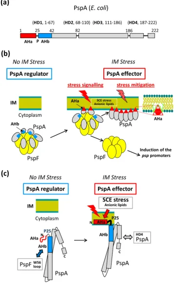
force (pmf)[1–4]. By an unknown mechanism, the Psp response rescues the proton gradient and conserves the pmf by protecting the plasma membrane integrity. The central component of the Psp system is the pe-ripheral plasma membrane binding protein PspA belonging to the IM30 family of proteins found in many organisms. In bacteria, the Psp re-sponse and PspA-like proteins are implicated in protein translocation, virulence and resistance to antimicrobials that target the cell wall or reorganise the membrane architecture[3,5–9].
In enterobacteria, PspA is a dual function protein responsible for both the negative regulation and effector function of the Psp response. Under non-stress conditions PspA directly interacts with the subunits of thepsptranscription activator, the hexameric bacterial enhancer binding protein PspF, imparting negative regulation by inhibiting PspF's ATPase activity and the subsequent sigma54-dependent transcription of
pspgenes[3,10]. PspA-PspF interactions occurviathe PspF W56 loop, a surface exposed hydrophobic region on each PspF subunit[11–13]. Under inner membrane (IM) stress conditions, PspF is released from the PspA-F inhibitory complex leading to induction of the Psp response. Stresses such as defects in protein translocation systems or mislocalisation of outer membrane secretins into the IM are sensed by the IM proteins PspB and PspC[13,14]resulting in a direct interactions
Abbreviations:Psp, Phage shock protein; pmf, proton motive force; IM, inner membrane; TLE, total lipid extract; PG, phosphatidylglycerol; PS, phosphatidylserine; SCE, stored curvature elastic; AH, amphipathic helix; ITC, isothermal titration calorimetry; CD, circular dichroism; SUV, small unilamellar vesicle; DOPC, 1,2-dioleoyl-sn-glycero-3-phosphocholine; DOPG, 1,2-dioleoyl-sn-glycero-3-phospho-(1′-rac-glycerol); LUV, large unilamellar vesicle; DMPC, 1,2-dimyristoyl-sn-glycero-3-phosphocholine; DOPE, glycero-3-phosphoethanolamine; DOPS, 1,2-dioleoyl-sn-glycero-3-phosphoserine; TFE, trifluoroethanol.
⁎ Corresponding author at: Department of Life Sciences, SAFB, Imperial College London, London, SW7 2AZ, UK.
E-mail address:[email protected](M. Buck).
http://dx.doi.org/10.1016/j.bbamem.2016.10.018
0005-2736/© 2016 The Author(s). Published by Elsevier B.V. This is an open access article under the CC BY license (http://creativecommons.org/licenses/by/4.0/).
Contents lists available atScienceDirect
Biochimica et Biophysica Acta
between PspB-C and the PspA subunits of the PspA-F inhibitory com-plex that release PspF. The PspB-C dependent signalling is condition-al and severe stresses such as extreme temperature, hyperosmotic or ethanol shock cause partial or complete PspB-C-independent induc-tion of the Psp response[1,3,4]. Following release of PspF, the PspA binds to the IM and forms high-order oligomeric (up to 36mer) effec-tors complexes[13,15,16]able to repair IM damage and conserve the pmf[17]but unable to stably interact with PspF to impose negative control[18].
Characterising PspA IM-binding is key to understanding the mecha-nism by which PspA repairs the membrane. PspA as a high-order oligo-mer binds to vesicles made fromE. colitotal lipid extracts (TLE) and vesicles containing the anionic lipids phosphatidylglycerol (PG) or phosphatidylserine (PS). PspA was also shown to prevent proton leak-age formE. coliTLE vesiclesin vitro[17]. McDonald et al.[18]showed that both anionic lipids and accumulation of the membrane Stored Cur-vature Elastic (SCE) stress from type II lipids drive vesicle association of PspA.Psp-inducing extracytoplasmic stress stimuli may well lead to the accumulation of SCE stress and associated lipid packing defects within the IM which disrupt membrane integrity and so stimulate PspA bind-ing. Anionic lipids promote PspA binding to vesicles with low mem-brane SCE stress; the higher the memmem-brane SCE stress, the less anionic lipids contribute to membrane association[18]suggesting the SCE stress may be the primary signature of the damaged membrane to be repaired by PspA.
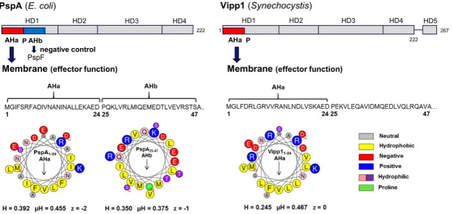
The N-terminal region of PspA consists of two putative amphi-pathic helixes (AHs) (seeFig. 1), AHb required for negative control, and AHa required for effector function[19]. The N-terminal AHa (ahA; residues 2–19) is responsible for IM binding and effector func-tionin vivo. The lack of AHa or amino acid substitutions in the hydro-phobic face of the helix abolishes PspA IM-binding and high-order oligomer formationin vivoandin vitro. The adjacent AHb (ahB; residues 25–42) is implicated in PspA negative control. The lack of AHb or amino acid substitution on the hydrophobic face of the helix abolishes PspA-PspF interaction and PspA negative control function but does not affect the IM-binding and effector function of PspA[19]. Notably, in the absence of PspF and lipids, PspA is able to
form high-order oligomersin vitro[10,20]suggesting interaction with PspFviaAHb is critical for preventing PspA to oligomerise. The conserved P25 helix-breaking residue separates AHa and AHb and is important for both the negative regulatory and effector func-tion of PspA and thus might establish a mutually exclusive use of two AHs[19]. A monomeric PspA fragment 1–144 (PspA1–144) is suf-ficient for PspF negative control, dependent upon residue E37 within the AHb. Importantly, in the crystal structure of PspA1–144the puta-tive AHa region is unordered[21]. A transition from unordered-to-ordered alpha-helical structure upon membrane association would establish the region as abonafideAH. If the PspA AHa is indeed a typ-ical membrane-sensing AH, a structural transition following binding to the IM may cause the switch in function of the PspA. Indeed, a con-formational change of PspA AHa upon IM interaction has been in-ferred from other functional and structural studies[19,21].
The PspA homologue Vipp1 which is implicated in thylakoid membrane biogenesis and protection in cyanobacteria, green algae and higher plants[22–25]also carries a putative N-terminal AHa (seeFig. 1). Notably, Vipp1 can substitute for PspA inE. coli[26,27] and the presence of its N-terminal region is required for binding to lipid vesicles and high-order oligomer formation[18,28]. Vipp1’s functional similarity to PspA for vesicle stress recognition is high, ex-cept that the role of anionic lipids in Vipp1-membrane binding is more pronounced[18]in accordance with suggested function in thy-lakoid membrane fusion[29].
[image:3.595.77.532.469.685.2]It appears that the interactions of the putative AH regions of PspA and Vipp1 are a critical aspect of the mechanisms by which the Psp re-sponse is regulated and IM or thylakoid membrane stress is ameliorat-ed. However, there is currently no experimental evidence to show the direct functionality of these regions or characterise their behaviour as typical AHs. In this work we used synthetic peptides based on PspA and Vipp1 N-terminal AHs and vesicles of well-defined size and lipid composition to determine a direct AHa-membrane interaction and inhi-bition of PspF ATPase activity by PspA AHb. The quantifications of AHa membrane-binding and structural transition as well as its effect on ves-icles stability offer novel insight to a possible mechanism of stress miti-gation by the PspA and related effector complexes.
2. Materials and methods
2.1. Peptides
All peptides used in this study were purchased from Insight Biotech-nology at≥95% specific peptide purity (confirmedviaHPLC and MS by the manufacturer). The peptides were supplied in powder form (includ-ing some counter ions and impurities) and the exact peptide content was determined via CHN analysis (Medac LTD) to enable preparation of peptide solutions with accurate concentrations. A table of the pep-tides used in this study detailing their amino acid sequences and purity is shown is Supplementary Table 2.
2.2. Preparation of lipid vesicles
Lipids were purchased from Avanti Polar Lipids (Alabastar, AL, USA). Chloroform stocks of lipids were mixed in glass vials and the chloroform was then evaporated under nitrogen. Then lipids were left in a freeze dryer overnight. Buffer was added to a 5 mMfinal phospholipid concen-tration and the suspension left above the TMof the lipids for 1 h. The sus-pension was then subject tofive freeze-thaw-vortex cycles. 100 nm vesicles were produced by extruding the suspension through polycar-bonatefilters of 100 nm pore sizes using a miniextruder (Avanti Polar Lipids). Vesicles for Isothermal Titration Calorimetry (ITC) experiments were prepared by probe sonication. All vesicles were used immediately or stored at 4 °C and used within 72 h. Vesicle sizes were characterized
viadynamic light scattering (DLS) to ensure consistency.
2.3. Dynamic light scattering (DLS)
DLS on vesicles was undertaken as previously published (17). For measurements in the presence of Vipp11–24, 100 nm extruded DOPC/ DOPG 6:4 vesicles (1 mM) were incubated with Vipp11–24(500μM) for 30 min before sizing.
2.4. Isothermal titration calorimetry (ITC)
Experiments were performed using a MicroCal iTC200 (Malvern In-struments Ltd). SUVs were prepared in PBS (pH 7.4). To obtain peptide binding curves, SUVs were injected into the sample cell (volume, 0.2 ml) containing peptides (in the same buffer). For experiments deter-mining the molar enthalpy of peptide binding, peptides were injected into the sample cell containing vesicles at 25 °C. The enthalpy of dilution was determined in control experiments by injecting either peptide or vesicle samples into buffer (PBS, pH 7.4) and subtracted from the corre-sponding enthalpies determined in peptide-lipid binding experiments. The results were analysed using Origin software. The experiments were performed in triplicate.
2.5. Circular dichroism (CD) spectroscopy
Peptides and SUVs were dissolved or suspended in 20 mM Tris-HCl (pH 7.4). Suprasil demountable cells (Hellma UK Ltd.) with pathlengths of 0.01 and 0.005 cm were used and three spectra were collected for each sample on an Aviv 62DS spectropolarimeter over the wavelength range from 190 to 260 nm, with a 1 s dwell time at 25 °C. The replicates were averaged, and the averaged baselines (consisting of the buffer with corresponding vesicles) were subtracted from the averaged sam-ple spectra. Data processing was carried out with CDtool software [30]. Data was analysed with the DichroWeb analysis server[31]; the re-ported values were the averaged results using the CONTINLL algorithm [32,33]and reference dataset 7 (chosen specifically as this reference data set includes unfolded proteins with a significant amount of unor-dered secondary structure). Trifluoroethanol (TFE) samples consisted of 300μM peptide dissolved in 2,2,2-trifluoroethanol (TFE).
2.6. Calcein leakage assay
Vesicles loaded with calcein were preparedviaextrusion of lipids hydrated in PBS pH 7.4 with 50 mM calcein (described above) followed by gelfiltration through a G50 Sephadex column. Vesicles were diluted to a 1 mM lipid concentration and calcein leakage was monitored by measuringfluorescence intensity at 520 nm (excitation at 485 nm) dur-ing peptide titrations usdur-ing a FLUOstar Omega microplate reader (BMG LABTECH GmbH). The experiments were undertaken in PBS pH 7.4 buff-er and at 25 °C. Aftbuff-er peptide titrations, vesicles wbuff-ere burstviaaddition of 0.2 M Octaethylene glycol monododecyl ether (C12E8) (Sigma) and fluorescence was measured to give a value for a complete dye release event. The percentage of calcein leakage was calculated according to the equation; % of calcein efflux = (Ft−F0)/(F∞−F0) × 100, where Ft was thefluorescence at time t, F0was thefluorescence at time t0, and F∞was the maximumfluorescence (100%) after the addition of C12E8.
2.7. Electron microscopy
Negative Stain Electron Microscopy was used to visualise the effect Vipp11–24peptide had on vesicles in a calcein dye leakage assay. 3μl samples were incubated on copper grids layered with thin carbon, washed with 3 drops of MilliQ water, and stained with 2% uranyl ace-tate. Images were taken on an FEI Tecnai T12 Spiritfitted with an FEI Eagle 2 K CCD.
2.8. ATPase activity
PspF1–275WT and PspF1–275W56A proteins were purified as de-scribed in Joly et al.[34]. The steady-state ATP hydrolysis activity of the PspF1–275proteins in the presence of peptides was measured using an NADH-coupled regeneration system[35]. ATPase activity was measured at 37 °C in afinal volume of 100μl containing: 25 mM Tris-HCL (pH 8.0), 100 mM KCl, 10 mM MgCl2, 1 mM DTT, 1 mM NADH, 10 mM phosphoenolpyruvate, 10 U ml−1pyruvate kinase, 20 U ml−1 lactate dehydrogenase, 50 mM ATP, PspF1–275(0–5 mM) and peptides (10–500μM). Absorbance at 340 nm was monitored every 60 s using a FLUOstar Omega microplate reader (BMG LABTECH GmbH).
3. Results
3.1. Peptide binding to small unilamellar vesicles (SUVs)
Synthetic peptides consisting of thefirst 24 residues ofE. coliPspA (PspA1–24) andSynechocystisVipp1 (Vipp11–24) were used to probe
the membrane binding properties of the putative AHa region (Fig. 1). Removal of residues 2–19 of PspA[19]and residues 1–21 of Vipp1 [28]had previously been shown to result in loss of the PspA and Vipp1 membrane binding functions. However, additional residues up to the helix-breaking P25 were included in this study as charged flanking regions have been shown to contribute to AHs membrane sens-ing[36]and should provide a better comparison of binding with that of the full length proteins[18].Fig. 1and SupplementaryFig. 1show heli-cal wheel projections, net hydrophobicity, hydrophobic moment and charge of the peptides used.
−0.4 Kcal mol−1(Supplementary Fig. 3a), yielding a reaction enthalpy of−6.2 (±0.28) Kcal mol−1. Injecting Vipp11
–24into DOPC SUVs (Sup-plementary Fig. 2c) resulted in a net molar enthalpy of−3.9 (±0.18) Kcal mol−1(with buffer control of 0.1 Kcal mol−1, Supplementary Fig. 3b). These values imply that both PspA1–24and Vipp11–24are able to associate with SUVs in an exothermic binding event.
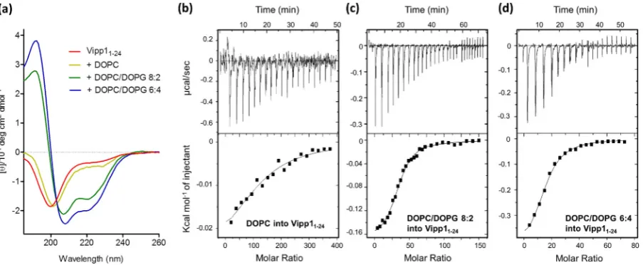
Circular dichroism (CD) spectroscopy was used to examine the sec-ondary structures of PspA1–24and Vipp11–24. Peptides tend to have low
spectral magnitudes as they adopt multiple conformations in equilibri-um rather than a single structure. The precise values of the calculated secondary structures determined using reference databases derived from globular soluble proteins should therefore be treated with a degree of caution, although the relative values provide valuable comparisons [37]. The CD spectra and calculated helical contents of PspA1–24and Vipp11–24(Figs. 2a,3a andTable 1) show that in the absence of phos-pholipids, both peptides were largely unstructured (~10% helical con-tent). When DOPC SUVs (14:1 lipid to peptide -L/P- ratio) were added to PspA1–24, a spectrum more reminiscent of a helical structure with
an increased 224 nm peak was observed (Fig. 2a) and the calculated α-helix content more than doubled. For Vipp11–24the spectral change was smaller (Fig. 3a) and corresponded to a ~4% increase in helix con-tent. Although the net increase in helix content is relatively small, the CD spectra result from an ensemble of the free and bound peptide struc-tures. Therefore the helical contents may represent either partially folded peptides in the bound state or folded bound peptides in the pres-ence of disordered unbound peptide. The peptide-lipid binding equilib-rium can be described byP+Lc=PLc, wherePis the peptide andLcis theNlipids associated with the peptide[37]. The % of membrane-bound peptide,PLc, in the CD experiments can therefore be calculated with knowledge ofNand the associated macroscopic binding constant
Kb[38]. To obtain these values, ITC titrations were used to derive bind-ing isotherms (Figs. 2b,3b and Supplementary Fig. 2b, d). PspA1–24 was found to haveN= 76 (±4) DOPC molecules associated per peptide and Vipp11–24hadN= 122 (± 17). Binding constants,Kb,of 9.1 × 10⁻⁴M−1and 9.2 × 10⁻⁴M−1for PspA1–24and Vipp11–24were obtain-ed, respectively. Using these parameters the amounts of membrane-bound peptide in the CD samples were calculated to be 17% for PspA1–
24and 10% for Vipp11–24. Thus, a large fraction of the peptides are not
membrane-associated. The helicities of the membrane-bound peptides were calculated to be 78% for PspA1–24and 42% for Vipp11–24.
Mem-brane association is clearly accompanied by significant α-helix
formation in both peptides, confirming their behaviour as typical of membrane-binding amphipathic helixes.
3.2. Impact of anionic lipids on binding
Both PspA and Vipp1 proteins have increased membrane-binding capabilities in the presence of anionic lipids[18,28,29]. To determine if the AHa peptides properties are similar, SUVs containing 8:2 and 6:4 (M:M) DOPC/DOPG were used in the CD spectroscopic and ITC studies (Fig. 2b-d). For PspA1–24, addition of anionic DOPG resulted in a change in the CD spectrum corresponding to a higher helical content compared with zwitterionic DOPC alone (Fig. 2a). DOPC/DOPG 8:2 SUVs (14:1 L/P) resulted in 34% calculated total helix content and DOPC/DOPG 6:4 SUVs (14:1 L/P) produced a further increase to 45% helicity (Table 1). ITC studies showed that this corresponded with a decrease in the number of lipid molecules per peptide binding site,N, to 39 for DOPC/DOPG 8:2 and 25 for DOPC/DOPG 6:4 (Fig. 2c, d). When this data was used to calculate the helical content of the PspA1–24membrane-bound frac-tion (as above), results very similar to those observed for DOPC SUVs were obtained (Table 1). Therefore, it appears that anionic lipids do not have a significant effect on the helicity of the membrane-bound PspA but increase the number of peptide binding sites per SUV.
In the case of Vipp11–24, addition of DOPG resulted in even more
pro-nounced differences in membrane binding. Calculated total helical con-tents of 39% and 50% were obtained with DOPC/DOPG 8:2 and DOPC/ DOPG 6:4 SUVs, respectively (Fig. 3a andTable 1). The number of lipid molecules per peptide binding site obtained from ITC studies (Fig. 3b-d an3b-dTable 1) dramatically reduce fromN = 122 toN = 35 for DOPC/DOPG 8:2 andN= 21 for and DOPC/DOPG 6:4. 91% of the resi-dues in membrane-bound Vipp11–24were calculated to be helical for DOPC/DOPG 8:2, more than double that of DOPC. A helical content of 77% for Vipp11–24bound to DOPC/DOPG 6:4 still indicated significantly more helical residues than in DOPC. DOPG therefore appears to promote further helix formation of membrane-bound Vipp11–24as well as in-creasing the number of available binding sites on the SUVs.
For both PspA1–24and Vipp11–24a two-fold increase inKbis also ob-served when 20% DOPG is present in the membrane (Table 1) showing that the peptides have a higher affinity for membranes containing the anionic lipid. The enthalpy of reaction for PspA1–24binding to DOPC/ DOPG 8:2 is only slightly more exothermic than to DOPC [−8.2 (±0.22) kcal/molversus−6.2 (±0.28) kcal/mol] while for Vipp11–24it
almost doubles [−7.6 (±0.21) kcal/molversus−3.9 (±0.18) kcal/mol]. This is expected as most of the enthalpy released in AH-membrane bind-ing interactions arises from peptide foldbind-ing. Increasbind-ing the amount of an-ionic lipids in the membranes from 20% to 40% only appears to have a significant effect on the number of peptide binding sites while other bind-ing parameters are largely unaffected.
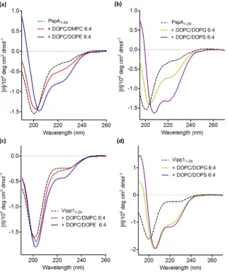
3.3. CD studies with LUVs
SCE stress promotes membrane-binding of both PspA and Vipp1 proteins in an AHa-dependent manner[18]. Thus SCE stress might also affect the PspA1–24and Vipp11–24peptides binding properties if these sequences are the major membrane binding determinants and able to function independently. Large unilamellar vesicles (LUVs) were therefore produced for the SCE stress studies. The compositions of the vesicles to test the effect of SCE stress on PspA1–24and Vipp11–24peptides binding and structure were chosen as two extremes shown to affect the interactions of PspA and Vipp1 proteins[18]. Varying membrane SCE stress has a significant effect on the helical content of PspA1–24(Fig. 4a and Supplementary Table 1). The low SCE stress vesicles (LUVs com-posed of DMPC/DOPC 4:6) caused a slight helical increase of 3% as determined from the CD spectra of PspA1–24. However, the high
SCE stress vesicles (DOPE/DOPC 4:6) had a larger effect with helix content increasing by 15%. The addition of DOPC LUVs containing 40% anionic DOPG or DOPS resulted in increases in calculated helical contents of 13% and 19%, respectively (Fig. 4b and Supplementary
Table 1). The LUVs with high SCE stress increased the helicity of Vipp11–24by 6% (Fig. 4c and Supplementary Table 1) suggesting a lower level of AH mediated binding than PspA1–24. Notably, LUVs
con-taining DOPC alone also had an effect on the structure of PspA1–24and Vipp11–24peptides (Supplementary Fig. 4a). Although the lipid to pep-tide ratio slightly differs to the DOPC/DOPE and DOPC/DMPC LUVs, preventing direct comparison, empirically the effect appears to lie somewhere between that of the LUVs possessing the two extremes of SCE stress. A large increase in helical content of Vipp11–24is observed upon addition of DOPC LUVs with 40% mole fraction anionic lipid content (increase of 27% with DOPG and 38% with DOPS) (Fig. 4d and Supplementary Table 1). The use of two different anionic lipid species (DOPG and DOPS) may rule out any specific head group interac-tions mediating AH formation, hence, the effect of anionic lipids on PspA1–24and Vipp11–24AHs are likely due to electrostatic interactions. It might be that, rather than exclusively SCE stress, the individual nega-tive charge present within PE in DOPE/DOPC LUVs could trigger the interactions of PspA1–24peptides as well. However, although formally possible this is less likely since Vipp11–24, whose interactions are
strongly triggered with anionic lipids, in this case was clearly less re-sponsive than PspA1–24.
[image:6.595.70.522.52.238.2] [image:6.595.33.554.657.744.2]The Vipp11–24peptide binding to lipids and accompanying increase in helical structure is sensitive to the amount of anionic lipids: a gradual increase in the LUVs anionic lipid content using 10%, 20%, 30% and 40% mole fractions of DOPG resulted in a corresponding increase in the net helical content of Vipp11–24at a 14:1 L/P ratio (Supplementary Fig. 4b Fig. 3.Binding of Vipp11–24to SUVs monitoredviaCD spectroscopy and ITC. (a) CD spectra of Vipp11–24in the absence and presence of DOPC, DOPC/DOPG 8:2 and DOPC/DOPG 6:4 SUVs. Peptide concentration was 300μM and the lipid concentration was 4.2 mM giving a 14:1 lipid to peptide ratio. (b–d) ITC tracings (top) and heat of reaction per injection number (bottom) for experiments undertaken in buffer are shown for: (b) injection of 2μl aliquots of 40 mM DOPC into 25μM Vipp11–24; (c) injections of 1.5μl (injections 1–20) and 3μl (injections 21–30) aliquots of 40 mM DOPC/DOPG 8:2 into 50μM Vipp11–24; (d) injections of 1.5μl aliquots of 40 mM DOPC/DOPG 6:4 into 50μM Vipp11–24. All experiments were performed in triplicate and the raw heats shown are a representative example. The heats of dilution, measured in separate control experiments, were subtracted from the heats of reaction in each trace. The buffer controls for (b–d) are presented in Supplementary Fig. 3c–e.
Table 1
Parameters of membrane-binding of PspA1–24and Vipp11–24.aΔHare directly measured binding enthalpies estimated from peptide into lipid titrations.bThe single macroscopic binding constantsKbwere derived using the model described in the text.cThe number of lipid molecules per peptide binding siteNwere also derived using the model described in the text.dTotal
helicity refers to the netα-helix content of peptides incubated with SUVs at a 14:1 lipid-to-peptide ratio.e
NRMSD is a goodness-of-fit parameter between the data and calculated structures.f
Bound helix content is the calculated percentage of residues in anα-helix conformation when the peptide is membrane-bound. N/A, not applicable.
Peptide SUV composition aΔH (kcal/mol) bK
b(M−1) cN dTotal helix content (%) eNRMSD fBound helix content (%)
PspA1–24 N/A 9 0.03
DOPC −6.2 ± 0.28 9.1 × 10⁴
76 21 0.05 78
DOPC/DOPG 8:2 −8.2 ± 0.22 2.6 × 10⁵
39 34 0.02 80
DOPC/DOPG 6:4 −8.0 ± 0.13 1.7 × 10⁵ 25 45 0.04 77
Vipp11–24 N/A 8 0.06
DOPC −3.9 ± 0.18 9.2 × 10⁴
125 12 0.04 42
DOPC/DOPG 8:2 −7.6 ± 0.21 2.1 × 10⁵
35 39 0.05 91
DOPC/DOPG 6:4 −8.0 ± 0.17 2.3 × 10⁵
and Supplementary Table 2). The trend is not linear, with modest in-creases observed from 10% to 30% DOPG before a large increase is seen between 30% and 40%. A similar trend was observed with DOPC LUVs containing 20% or 40% DOPS (Supplementary Fig. 4c and Supple-mentary Table 2). Considering the ITC results with Vipp11–24(see above), this increase in netα-helix content is likely due to both more peptides being associated with the membrane and a higher helical con-tent of those membrane-bound.
The inference that anionic lipid charge density and electrostatic in-teractions affect PspA1–24and Vipp11–24membrane binding and struc-ture was also tested by usingE. coliTLE LUVs. These have high anionic lipid content (over 30%) and more closely represent the native lipid en-vironment of PspA1–24than DOPC-based synthetic vesicles. Notably, Vipp1 N-terminal region comprising Vipp11–24is required for binding
E. coliTLE vesicle[18,28]. The CD spectra for both peptides gradually change from an appearance of unordered structure to that of a peptide with increasing helical content as the L/P ratio increases (Fig. 5and Sup-plementary Table 1).
A V11E substitution in the hydrophobic face of AHa abolishes PspA IM-binding and formation of high-order oligomeric effectors[19]. Ac-cordingly, a PspA1–24V11E peptide (see Supplementary Fig. 1) did not bind DOPC SUVs in ITC studies (Supplementary Fig. 5a) nor did it display any transition from unordered to helical structure in the presence of LUVs with either high level SCE stress, 40% DOPG orE. coliTLE in CD
[image:7.595.143.466.52.440.2]studies (Supplementary Fig. 5b and Supplementary Table 2). The addi-tion of trifluoroethanol (TFE) caused a transition of PspA1–24V11E to a helical structure (Supplementary Fig. 5c). The inability of PspA1–24 V11E to gain helicity in the presence of vesicles is therefore not a Fig. 4.CD spectra showing spectral effects associated with a disordered-to-helix transition of PspA1–24and Vipp11–24when incubated with LUVs of different phospholipid compositions. (a) PspA1–24incubated with DOPC/DMPC 6:4 (low SCE stress) and DOPC/DOPE 6:4 (high SCE stress) LUVs. (b) PspA1–24incubated with LUVs containing anionic lipids DOPC/DOPG 6:4 and DOPC/DOPS 6:4. (c) Vipp11–24incubated with DOPC/DMPC 6:4 (low SCE stress) and DOPC/DOPE 6:4 (high SCE stress) LUVs. (d) PspA1–24incubated with LUVs containing anionic lipids DOPC/DOPG 6:4 and DOPC/DOPS 6:4. The peptide concentration was 300μM and phospholipid concentration was 4.2 mM.
Fig. 5.CD spectra of PspA1–24and Vipp11–24when incubated with increasing amounts of
[image:7.595.314.558.550.702.2]consequence of the V11E substitution itself making a helical structure unfavourable, but rather through failing to bind to vesicles.
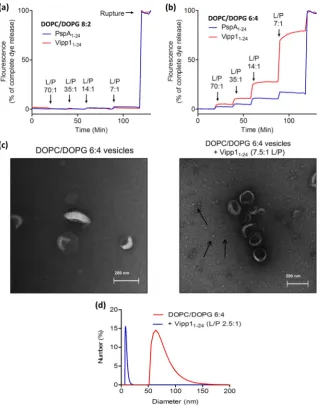
3.4. Effect of peptides on LUVs stability
To examine whether PspA1–24and Vipp11–24peptide binding exerts an effect on membrane stability of vesicles a calcein efflux assay was used to probe leakage. Previously described vesicle compositions (see above) were used to explore if any peptide effects on calcein dye release are modulated by bilayer content. The vesicles bilayer stability was monitored as a function of peptide concentration. The PspA and Vipp1 AHa peptides had no effect on the stability of DOPC vesicles (data not shown) or those with 20% mole fraction DOPG (Fig. 6a). However, at 40% mole fraction DOPG dye leakage was observed starting from a 70:1 L/P ratio (Fig. 6b). The results revealed that PspA and Vipp1 AHa peptides can affect the vesicle structure upon binding and that Vipp11–24was markedly more effective at causing dye release than PspA1–24. PspA1–24V11E did not cause dye release from any vesicle
composition (Supplementary Fig. 6a, b). Samples of 40% DOPG vesicles incubated with Vipp11–24were submitted for EM analysis alongside non-treated vesicles to see if a detrimental effect on the membrane upon peptide binding could be visualised (Fig. 6c). In the presence of Vipp11–24multiple small spheres measuring around 10–20 nm in diam-eter are seen. These spheres were not seen in samples containing either vesicles or Vipp11–24alone (data not shown), thus they are unlikely to result from an intrinsic vesicle instability or peptide aggregation. In-stead, they may be micelles caused by detergent like disintegration of the vesicles by the Vipp11–24. To further test this possibility, DLS studies
were undertaken on 40% DOPG vesicles in the absence and presence of Vipp11–24(Fig. 6d). The measured vesicles size distributions showed that 40% DOPG vesicles are all over 50 nm in diameter but when they are incubated with Vipp11–24a shift to particles with diameters around 10–20 nm is observed, similar in size to the particles seen in the EM images.
[image:8.595.135.456.268.677.2]The stability of vesicles with low (DMPC/DOPC 4:6) and high (DOPE/DOPC 4:6) SCE stress were probed with PspA1–24 and
Vipp11–24peptides. The addition of PspA1–24caused moderate dye release exclusively from vesicles with high SCE stress starting from 14:1 L/P ratio (Supplementary Fig. 6c). The Vipp11–24showed no ef-fect on vesicles stability irrespective of low or high SCE stress (Sup-plementary Fig. 6d).
Taken together with the results obtained in ITC and CD experiments we now establish that AHa of PspA and Vipp1 is able to sense SCE stress and anionic lipids to directly bind the membrane, affecting both protein and (at least in certain cases) membrane structure.
3.5. Inhibition of PspF activity
The AHb of PspA (residues 25–42) is a putative AH adjacent to AHa (Fig. 1). Notably, the crystal structure of the monomeric PspA1–144 frag-ment which binds to PspF and acts as a negative regulator, includes AHb as a part of the coiled coil[21]. Previous studies strongly suggested a hy-drophobic interaction between AHb and the W56 loop of PspF1–275is needed for PspF negative regulation[12,19]. However, the specific na-ture of the interaction is not characterised and a direct effect of the AHb upon PspF activity was not demonstrated. We used the NADH-coupled ATPase assay to determine if a PspA AHb-derived peptide, PspA25–47(Fig. 1and Supplementary Fig. 1), can negatively regulate the AAA domain of PspF1–275. A 3-fold excess of full length PspA is
re-quired for complete inhibition of the PspF1–275ATPase activity[10] and in agreement, equimolar and 2:1 ratios of PspA25–47to PspF1–275 had no apparent effect on the ATPase activity of PspF1–275(data not shown). However, at a 5-fold excess of PspA25–47inhibition of ATPase was evident with activity dropping to around 80% that of PspF1–275 alone (Fig. 7a). Further increases in PspA25–47concentration gave a lin-ear decrease in ATPase activity until nlin-ear complete inhibition was achieved at around a 24:1 PspA25–47to PspF1–275M ratio (Fig. 7a, b). However, even at a 50-fold molar excess, PspA25–47was unable to inhib-it ATPase activinhib-ity of PspF1–275W56A (Fig. 7a). To see if inhibition is spe-cific to PspA25–47, other PspA peptides were also assayed. PspA1–24is also able to impart a degree of inhibition dependent on the W56 residue of PspF (Fig. 7a), and at a 24-fold excess of PspA1–24over PspF1–275the ATPase activity is reduced to around 40% of PspF1–275alone (Fig. 7a, b). The V11E mutation on the hydrophobic face of PspA1–24strongly di-minished the inhibition of PspF1–275ATPase activity (Fig. 7b). Vipp11–24 also repressed the ATPase activity of PspF1–275showing over 20% reduc-tions at a 24-fold excess (Fig. 7b). It appears that a degree of ATPase re-pression can be obtained by all AH peptides, yet the AHb containing PspA25–47is noticeably the most potent.
4. Discussion
4.1. Influence of membrane stress and composition on AHa helical structure
The results obtained in this work establish that the N-terminal AH of PspA and Vipp1, AHa, is an IM-binding determinant which may act as a membrane stress-sensing AH. The AH mediated membrane sensing trends of the PspA1–24and Vipp11–24peptides coincide with comparative vesicle-binding levels seen in PspA WT and Vipp1 WT proteins[18]. This provides strong evidence for a direct lipid interaction between the N-terminal AHa of PspA and Vipp1 being responsible for the specific mem-brane binding. Direct vesicle-binding of both PspA1–24and Vipp11–24is observed in ITC experiments. The results indicate that increases in the anionic lipid content increase the number of PspA1–24and Vipp11–24interactions per lipid vesicle. CD spectroscopic studies in-dicate that the PspA and Vipp1 N-terminal peptides behave similarly to a typical membrane sensing AH[39,40], being unordered in solu-tion and then folding into more helical structures upon membrane association. A V11E mutation of PspA1–24prevents any membrane binding and helix formation irrespective of the tested vesicle compo-sition. Most likely the conserved amphiphilicity of PspA1–24is re-quired for AHa mediated membrane-binding and V11 is part of this helix once membrane associated.
Electrostatics and SCE stress both play a role in the membrane asso-ciation of PspA1–24and Vipp11–24AHa. The membrane SCE stress-dependent AHa binding is much more apparent for PspA1–24than Vipp11–24. The same binding trends are seen for PspA and Vipp1
pro-teins indicating that SCE stress sensing must be directly mediated by the corresponding N-terminal AHa region. In contrast to the SCE stress-sensing trends, Vipp11–24's membrane association is strongly
modulated by anionic lipid content, but the effects on PspA1–24are less pronounced. A large increase in the helix content of membrane-bound Vipp11–24is also observed when anionic lipids are included in the membrane. Similar increased specificity for negatively charged membranes has been seen for a number of AH peptides including those from N-BAR domains and the N-terminal region of GTPase activat-ing protein RGS4[41,42].
The differences in SCE stress and anionic lipids sensing between PspA1–24and Vipp11–24could be explained by their distinct physicochem-ical properties. PspA1–24 has a higher average hydrophobicity than Vipp11–24(PspA1–24—0.392 H and Vipp11–24—0.245 H). PspA1–24should therefore have a higher avidity for the hydrophobic cavities created by lipid packing defects induced SCE stress, increasing its stress sensing abil-ity in neutral bilayers. AHs such as the ALPS motif that purely sense lipid
packing defects caused by SCE stress are typified by their lack of charged residues on the hydrophilic face[39]. Increasing the number of charged residues on the polar face decreases packing defect sensitivity of the AH but increases membrane association through electrostatic interactions [43]. The lower number of charged residues on the polar face of PspA AHa can further explain its enhanced sensitivity for SCE stress dependent binding compared with the Vipp1 AHa. Positively charged residues on the AH encourage interactions with negatively charged lipids, therefore Vipp1’s two extra cationic residues will promote its interaction with an-ionic lipids. This is in full agreement with results obtained with PspA and Vipp1 proteins[18].
4.2. Effect of AHa on membrane stability
The vesicle destabilising properties of the peptides show, for thefirst time, that binding of the AHa region of PspA and Vipp1 has an effect on the physical state of the membrane. However, the detrimental nature of this interaction seems counterintuitive for a membrane maintenance role of PspA and Vipp1 proteinsin vivo. Potentially, the PspA and Vipp1 high-order oligomers may function with AHa's regularly and pre-cisely spaced to regularise and limit the number of the contacts that can be made with the membrane. PspA forms a range of oligomers, up to 36-mer, following membrane stressin vivo[13,16]and is arranged as a 9-fold symmetry 36-mer ring structurein vitro[15]. Indeed, PspA mono-mers have a high affinity for the IM but no effector function in contrast to the PspA high-order oligomers which do act as effectors[17,19]. Re-cent data also suggests that although low-order oligomers of Vipp1 have a high affinity for the membrane, it is the high-order oligomeric ring, with lower membrane affinity, that performs as an effector
in vitro[44]. Notably, the C-terminal tail (HD5) in Vipp1 (seeFig. 1) neg-atively controls a non-productive association between the Vipp1 high-order oligomeric effectors increasing tolerance against thylakoid mem-brane stress and suggesting single Vipp1 ring structure are a functional unit[45]. Therefore, an upper size limited and likely ring shaped high-order assembly of PspA or Vipp1 could provide a sufficient density of membrane binding AHa's to insert into the hydrophobic cavities arising from lipid packing defects and relax the resulting membrane SCE stress (seeFig. 8a, b). This mechanism could act to stabilise stressed mem-branes while preventing bilayer carpeting and the resulting detergent like disintegration through an overwhelming of the membrane with AHa's. Lipid packing defects and accumulation of membrane SCE stress can specifically influence the structure and activity of peripheral brane proteins by providing energetically favourable sites for mem-brane binding often changing conformation and the oligomeric state of a protein[46]. The insertion of AHs into a lipid bilayer can then result in a decrease of SCE stress[46,47]and so this may be one function of the AHa's of PspA and Vipp1 when acting as high-order oligomeric effectors.
4.3. Role of AHb in negative control
Here we also demonstrated that the PspA25–47peptide containing AHb specifically represses PspF1–275ATPase activity. Results showing that the ATPase activity of a PspF1–275W56A mutant cannot be sup-pressed by PspA25–47demonstrate that the peptide is likely using the same inhibition mechanism as the full length PspA through contacting the W56 loop of PspF[3,10,12]. Although the PspA AHa and even the Vipp1 AHa peptide are able to decrease the ATPase activity, PspA25–47 is by far the most effective inhibitor suggesting that an AHb conserved sequence might be specifically required for this functionality. The tested peptides share an amphipathic nature when in anα-helical conforma-tion so hydrophobic interacconforma-tions between the apolar face of the AHb and the W56 loop of PspF could be a prime interaction candidates for the ATPase inhibition. Indeed, the substitution of V29 with charged res-idue on the hydrophobic face of AHb abolishes interactions and negative control of PspF[19]while an E37A substitution of the charged residue on the opposite face of AHb, has only moderate effect on ATPase
inhibition and no effect on the binding interaction with PspF[21]. Full length PspA is much more effective at repressing PspF1–275than the PspA25–47peptide. A 24-fold molar excess of PspA25–47is required for complete PspF1–275ATPase inhibition, but, in similar assays only a three-fold excess of full length PspA can cause the same level of inhibi-tion[10]. PspA and PspF1–275demonstrate a strong propensity to self-assemble into a single defined heteromeric regulatory complex but iso-lated PspA helical domains (HDs) have a low affinity for PspF1–275[10]. Considering that PspA residues 69–186 also contribute to negative con-trol of PspF1–275[10], it is likely that PspA makes multiple interactions with PspF1–275which co-operate to form a tightly bound complex where the AHb region is in the correct location to directly associate with the W56 loop. This is clearly not the case for the PspA25–47peptide where only a single interaction with the W56 loop is possible. The bind-ing affinity of the peptide for PspF1–275will therefore be much lower than PspA WT and a much higher concentration of the peptide will be required for a similar level of ATPase inhibition.
4.4. An integrated perspective
Taken together, our results imply that the PspA N-terminal AHa is di-rectly responsible for membrane association and effector function while the AHb directly imposes inhibition of PspF's ATPase activity and nega-tive control. The AHa region is unordered in a monomeric form of PspA acting as a negative regulator[21]and we show here that upon mem-brane interaction the PspA AHa peptide undergoes a transition to an or-dered helical structure. Considering that AHa and AHb are located adjacent to one another separated by a conserved proline residue in the primary sequence of full length PspA, some steric hindrance or structural remodelling following membrane binding could prevent con-current association to PspF and the bilayer (seeFig. 8a, c). A molecular structure of membrane-bound PspA would help to address the cause for the inferred discordant use of AHa or AHb.
We propose that the SCE stress which elicits PspA vesicle binding via AHa relates a physical membrane state to the physiological membrane stressin vivo. It is challenging to quantitatively measure the changes in membrane SCE stressin vivo, still, severepsp-inducing stimuli such as hyperosmotic, ethanol and extreme temperature shocks can affect the lipid packing and so may induce accumulation of SCE stress. The same but to a lesser extent may be true for the PspB-C-dependent
psp-inducing stimuli such as defects in protein translocation or mislocalisation of type II or III systems secretins.
Maintaining particular membrane mechanical properties upon extra-cytoplasmic stress may be vital for PspA to support pmf-dependent cellular processes as well as for the roles of PspA in the virulence and an-timicrobial resistance of bacteria. The Psp system and PspA proteins de-terminants such as AHa are potential targets for the development of antimicrobials and-or attenuated vaccines against enterobacteria and other pathogenic Gram-negative or Gram-positive bacteria and Mycobac-terium tuberculosis. In addition, the PspA AHb-based peptide(s) might di-rectly inhibit the Psp response in enterobacteria.
high density of AHa insertions of a Vipp1 high-order oligomer could in-duce local membrane disruption resulting in formation of Vipp1-lipid complexes enriched in PG. Vipp1 could then deliver these lipids by interacting with Alb3.2 located in the thylakoid membrane[49]. Poten-tially Vipp1 could undertake a membrane maintenance function based on its SCE stress sensing properties, and a role in thylakoid biogenesis through its anionic lipid binding determinant.
Competingfinancial interests
The authors declare no competingfinancial interests.
Authors contributions
CM, GJ, OC and MB conceived and designed experiments. CM per-formed experiments. CM, GJ, OC and MB analysed the experiments. CM and BAW designed, performed and analysed the CD experiments. GJ, CM and MB wrote the paper. CM prepared thefigures. All authors reviewed the results and approved the submitted version of the manuscript.
Conflict of Interest
The authors declare that they have no conflicts of interest.
Transparency document
TheTransparency documentassociated with this article can be found, in the online version.
Acknowledgements
This work was funded by the Leverhulme Trust Research project grant RPG-2012-705, the EPSRCviagrant EP/G00465X/1, by grant J019135 from the UK BBSRC to BAW, and by an EPSRC Centre for Doctor-al Training Studentship from the Institute of ChemicDoctor-al Biology (ImperiDoctor-al College London) awarded to CM. We would like to thank Dr. Harry H. Low (Imperial College London) for the help with EM experiments. We would like to thank members of the M. Buck laboratory for critical read-ing and comments on the manuscript.
Appendix A. Supplementary data
Supplementary data to this article can be found online atdoi:10. 1016/j.bbamem.2016.10.018.
References
[1] P. Model, G. Jovanovic, J. Dworkin, TheEscherichia coliphage-shock-protein (psp) operon, Mol. Microbiol. 24 (1997) 255–261.
[2] A.J. Darwin, The phage-shock-protein response, Mol. Microbiol. 57 (2005) 621–628. [3] N. Joly, C. Engl, G. Jovanovic, M. Huvet, T. Toni, X. Sheng, M.P. Stumpf, M. Buck, Man-aging membrane stress: the phage shock protein (Psp) response, from molecular mechanisms to physiology, FEMS Microbiol. Rev. 34 (2010) 797–827.
[4] J. Flores-Kim, A.J. Darwin, The phage shock protein response, Annu. Rev. Microbiol. 70 (2016) 83–101.
[5] G. Rowley, M. Spector, J. Kormanec, M. Roberts, Pushing the envelope: extracytoplasmic stress responses in bacterial pathogens, Nat. Rev. Microbiol. 4 (2006) 383–394. [6] A.J. Darwin, Stress relief during host infection: the phage shock protein response
supports bacterial virulence in various ways, PLoS Pathog. 9 (2013), e1003388. [7] P. Datta, J. Ravi, V. Guerrini, R. Chauhan, M.B. Neiditch, S.S. Shell, S.M. Fortune, B.
Hancioglu, O.A. Igoshin, M.L. Gennaro, The Psp system ofMycobacterium tuberculosis
integrates envelope stress-sensing and envelope-preserving functions, Mol. Microbiol. 97 (2015) 408–422.
[8] R.M. Armstrong, K.L. Adams, J.E. Zilisch, D.J. Bretl, H. Sato, D.M. Anderson, T.C. Zahrt, Rv2744c is a PspA ortholog that regulates lipid droplet homeostasis and non-replicating persistence inMycobacterium tuberculosis, J. Bacteriol. 198 (2016) 1645–1661.
[9] N. Zhang, G. Jovanovic, C. McDonald, O. Ces, X. Zhang, M. Buck, Transcripiton regu-lation and membrane stress management in enterobacterial pathogens, in: M.C. Leake (Ed.), Biophysics of Infection, Advances in Experimental Medicine and
Biolo-gy, Springer International Publishing, Switzerland 2016, pp. 207–230.
[10] N. Joly, P.C. Burrows, C. Engl, G. Jovanovic, M. Buck, A lower-order oligomer form of phage shock protein A (PspA) stably associates with the hexameric AAA(+) tran-scription activator protein PspF for negative regulation, J. Mol. Biol. 394 (2009) 764–775.
[11]P. Mehta, G. Jovanovic, T. Lenn, A. Bruckbauer, C. Engl, L. Ying, M. Buck, Dynamics and stoichiometry of a regulated enhancer-binding protein in liveEscherichia coli
cells, Nat. Commun. 4 (2013) 1997.
[12] N. Zhang, T. Simpson, E. Lawton, P. Uzdavinys, N. Joly, P.C. Burrows, M. Buck, A key hydrophobic patch identified in an AAA(+) protein essential for itsin trans inhibi-tory regulation, J. Mol. Biol. 425 (2013) 2656–2669.
[13] G. Jovanovic, P. Mehta, L. Ying, M. Buck, Anionic lipids and the cytoskeletal proteins MreB and RodZ define the spatio-temporal distribution and function of membrane stress controller PspA inEscherichia coli, Microbiol.-sgm 160 (2014) 2374–2386. [14] S. Yamaguchi, D.A. Reid, E. Rothenberg, A.J. Darwin, Changes in Psp protein binding
partners, localization and behaviour upon activation of theYersinia enterocolitica
phage shock protein response, Mol. Microbiol. 87 (2013) 656–671.
[15] B.D. Hankamer, S.L. Elderkin, M. Buck, J. Nield, Organization of the AAA(+) adaptor protein PspA is an oligomeric ring, J. Biol. Chem. 279 (2004) 8862–8866. [16] T. Lenn, C.N. Gkekas, L. Bernard, C. Engl, G. Jovanovic, M. Buck, L. Ying, Measuring the
stoichiometry of functional PspA complexes in living bacterial cells by single mole-cule photobleaching, Chem. Commun. 47 (2011) 400–402.
[17]R. Kobayashi, T. Suzuki, M. Yoshida,Escherichia coliphage-shock protein A (PspA) binds to membrane phospholipids and repairs proton leakage of the damaged membranes, Mol. Microbiol. 66 (2007) 100–109.
[18] C. McDonald, G. Jovanovic, O. Ces, M. Buck, Membrane stored curvature elastic stress modulates recruitment of maintenance proteins PspA and Vipp1, MBio 6 (2015), e01188–15.
[19] G. Jovanovic, P. Mehta, C. McDonald, A. Davidson, P. Uzdavinys, L. Ying, M. Buck, The N-terminal amphipathic helices determine regulatory and effector functions of Phage shock protein A (PspA) inEscherichia coli, J. Mol. Biol. 426 (2014) 1498–1511. [20]S.L. Elderkin, P. Bordes, S. Jones, M. Rappas, M. Buck, Molecular determinants for PspA-mediated repression of the AAA transcriptional activator PspF, J. Bacteriol. 187 (2005) 3238–3248.
[21] H. Osadnik, M. Schopfel, E. Heidrich, D. Mehner, H. Lilie, C. Parthier, H.J. Risselada, H. Grubmuller, M.T. Stubbs, T. Bruser, The PspF-binding domain PspA and the PspA.F complex: new insights into the coiled-coil dependent regulation of AAA+ proteins, Mol. Microbiol. 98 (2015) 743–759.
[22] U.C. Vothknecht, S. Otters, R. Hennig, D. Schneider, Vipp1: a very important protein in plastids?! J. Exp. Bot. 63 (2012) 1699–1712.
[23] L. Zhang, W. Sakamoto, Possible function of VIPP1 in thylakoids: protection but not formation? Plant Signal. Behav. 8 (2013), e22860.
[24] S.J. Bryan, N.J. Borroughs, D. Shevela, J. Yu, L.-N.L. Rupprecht, G. Mastroianni, Q. Xue, I. Llorente-Garcia, M.C. Leake, L.A. Eichacker, D. Schneider, P.J. Nixon, C.W. Mullineaux, Localisation and interactions of the Vipp1 protein in cyanobacteria, Mol. Microbiol. 94 (2014) 1179–1195.
[25] L. Zhang, W. Sakamoto, Possible function of Vipp1 in maintaining chloroplast mem-branes, Biochim. Biophys. Acta 1847 (2015) 831–837.
[26] E. Aseeva, F. Ossenbühl, L.A. Eichacker, G. Wanner, J. Soll, U.C. Vothnecht, Complex formation of Vipp1 depends on it'sα-helical PspA-like domain, J. Biol. Chem. 279 (2004) 35535–35541.
[27] M.P. DeLisa, P. Lee, T. Palmer, G. Georgiou, Phage shock protein PspA ofEscherichia colirelieves saturation of protein exportviathe Tat pathway, J. Bacteriol. 186 (2004) 366–373.
[28] S. Otters, P. Braun, J. Hubner, G. Wanner, U.C. Vothknecht, F. Chigri, Thefirst alpha-helical domain of the vesicle-inducing protein in plastids 1 promotes oligomeriza-tion and lipid binding, Planta 237 (2013) 529–540.
[29] R. Hennig, J. Heidrich, M. Saur, L. Schmuser, S.J. Roeters, N. Hellmann, S. Woutersen, M. Bonn, T. Weidner, J. Markl, D. Schneider, IM30 triggers membrane fusion in cyanobacteria and chloroplasts, Nat. Commun. 6 (2015) 7018.
[30] J.G. Lees, B.R. Smith, F. Wien, A.J. Miles, B.A. Wallace, CDtool-an integrated software package for circular dichroism spectroscopic data processing, analysis, and archiv-ing, Anal. Biochem. 332 (2004) 285–289.
[31]L. Whitmore, B.A. Wallace, Protein secondary structure analyses from circular di-chroism spectroscopy: methods and reference databases, Biopolymers 89 (2008) 392–400.
[32]S.W. Provencher, J. Glöckner, Estimation of globular protein secondary structure from circular dichroism, Biochemistry 20 (1981) 33–37.
[33] I.H. van Stokkum, H.J. Spoelder, M. Bloemendal, R. van Grondelle, F.C. Groen, Estima-tion of protein secondary structure and error analysis from circular dichroism spec-tra, Anal. Biochem. 191 (1990) 110–118.
[34]N. Joly, J. Schumacher, M. Buck, Heterogeneous nucleotide occupancy stimulates functionality of phage shock protein F, an AAA+ transcriptional activator, J. Biol. Chem. 281 (2006) 34997–35007.
[35] J.G. Nørby, Coupled assay of Na +, K+ ATPase activity, Methods Enzymol. 156 (1988) 116–119.
[36] S.S. Chong, S.G. Taneva, J.M. Lee, R.B. Cornell, The curvature sensitivity of a membrane-binding amphipathic helix can be modulated by the charge on a
flanking region, Biochemistry 53 (2014) 450–461.
[37] B. Nucher, Alpha-synuclein has a high affinity for packing defects in a bilayer mem-brane: a thermodynamics study, J. Biol. Chem. 279 (2004) 21966–21975. [38] T. Wieprecht, M. Beyermann, J. Seelig, Thermodynamics of the coil-alpha-helix
tran-sition of amphipathic peptides in a membrane environment: the role of vesicle cur-vature, Biophys. Chem. 96 (2002) 191–201.
[40] G. Drin, J.F. Casella, R. Gautier, T. Boehmer, T.U. Schwartz, B. Antonny, A general am-phipathic alpha-helical motif for sensing membrane curvature, Nat. Struct. Mol. Biol. 14 (2007) 138–146.
[41]F. Fernandes, L.M. Loura, F.J. Chichon, J.L. Carrascosa, A. Fedorov, M. Prieto, Role of helix 0 of the N-BAR domain in membrane curvature generation, Biophys. J. 94 (2008) 3065–3073.
[42] L.S. Bernstein, A.A. Grillo, S.S. Loranger, M.E. Linder, RGS4 binds to membranes through an amphipathic alpha-helix, J. Biol. Chem. 275 (2000) 18520–18526. [43] M.B. Jensen, V.K. Bhatia, C.C. Jao, J.E. Rasmussen, S.L. Pedersen, K.J. Jensen, R. Langen, D.
Stamou, Membrane curvature sensing by amphipathic helices: a single liposome study using alpha-synuclein and annexin B12, J. Biol. Chem. 286 (2011) 42603–42614. [44] J. Heidrich, V. Wulf, R. Hennig, M. Saur, J. Markl, C. Sönnichsen, D. Schneider,
Orga-nization into higher-ordered ring structures counteracts membrane binding of IM30, a protein associated with inner membranes in chloroplasts and cyanobacteria, J. Biol. Chem. (2016),http://dx.doi.org/10.1074/jbc.M116.722686.
[45] L. Zhang, H. Kondo, H. Kamikubo, M. Kataoka, W. Sakamoto, VIPP1 has a disordered C-terminal tail necessary for protecting photosynthetic membranes against stress in Arabidopsis, Plant Physiol. (2016),http://dx.doi.org/10.1104/pp.16.00532.
[46] E. van den Brink-van der Laan, J.A. Killian, B. de Kruijff, Nonbilayer lipids affect pe-ripheral and integral membrane proteinsviachanges in the lateral pressure profile, Biochim. Biophys. Acta 1666 (2004) 275–288.
[47] L. Iversen, S. Mathiasen, J.B. Larsen, D. Stamou, Membrane curvature bends the laws of physics and chemistry, Nat. Chem. Biol. 11 (2015) 822–825.
[48]A. Nordhues, M.A. Schottler, A.K. Unger, S. Geimer, S. Schonfelder, S. Schmollinger, M. Rutgers, G. Finazzi, B. Soppa, F. Sommer, T. Muhlhaus, T. Roach, A. Krieger-Liszkay, H. Lokstein, J.L. Crespo, M. Schroda, Evidence for a role of VIPP1 in the struc-tural organization of the photosynthetic apparatus in Chlamydomonas, Plant Cell 24 (2012) 637–659.
[49] V. Gohre, F. Ossenbuhl, M. Crevecoeur, L.A. Eichacker, J.D. Rochaix, One of two Alb3 proteins is essential for the assembly of the photosystems and for cell survival in Chlamydomonas, Plant Cell 18 (2006) 1454–1466.