3 D s c a n n e r fo r a s s e s s m e n t of t h e
s h a p e a n d vol u m e of a m p u t e e s’
r e si d u a l li m b m o d e l s
S e m i n a ti, E , C a n e p a Tal a m a s , D, You n g , M , Twi s t e , M , D h o ki a, V
a n d Bilzo n , J
h t t p :// dx. d oi.o r g / 1 0 . 1 3 7 1 /jo u r n a l. p o n e . 0 1 8 4 4 9 8
T i t l e
Validi ty a n d r eli a bility of a n o v el 3 D s c a n n e r fo r
a s s e s s m e n t of t h e s h a p e a n d vol u m e of a m p u t e e s’ r e si d u al
li m b m o d el s
A u t h o r s
S e m i n a ti, E , C a n e p a Tal a m a s , D, You n g , M , Twi s t e , M ,
D h o ki a, V a n d Bilzo n , J
Typ e
Ar ticl e
U RL
T hi s v e r si o n is a v ail a bl e a t :
h t t p :// u sir. s alfo r d . a c . u k /i d/ e p ri n t/ 4 3 6 6 2 /
P u b l i s h e d D a t e
2 0 1 7
U S IR is a d i gi t al c oll e c ti o n of t h e r e s e a r c h o u t p u t of t h e U n iv e r si ty of S alfo r d .
W h e r e c o p y ri g h t p e r m i t s , f ull t e x t m a t e r i al h el d i n t h e r e p o si t o r y is m a d e
f r e ely a v ail a bl e o nli n e a n d c a n b e r e a d , d o w nl o a d e d a n d c o pi e d fo r n o
n-c o m m e r n-ci al p r iv a t e s t u d y o r r e s e a r n-c h p u r p o s e s . Pl e a s e n-c h e n-c k t h e m a n u s n-c ri p t
fo r a n y f u r t h e r c o p y ri g h t r e s t r i c ti o n s .
Validity and reliability of a novel 3D scanner
for assessment of the shape and volume of
amputees’ residual limb models
Elena Seminati1,2, David Canepa Talamas3, Matthew Young2, Martin Twiste4,5, Vimal Dhokia3, James L. J. Bilzon1,2*
1 Department for Health, University of Bath, Bath, United Kingdom, 2 CAMERA Centre, University of Bath,
Bath, United Kingdom, 3 Department of Mechanical Engineering, University of Bath, Bath, United Kingdom,
4 School of Health Sciences, University of Salford, Salford, United Kingdom, 5 United National Institute for
Prosthetics & Orthotics Development (UNIPOD), University of Salford, Salford, United Kingdom
Abstract
Background
Objective assessment methods to monitor residual limb volume following lower-limb ampu-tation are required to enhance practitioner-led prosthetic fitting. Computer aided systems, including 3D scanners, present numerous advantages and the recent Artec Eva scanner, based on laser free technology, could potentially be an effective solution for monitoring residual limb volumes.
Purpose
The aim of this study was to assess the validity and reliability of the Artec Eva scanner (prac-tical measurement) against a high precision laser 3D scanner (criterion measurement) for the determination of residual limb model shape and volume.
Methods
Three observers completed three repeat assessments of ten residual limb models, using both the scanners. Validity of the Artec Eva scanner was assessed (mean percentage error
<2%) and Bland-Altman statistics were adopted to assess the agreement between the two
scanners. Intra and inter-rater reliability (repeatability coefficient<5%) of the Artec Eva scanner was calculated for measuring indices of residual limb model volume and shape (i.e. residual limb cross sectional areas and perimeters).
Results
Residual limb model volumes ranged from 885 to 4399 ml. Mean percentage error of the Artec Eva scanner (validity) was 1.4% of the criterion volumes. Correlation coefficients between the Artec Eva and the Romer determined variables were higher than 0.9. Volume intra-rater and inter-rater reliability coefficients were 0.5% and 0.7%, respectively. Shape percentage maximal error was 2% at the distal end of the residual limb, with intra-rater a1111111111 a1111111111 a1111111111 a1111111111 a1111111111 OPEN ACCESS
Citation: Seminati E, Canepa Talamas D, Young M,
Twiste M, Dhokia V, Bilzon JLJ (2017) Validity and reliability of a novel 3D scanner for assessment of the shape and volume of amputees’ residual limb models. PLoS ONE 12(9): e0184498.https://doi. org/10.1371/journal.pone.0184498
Editor: Steven Allen Gard, Northwestern University,
UNITED STATES
Received: June 27, 2017
Accepted: August 24, 2017
Published: September 8, 2017
Copyright:©2017 Seminati et al. This is an open access article distributed under the terms of the
Creative Commons Attribution License, which permits unrestricted use, distribution, and reproduction in any medium, provided the original author and source are credited.
Data Availability Statement: Data is available at
https://doi.org/10.15125/BATH-00417.
Funding: This study was funded by the Engineering
and Physical Sciences Research Council UKs Impact Acceleration Account (RC-FH1136).
Competing interests: The authors have declared
reliability coefficients presenting the lowest errors (0.2%), both for cross sectional areas and perimeters of the residual limb models.
Conclusion
The Artec Eva scanner is a valid and reliable method for assessing residual limb model shapes and volumes. While the method needs to be tested on human residual limbs and the results compared with the current system used in clinical practice, it has the potential to quantify shape and volume fluctuations with greater resolution.
Introduction
The post-operative phase following limb amputation is characterised by rapid residual limb volume reduction due to decreased post-surgical oedema and muscle atrophy. The range of volume reduction, 6-months post-surgery, varies between 17% and 40% of the original volume
and appears to be characterised by a negative exponential function of time [1–3]. The
transi-tion from acute clinical rehabilitatransi-tion to stable long-term recovery occurs 12–18 months
post-operatively [4] and currently there is no definitive method for establishing when the residual
limb volume has stabilised [2,3]. In addition, mature residual limbs (i.e.>18-months post
amputation) are still subject to short term volume changes (diurnal changes range between
-3.5% and +10.9% [5–7]). Prosthetists engage in labour-intensive processes to modify sockets
using manual methods, and fitting problems can eventually result in expansive and time
con-suming socket-prosthesis adjustments/replacements and low quality of life [8]. Management
and assessment of residual limb volume is important because it affects decisions regarding: i) timing of fit of the first prosthesis (i.e. when to switch from a temporary to a definitive pros-thetic socket); ii) design of a prospros-thetic socket/liner that can best transmit loading forces, mini-mising discomfort through offering an ‘ideal fit’ without compromini-mising easy donning/doffing of the prosthesis (if high stresses develop where the skin is of low load-tolerance, then the nor-mal, shear, and frictional forces can compound and lead to pain, surface abrasions, deep skin breakdown or even deep tissue injury) and; iii) prescription of accommodation strategies for daily volume fluctuations. Furthermore, by building a database of residual limb volume changes, which include details of patient background and lifestyle behaviours, predictive modelling work can be undertaken to help streamline the process of prosthetic refitting for individuals undergoing routine volume change.
Many techniques for the measurement of residual limb volume have been described, using patient residual limbs and models. Not all methods are suitable for clinical use, either because they lack necessary resolution for volume measurement and/or they are unable to detect
changes in residual limb shape [4]. Anthropometric measures [9,10] are practical and
accept-able for macroscopic residual limb changes, but they have a poor inter- and intra-observer repeatability and lack the precision necessary to establish the threshold for stabilisation. Water
immersion methods [11] have lower variability, but cannot be used when open wounds are
present or on patients with bilateral leg amputations. MRI [12], ultrasound measurement [13,
14] and computed tomography (CT) scans [15,16] have been described in the literature, and
scanning time). Laser scanning methods, including CAD/CAM systems [1,9,17–21], have been introduced to aid the manufacture of prosthetic sockets, reducing fitting errors, fabrica-tion time and overall costs. Some of these techniques have shown promise, with the Omega
Tracer scanner being the method of choice for current clinical practice in the UK [2,9].
Repeatability coefficients for this system appear relatively high and range from 45 ml (~5% of
volume) when scanning residual limb models [9] to 129 ml (~13% of volume) when scanning
human transtibial residual limbs [22]. A recent study evaluated the accuracy of the imaging/
acquisition process of three new surface 3D scanners and suggested that the VIUScan marker assisted laser scanner was the most accurate for determining residual limb model volume and shape. The systematic bias/error reported between the VIUScan scanner and the criterion was lower than 1%, but they used a single 3D printed transtibial model of known geometry as a
gold standard, without considering the effect of different model sizes [23]. The method
revealed estimated repeatability coefficients<45 ml for within and between observer
assess-ments. However, a full comparison is limited because of the statistical methods adopted. Fur-thermore, as with many other similar studies, this method has not yet been tested on human residual limbs.
New CAD/CAM technologies are emerging, with new features that can help prosthetists and practitioners in decision-making regarding the timing and design of prosthetic sockets. The Artec Eva scanner (Artec Group, Luxembourg, Luxembourg) is a state-of-the art technol-ogy for 3D surface scanning. It uses laser free structured light scanning technoltechnol-ogy and has
already been successful in human applications [24–26], but it has not been used for the
assess-ment of amputee residual limb shape or volume. It is quick to use, can accommodate some tar-get movement and is able to capture geometry and texture/colour information, facilitating anatomical features detection, which eliminates the need for reference targets or markers to be placed on the limb. Before adopting this new system in clinical practice, it is necessary to deter-mine its accuracy in terms of validity and reliability with residual limb models. As residual limb models are not prone to movement artefact, the technical measuring error of the equip-ment and of the observer can be independently assessed.
The aim of this study was therefore to assess the accuracy (validity and reliability) of the Artec Eva scanner for estimating the volume, shape and size of transtibial and transfemoral residual limb models. In order to be clinically meaningful, the Artec Eva scanner would need
to demonstrate a mean percentage error (validity) of<2% compared to the Romer scanner
(criterion measure). In order to be considered reliable, it would need to elicit intra and
inter-rater repeatability coefficients of<5%. Validity threshold has been determined, after
consider-ing the most accurate methods presented in the literature [4,23]. The repeatability threshold
has been set, in part, based on the methods currently used in clinical practice [9] and, also, the
desire to detect smaller but meaningful acute residual limb changes (short term changes, diur-nal changes or changes due to postdoffing).
Materials and methods
In this study, ten residual limb models were scanned by three independent observers, each on three separate occasions, using two different scanners (i.e. 180 scans), over a 4 months’ period (May—August 2016). The models were selected from anonymous transtibial (n = 5) and
trans-femoral (n = 5) amputees (Table 1) to evaluate a large range of representative shapes and
Measurements systems
The use of high precision and resolution laser scanner has been suggested for evaluation of
new scanning systems [4]. For this reason, we used the Romer high precision and resolution
scanner (Romer scanner, CMS108, Hexagon, UK) as the criterion measure to validate the ‘trueness’ of the Artec Eva scanner (practical measure). The Romer scanner is a powerful tool integrated with a Romer coordinate measuring arm that comprises different rotation axes to
allow freedom of movement. It uses a laser line to reconstruct the 3D model (Fig 1B) with an
accuracy of about 0.04 mm [27]. In contrast, the Artec Eva (practical measure) is relatively
small and uses regular flash bulb technology, illuminating the object with patterns of stripes by normal visible light to reconstruct 3D data from the surface with a reported accuracy of 0.5
mm [25].
Experimental design and data collection
Three independent observers were trained to use both the Artec Eva and Romer scanners. Prior to data collection, they completed two 2-hour familiarisation sessions, using both scan-ners. Ten different sessions were organised to measure each of the ten selected residual limb models. During each session, each model was measured three times by each observer with the
two different scanners. This resulted in a total of 18 measurements (2 scanners×3 observers×3
repetitions) per model/session. The observers performed the measurements in randomised order (both observer sequence and scan sequence), with a 10-min break between each scan. Time per scan was between 1 and 3 minutes for the Artec Eva and Romer scanners, respectively.
Prior to scanning, three 4 mm diameter hemispherical adhesive markers made of soft rub-ber were placed on the 3D surface of each residual limb model to approximately identify three anatomical landmarks for transfemoral (greater trochanter, Scarpa’s triangle and ischial
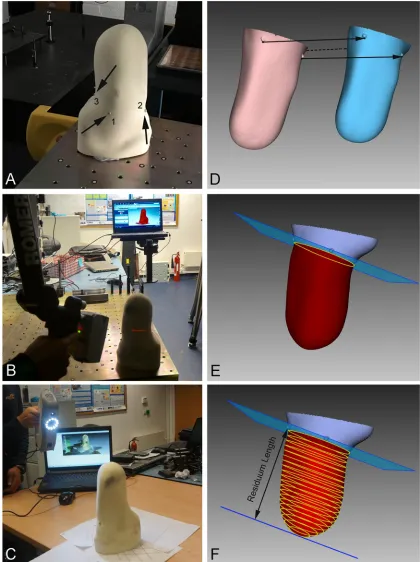
tuber-osity) and transtibial (tibial crest, fibula head and popliteal fossa) models (Fig 1A). The distal
borders of these markers were used to determine a plane used as the proximal end of each scan. Each model was placed on a metrology table for the Romer scans and on a normal table
for the Artec Eva scans, with the distal end of the residual limb pointing upward (Fig 1B and
1C). Table 1. Residual limb models characteristics.
Model Level Material Romer scanner (ml) Artec scanner (ml)
1 TF foam 4277 (3) 4316 (7)
2 TF foam 2332 (4) 2362 (10)
3 TT foam 1782 (1) 1807 (7)
4 TF plaster 4019 (3) 4053 (12)
5 TF plaster 3003 (2) 3030 (5)
6 TF foam 2930 (3) 2969 (8)
7 TT foam 2606 (4) 2643 (5)
8 TT foam 1326 (1) 1352 (3)
9 TT foam 1529 (2) 1555 (2)
10 TT foam 869 (1) 887 (4)
Level of amputation (transfemoral—TF or transtibial—TT), type of material (foam or plaster) and mean volumes calculated with the Romer and the Artec scanners. The values reported in column 4 and 5 represent the mean volume of 9 trials (three operators and three trials for each scanner). Standard deviation is indicated in brackets.
Fig 1. Residual limb scanning and processing procedures. A) Example of transtibial residual limb with the three
anatomical markers; B) Romer scanning conditions; C) Artec Eva scanning conditions; D) Romer (pink) and Artec Eva (blue) 3D models prior to alignment; E) aligned models with the plane 0, defined as the plane passing through the distal border of the three reference points; F) CSAs along the residual limb.
Data processing
Artec Eva and Romer data files were processed using the same software used for data collec-tion: Artec Studio 9.2 (Artec Group, Luxembourg, Luxembourg) and Geomagic Studio 2014 (Geomagic—3D Systems, USA). Both the Artec Eva and Romer mesh models were exported and aligned manually in the same reference system x, y and z, using a graphical user interface
according to the positions of the anatomical markers on the model (Fig 1D). The volume of
each residual limb model was calculated using the distal end to the proximal end of the residual
limb as indicated by the plane and defined by the three anatomical markers (Fig 1E). Parallel
to this first plane 19 other planes were defined across the residual limb volume, obtaining a set of 20 parallel different sections at intervals of 5% across the residual limb length, with the first section (i.e. 0%) indicating the first proximal section of the residual limb model. For each sec-tion created by the 20 planes the relative Cross Secsec-tional Area (CSA) and the perimeter (PE)
were calculated (Fig 1F).
To assess residual limb model geometrical differences between the two scanners, the Root Mean Square Error (RMSE) between each pair of aligned scans was calculated. In addition, assuming the residual limb to be confined in a bounding box, residual limb sizes (width, depth and length) along three axes (x, y, and z, respectively) were calculated. Body Centre of Mass (BCOM) coordinates were calculated assuming the material of the models to be homogenous. For these calculations, each volume was processed using the Compute geometric measures
fil-ter [28] in Meshlab software.
Statistical analysis
Accuracy of the Artec Eva scanner was assessed in terms of validity (trueness) and reliability (precision) using the statistical approach suggested by Hopkins, which was used to assess the
validity and reliability of DEXA imaging methods [29,30]. The following scanning variables
were considered: residual limb volume, residual limb sizes (width, depth and length) along the three different axes (x, y, and z, respectively), BCOM coordinates, CSA and PE for each of the 20 levels of the residual limb length, where level 0 was defined as the plane passing through the distal border of the three anatomical/reference points. To ensure normality of the sampling distribution, each measurement was log transformed before analysis and back transformed
after analysis [31,32]. Log transformation was necessary to ensure uniformity of error,
particu-larly where larger values of the original variable have greater absolute but a similar relative (%) error. Log transformation was not applied to the BCOM variables, since they were expressed in the relative Romer reference system.
Validity. Validity of the Artec Eva scan was assessed using a previously published method
[33], where the Romer scanner was considered as the criterion and the Artec Eva scanner as
the practical measurement system. Limits of agreement were calculated using methods
described by Bland-Altman [30]. The overall bias and 95% limits of agreement were calculated
and Pearson correlation coefficient and Coefficient of Variation (CV) were determined. Modi-fied Bland and Altman plots were used to show the Artec Eva validity as percentage change, when considering either only transtibial or only transfemoral residual limb models. We con-sidered using a mixed-model analysis of variance method to ensure that assumptions of sample independence were not violated, but the differences in results were negligible.
Reliability. To quantify the intra-rater variability (repeatability) of the Artec Eva scanner,
difference between the repeated scan results), typical error of the measurements (TEMs; standard
deviation of the difference scores of all the scans in the group divided byp2), and Intra-class
Correlation Coefficients (ICCs) for all scan parameters, using a published method [34], with a 95
percent Confidence Interval (CI). The type of ICC produced by Hopkins’ analysis of validity and
reliability [34] for intra- and inter-rater reliability, was ICC(3,1) [33]. According to Bland and
Altman [31] the within—subject standard deviation (or standard error of the measurement
[31]), represented by the TEMs, was used to calculate the intra-rater variability (repeatability
coefficients) and inter-rater volume variability (reproducibility coefficient) as 1.96p2TEMs as
reported in previous studies regarding residual limb volume repeatability [9,22,31].
Reporting. This article was prepared in accordance with the checklist for Strengthening
the Reporting of Observational Studies in Epidemiology (STROBE), which is provided as a
supplementary file (S1 Table).
Results
The three observers collected 60 scans each, during ten different sessions, and the measured
volumes ranged from 885 ml to 4399 ml (Table 1).
Validity
Volume measurements showed a consistent tendency for the volumes obtained with the Artec Eva scanner to exceed (~30 ml) values obtained with the Romer scanner. This bias corre-sponded to 1.4% of the actual volumes with most differences between measurements by two
methods falling within 1% of limits of agreements (Fig 2). Similarly, model size measurements
(i.e. width, depth and length), confirmed the overestimation of the Artec Eva scanner relative to the Romer, even though the bias was always lower than 1 mm and with 1% of Limits of
Agreement. BCOM coordinates estimation showed low bias values (<1mm) with the highest
variability observed for the vertical coordinate (z) (Table 2). The average RMSE values
calcu-lated in three dimensions between Artec Eva and Romer scans ranged from 0.23 to 0.65 mm,
with the Artec Eva scanner presenting higher values than the Romer scanner (Fig 3).
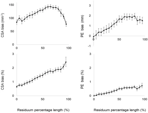
The bias of the CSAs and of the PE increased along the longitudinal length of the residual limb models, reaching differences close to 2% for the CSA and to 1% for the PE at the distal
end of the model (Fig 4).
Reliability
The Artec Eva scanner showed high levels of intra- and inter-rater reliability, with ICCs greater than 0.90 as it was for the criterion measurement. Intra- and inter-rater repeatability coeffi-cients for volume measurements were 13.9 and 18.6 ml for the Artec Eva scanner (respectively 0.5% and 0.7% of the average volumes), with TEMs slightly higher for the inter-rater variability (Table 3). Good reliability was also evident for model size and BCOM estimation with errors always lower than 0.2% for the model size and lower than 1 mm for the BCOM.
Results regarding the residual limb shape in term of CSA and PE revealed that the
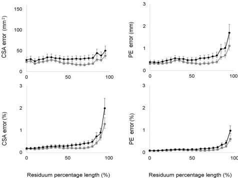
inter-observer error was always higher compared with the intra-inter-observer error. As shown inFig 5,
CSA and PE errors (TEMs) were always higher when considering inter-rater reliability, and they increased exponentially beyond 75% of the length of the residual limb.
Discussion
Fig 2. Scanners’ agreements. Top panel: Bland-Altman plots for model volumes calculated with the criterion
(Romer scanner) and the practical (Artec Eva scanner) measurement system; Bottom panel: modified Bland-Altman plots displaying the error of volumes measured with the practical (Artec Eva) scanner expressed as a percentage of the Romer scanner volumes (average between trials). The open circles represent transtibial models, while the full circles represent transfemoral models. The dashed lines indicate the upper and lower 95% limits of agreements.
Table 2. Validity results.
Overall Bias Limits of Agreements
Absolute Relative (%) Absolute Relative (%) Pearson CC CV (%) Volume 30.4 (28.3; 32.5) ml 1.40 (1.28; 1.51) 18.9 ml 1.01 0.99 0.34 (0.29; 0.40)
Residuum Size
Width (x) 0.49 (0.39; 0.59) mm 0.36 (0.29; 0.43) 0.91 mm 1.01 0.99 0.32 (0.28; 0.38)
Depth (y) 0.47 (0.38; 0.55) mm 0.35 (0.29; 0.41) 0.77 mm 1.01 0.99 0.24 (0.21; 0.28)
Length (z) 0.38 (0.26; 0.50) mm 0.14 (0.10; 0.19) 1.09 mm 1.00 0.99 0.20 (0.17; 0.23)
BCOM
x -0.01 (-0.04; 0.03) mm - 0.32 mm - 0.99
-y 0.02 (-0.04; 0.07) mm - 0.48 mm - 0.99
-z 0.23 (0.05; 0.41) mm - 1.63 mm - 0.99
-Validity results are presented in terms of overall bias (absolute and relative values), limits of agreements, Pearson Correlation Coefficient (CC) and Coefficient of Variation (CV). Values in brackets represent the 95% Confidence Limits.
https://doi.org/10.1371/journal.pone.0184498.t002
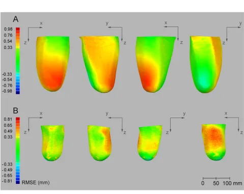
Fig 3. RMSE results. Example comparison of the same model collected with the 2 different scanners from the same observer for a
right-sided trans-femoral models (A) and a left-right-sided transtibial model (B). RMSE differences are indicated by the coloured scale on the left (red positive mean values indicate that the Artec Eva scanner measured a bigger volume). From left to right: anterior view, lateral view, medial view and posterior view of the residual limb model.
[image:10.612.96.575.280.661.2]design of prosthetic sockets. This study assessed the validity and reliability of a new structured light 3D scanner for measuring lower-limb residual limb volume and shape characteristics in a range of residual lower-limb models.
Validity and reliability determine the accuracy of a new measurement instrument and are
important in terms of the quality of the data and subsequent future clinical application [35].
Because no variance in the residual limb model volume occurs, the technical error of the Artec Eva scanner could be evaluated, before commencing further research on human residual limbs.
This investigation is the first to include both transtibial and transfemoral residual limb models to assess the accuracy of a new structured light 3D scanner for residual limb volume and shape monitoring. In addition, this study is particularly timely given the increased number of emerging CAD/CAM technologies socket volume accommodation.
The Artec Eva scanner showed a high degree of relative validity (<2%) in volume
[image:11.612.94.575.79.448.2]measure-ments with bias values of<1% when considering only transfemoral residual limb models
Fig 4. Residual limb shape validity results. The graphs represent the bias and limits of agreement for CSAs and PE calculated for each
section along the residual limb model, and expressed both in absolute units and as a relative percentage of the Romer scanner’s original measure (0% indicates the first proximal section of the residual limb model).
(Fig 2). These results are consistent with the literature investigating CAD systems for residual
limb volume measurements [18,19,23,36].
The use of high precision and resolution laser scanner has been suggested for evaluation of
new scanning systems [4], and in this study the Romer laser scanner (with an accuracy of one
tenth of a millimetre), was selected as the criterion measure to assess the validity of the Artec Eva 3D scanner on rigid residual limb models. Previous validity assessments have been limited by the fact that, in most cases, no gold standard more accurate than the instrument under
development was available [4]. One of the most accurate CAD systems investigated, the
CAPOD laser scanner reported variations in validity between 0.3 and 2.5% of water
displace-ment (criterion measure) [18,19]. Similar results were found for the VIUScan [23]
marker-assisted laser scanner, and the Go!SCAN 3D structured white light scanner, with 0.5% and 1.1% of the criterion (a single 3D printed transtibial model of known geometry). High levels of
accuracy (<2%) were also reported for the TracerCAD system in gross volume and lengths on
a cylindrical model [36]. However, this system was not as consistent when applied to the more
complex transtibial residual limb model [37].
A very small magnitude for RMSE (<1mm) was measured with the Artec Eva scanner.
When comparing models collected with the Artec Eva and Romer scanners, the average dis-tance between models was always lower than 1 mm (0.6% of a diameter of 160 mm), with the highest differences highlighted at the prominences of the residual limb models, including the tibial tuberosity and cut end of the tibia for the transtibial models and lateral aspect of cut end
[image:12.612.36.576.89.365.2]of the femur for the transfemoral models (seeFig 3). Results were similar to those previously
Table 3. Reliability results.
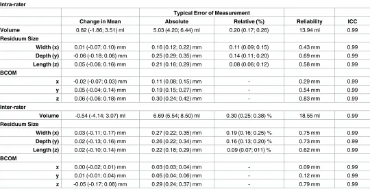
Intra-rater
Typical Error of Measurement
Change in Mean Absolute Relative (%) Reliability ICC
Volume 0.82 (-1.86; 3.51) ml 5.03 (4.20; 6.44) ml 0.20 (0.17; 0.26) 13.94 ml 0.99
Residuum Size
Width (x) 0.01 (-0.07; 0.10) mm 0.16 (0.12; 0.22) mm 0.11 (0.09; 0.15) 0.43 mm 0.99
Depth (y) -0.06 (-0.18; 0.06) mm 0.25 (0.29; 0.35) mm 0.14 (0.11; 0.20) 0.69 mm 0.99
Length (z) 0.05 (-0.06; 0.16) mm 0.21 (0.16; 0.29) mm 0.08 (0.06; 0.12) 0.58 mm 0.99
BCOM
x -0.02 (-0.07; 0.03) mm 0.11 (0.08; 0.15) mm - 0.29 mm 0.99
y 0.05 (-0.04; 0.14) mm 0.19 (0.15; 0.27) mm - 0.54 mm 0.99
z 0.06 (-0.06; 0.18) mm 0.30 (0.24; 0.42) mm - 0.83 mm 0.99
Inter-rater
Volume -0.54 (-4.14; 3.07) ml 6.69 (5.54; 8.50) ml 0.30 (0.25; 0.38) % 18.55 ml 0.99
Residuum Size
Width (x) 0.03 (-0.11; 0.17) mm 0.27 (0.22; 0.35) mm 0.19 (0.16; 0.25) % 0.75 mm 0.99
Depth (y) 0.02 (-0.13; 0.16) mm 0.26 (0.22; 0.34) mm 0.16 (0.13; 0.20) % 0.73 mm 0.99
Length (z) 0.02 (-0.10; 0.14) mm 0.22 (0.18; 0.29) mm 0.09 (0.07; 011) % 0.62 mm 0.99
BCOM
x 0.00 (-0.02; 0.01) mm 0.03 (0.03; 0.04) mm - 0.09 mm 0.99
y 0.01 (-0.01; 0.04) mm 0.05 (0.04; 0.06) mm - 0.12 mm 0.99
z -0.05 (-0.17; 0.08) mm 0.29 (0.24; 0.37) mm - 0.79 mm 0.99
Intra-rater reliability (upper panel) and inter-rater reliability (lower panel) results are presented in terms of Change in Mean, Typical Error of Measurement (absolute and relative values), Reliability Coefficients (Reliability) and Intra Correlation Coefficients (ICC). Values in brackets represent the 95% Confidence Limits.
reported [23] for the VIUScan and Go!SCAN systems, which both had a surface length error magnitude up to 0.20 mm and 0.33 mm, respectively. Similar values were observed in the trend for increasing bias of the CSAs along the residual limb (higher discrepancies at the proxi-mal end of the model up to 2% for the CSA and up to 1% for the PE).
Reliability results showed that the Artec Eva scanner is a very reliable instrument for lower-limb residual lower-limb volume and shape measurements. Correlation coefficients (ICCs) both for intra- and inter-rater repeatability exceeded the 0.90 threshold for clinically relevant reliability
[32,38]. The inter-observer error (between different observers) was higher than the
intra-observer error (within the same intra-observer). In fact, reliability coefficients for the Artec Eva scanner increased when different observers performed the scans. However, these coefficients were 58% (for inter-rater coefficient) and 69% (for intra-rater coefficient) lower compared to the ones reported when scanning residual limb models with the Omega Tracer scanner (45 ml, ~5%), currently considered as the most reliable scanner for residual limb volume monitoring
in applied clinical practice [2,22]. An intra-observer reliability coefficient of ~14 ml (0.5%)
[image:13.612.94.570.77.434.2]and inter-observer reliability coefficient of ~19 ml (0.7%) for the Artec Eva scanner indicates Fig 5. Residual limb shape reliability results. The graphs represent the TEMs and the lower and upper CL for CSAs and PE calculated
for each section along the residual limb model, and expressed in absolute units and as percentage of the Romer scanner’s original measure. Grey lines indicate intra-reliability results. Black lines indicate inter-reliability results (0% indicates the first proximal section of the residual limb model).
that there is a 95% chance that a next measurement will fall within 14 ml of the initial measure-ment, independent of session/occasion and within 19 ml of the initial measuremeasure-ment, indepen-dent of observer. Both values are important in terms of the ability to detect meaningful changes in residual limb volume and the flexibility to switch between clinical observers.
When expressing typical errors within and between observers as percentage of volumes (0.2 and 0.3% respectively), they were lower compared to other studies investigating different 3D
CAD systems (repeatability was 0.4% for the CAPOD system [18] and 3% for the TracerCAD
[39]). Similar to the Artec Eva scanner, the laser VIUScan estimated repeatability coefficient
was higher between observers, than within observers. However these results were restricted to transtibial models. The Artec Eva scanner was found to be reliable, not only for volume mea-surements, but also for residual limb shape assessment. Model CSA and PE errors were always lower than 1% for most of the length of the residual limb (both for intra and inter-assessment reliability), with future advantages for assessing complex traumatic residual limbs. In addition, model sizes and BCOM estimation showed errors always lower than 0.2%. This information can be useful for the estimation of inertial parameters and future biomechanical investigations related to patient gait characteristics.
Additional advantages of the Artec Eva scanner include the fact that it is considerably faster and less expensive than other imaging systems. Also, the Artec Eva scanner can detect colours, allowing the identification of anatomical reference points on the residual limb skin surface of the patient. This feature could be particularly useful during longitudinal assessment of residual limb change and alignment processes.
As previously stated, the Artec Eva scanner has some potentially useful features for moni-toring amputee residual limb shapes and volumes. However, some limitations need to be con-sidered. The definition of the residual limb proximal end is based on three landmarks that could generate sections, which are not transverse to the approximate axis of the residuum shape. Future tests on human amputees should be performed adopting the anatomical
land-marks suggested by Geil in 2007 [17] (including the mid patellar tendon) or by Bolt in 2010
[9], who adopted the knee joint reference points to define the proximal end of the residuum.
Although the scanning conditions were always the same and the observers were all trained the same to scan the models, the 3D models had to be manually aligned to be compared during the post-processing phase. This aligning could introduce some measurement artefacts and increase random errors or systematic errors in the RMSE, particularly at the distal end of the residual limb model. Therefore, the validity and the reliability coefficients obtained for the residual limb models may represent overestimations. Future studies should consider the
meth-ods suggested by Zachariah [40], which adopted automatic algorithms for aligning the residual
limb 3D models. In this study, only 5 transtibial and 5 transfemoral models were measured,
showing that the validity of the instrument possibly depends on the residual limb volumeper
se. Future studies should include different ranges of volumes and progress investigations to
human amputees. As demonstrated also in previous studies [9,22], it is therefore highly likely
that instrument validity and reliability coefficients will increase when the Artec Eva scanner is applied to amputee residual limbs, primarily because of movement artefacts, different charac-teristics of the patients (size, skin features), post-doffing changes and different skin colours, contours and surfaces.
Conclusion
reflects the results of the most accurate methods presented in the literature9and current scan-ning technologies. Furthermore, intra and inter-rater reliability coefficients were 0.5% and 0.7%, which are considerably less than the values reported current 3D scanners used in clinical practice (~5%), when scanning residual limb models. The Artec Eva scanner was also valid and reliable for assessing other residual limb model dimensions, centre of mass and cross sec-tional area. This measurement system still needs to be tested on human residual limbs and the results need to be compared with the current system used on amputees. However, the method has the potential to i) quantify smaller volume fluctuations (e.g. within the day fluctuations) ii) identify differences in the shrinking process between patient groups (transtibial/transfemoral —traumatic vs vascular causes) iii) provide shape information, which can help to understand where the residual limb changes are greatest and iv) provide texture/colour information, which might be useful to monitor the residual limb after surgery.
Supporting information
S1 Table. Strengthening the Reporting of Observational Studies in Epidemiology (STROBE)–STROBE checklist.
(DOC)
Acknowledgments
The authors acknowledge Luca Benedetti from the Department of Computer Science for his support in data collection/processing of the Artec Eva scans.
Author Contributions
Conceptualization: Elena Seminati, Martin Twiste, Vimal Dhokia, James L. J. Bilzon.
Data curation: Elena Seminati, David Canepa Talamas.
Formal analysis: Elena Seminati.
Funding acquisition: Matthew Young, Vimal Dhokia, James L. J. Bilzon.
Investigation: Elena Seminati, David Canepa Talamas, Matthew Young.
Methodology: Elena Seminati, David Canepa Talamas.
Project administration: Elena Seminati.
Resources: Matthew Young, Martin Twiste, Vimal Dhokia, James L. J. Bilzon.
Software: Elena Seminati, David Canepa Talamas.
Supervision: Martin Twiste, Vimal Dhokia, James L. J. Bilzon.
Validation: Elena Seminati, David Canepa Talamas.
Visualization: Elena Seminati, David Canepa Talamas.
Writing – original draft: Elena Seminati.
Writing – review & editing: Elena Seminati, David Canepa Talamas, Matthew Young, Martin
Twiste, Vimal Dhokia, James L. J. Bilzon.
References
2. Tantua AT, Geertzen JH, van den Dungen JJ, Breek JK, Dijkstra PU. Reduction of residual limb volume in people with transtibial amputation. J Rehabil Res Dev. 2014; 51(7): 1119–26.https://doi.org/10.1682/ JRRD.2013.11.0243PMID:25437771
3. Golbranson FL, Wirta RW, Kuncir EJ, Lieber RL, Oishi C. Volume changes occurring in postoperative below-knee residual limbs. J Rehabil Res Dev. 1988; 25(2): 11–8. PMID:3361456
4. Sanders JE, Fatone S. Residual limb volume change: systematic review of measurement and manage-ment. J Rehabil Res Dev. 2011; 48(8):949–86. PMID:22068373
5. Sanders JE, Harrison DS, Allyn KJ, Myers TR. Clinical utility of in-socket residual limb volume change measurement: case study results. Prosthet Orthot Int. 2009; 33(4): 378–90.https://doi.org/10.3109/ 03093640903214067PMID:19961297
6. Sanders JE, Zachariah SG, Jacobsen AK, Fergason JR. Changes in interface pressures and shear stresses over time on trans-tibial amputee subjects ambulating with prosthetic limbs: comparison of diurnal and six-month differences. J Biomech. 2005; 38(8): 1566–73.https://doi.org/10.1016/j. jbiomech.2004.08.008PMID:15958212
7. Zachariah SG, Saxena R, Fergason JR, Sanders JE. Shape and volume change in the transtibial resid-uum over the short term: preliminary investigation of six subjects. J Rehabil Res Dev. 2004; 41(5): 683– 94. PMID:15558398
8. Karmarkar AM, Collins DM, Wichman T, Franklin A, Fitzgerald SG, Dicianno BE et al. Prosthesis and wheelchair use in veterans with lower-limb amputation. J Rehabil Res Dev. 2009; 46(5): 567–76. PMID:
19882491
9. Bolt A, de Boer-Wilzing VG, Geertzen JH, Emmelot CH, Baars EC, Dijkstra PU. Variation in measure-ments of transtibial stump model volume: a comparison of five methods. Am J Phys Med Rehabil. 2010; 89(5): 376–84.https://doi.org/10.1097/PHM.0b013e3181d3ea94PMID:20216057
10. Kaulesar Sukul DM, den Hoed PT, Johannes EJ, van Dolder R, Benda E. Direct and indirect methods for the quantification of leg volume: comparison between water displacement volumetry, the disk model method and the frustum sign model method, using the correlation coefficient and the limits of agree-ment. J Biomed Eng. 1993; 15(6): 477–80. PMID:8277752
11. Fernie GR, Holliday PJ. Volume fluctuations in the residual limbs of lower limb amputees. Arch Phys Med Rehabil. 1982; 63(4): 162–5. PMID:7082139
12. Buis AW, Condon B, Brennan D, McHugh B, Hadley D. Magnetic resonance imaging technology in transtibial socket research: A pilot study. J Rehabil Res Dev. 2006; 43(7): 883–90. PMID:17436174
13. Zheng YP, Mak AF, Leung AK. State-of-the-art methods for geometric and biomechanical assessments of residual limbs: a review. J Rehabil Res Dev. 2001; 38(5): 487–504. PMID:11732827
14. He P, Xue K, Fan Y, Wang Y. Test of a vertical scan mode in 3-D imaging of residual limbs using ultra-sound. J Rehabil Res Dev. 1999; 36(2): 86–93. PMID:10661524
15. Commean PK, Smith KE, Cheverud JM, Vannier MW. Precision of surface measurements for below-knee residua. Arch Phys Med Rehabil. 1996; 77(5): 477–86. PMID:8629925
16. Commean PK, Brunsden BS, Smith KE, Vannier MW. Below-knee residual limb shape change mea-surement and visualization. Arch Phys Med Rehabil. 1998; 79(7): 772–82. PMID:9685090
17. Geil MD. Consistency, precision, and accuracy of optical and electromagnetic shape-capturing systems for digital measurement of residual-limb anthropometrics of persons with transtibial amputation. J Reha-bil Res Dev. 2007; 44(4): 515–24. PMID:18247248
18. Johansson S, Oberg T. Accuracy and precision of volumetric determinations using two commercial CAD systems for prosthetics: a technical note. J Rehabil Res Dev. 1998; 35(1): 27–33. PMID:9505250
19. Lilja M, Oberg T. Volumetric determinations with CAD/CAM in prosthetics and orthotics: errors of mea-surement. J Rehabil Res Dev. 1995; 32(2): 141–8. PMID:7562654
20. O¨ berg K, Kofman J, Karisson A, Lindstro¨m B, Sigblad G. The CAPOD System—A Scandinavian CAD/ CAM System for Prosthetic Sockets. JPO: J Prosthet Orthot. 1989; 1(3): 139–48.
21. Schreiner RE, Sanders JE. A Silhouetting Shape Sensor for the Residual Limb of a Below-Knee Ampu-tee. IEEE Trans Rehabil Eng. 1995; 3: 242–253.
22. de Boer-Wilzing VG, Bolt A, Geertzen JH, Emmelot CH, Baars EC, Dijkstra PU. Variation in results of volume measurements of stumps of lower-limb amputees: a comparison of 4 methods. Arch Phys Med Rehabil. 2011; 92(6): 941–6.https://doi.org/10.1016/j.apmr.2011.01.007PMID:21621671
23. Dickinson AS, Steer JW, Woods CJ, Worsley PR. Registering methodology for imaging and analysis of residual-limb shape after transtibial amputation. J Rehabil Res Dev. 2016; 53(2): 207–18.https://doi. org/10.1682/JRRD.2014.10.0272PMID:27148905
25. Modabber A, Peters F, Kniha K, Goloborodko E, Ghassemi A, Lethaus B et al. Evaluation of the accu-racy of a mobile and a stationary system for three-dimensional facial scanning. J Craniomaxillofac Surg: official publication of the European Association for Cranio-Maxillo-Facial Surgery 2016.
26. Yamamoto S, Miyachi H, Fujii H, Ochiai S, Watanabe S, Shimozato K. Intuitive Facial Imaging Method for Evaluation of Postoperative Swelling: A Combination of 3-Dimensional Computed Tomography and Laser Surface Scanning in Orthognathic Surgery. J Oral Maxil Surg: official journal of the American Association of Oral and Maxillofacial Surgeons 2016.
27. Paulus S, Shumann H, Kuhlmann H, Le´on J. High-precision laser scanning system for capturing 3D plant architecture and analysing growth of cereal plants. Biosyst Eng Journal. 2014; 121: 1–11.
28. Mirtich B. Fast and Accurate Computation of Polyhedral Mass Properties. Journal of Graphics Tools. 1996; 1(2): 31–50.
29. Nana A, Slater GJ, Hopkins WG, Burke LM. Effects of exercise sessions on DXA measurements of body composition in active people. Med Sci Sports Exerc. 2013; 45(1): 178–85.https://doi.org/10.1249/ MSS.0b013e31826c9cfdPMID:22895377
30. Colyer SL, Roberts SP, Robinson JB, Thompson D, Stokes KA, Bilzon JL et al. Detecting meaningful body composition changes in athletes using dual-energy x-ray absorptiometry. Physiol Meas. 2016; 37 (4): 596–609.https://doi.org/10.1088/0967-3334/37/4/596PMID:27027548
31. Bland JM, Altman DG. Measuring agreement in method comparison studies. Stat Methods Medical Res. 1999; 8(2): 135–60.
32. Hopkins WG. Measures of reliability in sports medicine and science. Sports Medicine (Auckland, NZ). 2000; 30(1): 1–15.
33. Hopkins WG. Spreadsheet for Analysis of Validity and Reliability 2015 (accessed 30 November 2016).
http://sportsci.org/2015/ValidRely.htm.
34. Hopkins WG. Precision of the measurement 2000 (accessed 22 November 2016)www.newstats.org/ precision.html.
35. Menditto A, Patriarca M, Magnusson B. Understanding the meaning of accuracy, trueness and preci-sion. Accredit Qual Assur. 2007; 12(1): 45–7.
36. McGarry T, McHugh B. Evaluation of a contemporary CAD/CAM system. Prosthet Orthot Int. 2005; 29 (3): 221–9.https://doi.org/10.1080/03093640500199497PMID:16466152
37. McGarry T, McHugh B, Buis A, McKay G. Evaluation of the effect of shape on a contemporary CAD sys-tem. Prosthet Orthot Int. 2008; 32(2): 145–54.https://doi.org/10.1080/03093640802015920PMID:
18569882
38. Portney LG, Watkins MP. Foundations of Clinical Research. Application to practice. Third ed.; 2014.
39. McGarry T, McHugh B. Comparison of the Results of Four Users of a Contemporary CAD/CAM Sys-tem. Prosthet Orthot Int. 2007; 31(1): 27–35.https://doi.org/10.1080/03093640600942101PMID:
17365882