0095-1137/03/$08.00⫹0 DOI: 10.1128/JCM.41.7.2894–2899.2003
Copyright © 2003, American Society for Microbiology. All Rights Reserved.
Rapid Screening and Identification of Methicillin-Resistant
Staphylococcus aureus
from Clinical Samples by
Selective-Broth and Real-Time PCR Assay
Hong Fang* and Go
¨ran Hedin
Department of Clinical Bacteriology, Huddinge University Hospital, SE-141 86 Stockholm, Sweden
Received 13 January 2003/Returned for modification 14 February 2003/Accepted 1 April 2003
A screening method for methicillin-resistant Staphylococcus aureus(MRSA) by using selective broth and
real-time PCR (broth-PCR) was developed and evaluated. The samples (nⴝ304) were cultured in the broth
overnight, followed bynucgene detection by real-time PCR.nuc-negative samples were further checked for the
presence of nuc amplification inhibitors by a PCR internal inhibitor assay. nuc-positive samples and
nuc-negative samples with PCR inhibitors were cultured onto plates and processed further. The diagnostic values for this MRSA screening method were 93.3% sensitivity, 89.6% specificity, 31.8% positive predictive value, and 99.6% negative predictive value. The application of the broth-PCR method will be able to report most of the negative samples (258 of 289 [89.3%]) on the next morning and can save as much as 84.9% (258 of 304) of the
labor and cost spent on processing thenuc-negative specimens on plates. In the study, all the samples were
processed in parallel by the broth enrichment method and the plating method for comparison. To identify
MRSA, the isolated oxacillin-resistantS. aureusstrains were tested by a duplex real-time PCR targeting the
mecAgene and thenucgene. A collection of MRSA, methicillin-susceptibleStaphylococcus aureus,
methicillin-resistantStaphylococcus epidermidis, and methicillin-susceptibleStaphylococcus epidermidisstrains and a panel
of standard strains of 11 bacterial species other thanS. aureuswere also tested by this method, which was
proved to be a valuable tool for MRSA identification in a routine microbiological laboratory.
Staphylococcus aureusis one of the most significant human pathogens, causing both nosocomial and community-acquired infections. Its main habitats are the nasal membranes and the skin of humans and warm-blooded animals.S. aureuscan cause a range of infectious diseases from mild conditions, such as skin and soft tissue infections, to severe, life-threatening de-bilitation (14, 23). Strains of methicillin-resistant S. aureus
(MRSA) were first detected in the early 1960s, shortly after methicillin came into clinical use. Resistance to methicillin is mediated by the presence of penicillin-binding protein 2a (PBP-2a), encoded by themecAgene (4). No available -lac-tam binds effectively to PBP-2a, and staphylococci resistant to methicillin or oxacillin should be generally regarded as resis-tant to all -lactams (13). Since the end of the 1970s, the occurrence of MRSA has increased steadily. Molecular epide-miological studies have shown that a limited number of MRSA strains have spread by clonal dissemination between different hospitals, cities, countries, and even continents and are now the cause of hospital infections worldwide (5, 15). MRSA strains are usually introduced into an institution by an infected or colonized patient or by a colonized health care worker. Thus, epidemiological surveys and control measures are par-ticularly important for MRSA. Rapid screening followed by accurate and timely identification of MRSA becomes an ele-mental procedure in preventive measures.
In the present study, a MRSA screening method using MRSA-selective broth and time PCR, and a duplex
real-time PCR assay for rapid identification of MRSA strains, were developed and evaluated.
MATERIALS AND METHODS
Clinical samples.Three hundred four consecutive clinical samples sent to our laboratory for MRSA screening were investigated. The samples were from wounds or abscesses (35.9%), the anterior nares (27.0%), the perineum (19.7%), urine (7.9%), catheter insertion sites (3%), skin and soft tissues (0.7%), sputum (0.3%), the trachea (0.3%), and other sites (5.2%). Most specimens were sam-pled by swabs.
Bacterial strains.Culture collection strains tested in the study included
Enterococcus faecalisATCC 29212,Enterococcus faeciumATCC 19434, Staph-ylococcus cohni ATCC 29974, Staphylococcus saprophyticus ATCC 15305,
Staphylococcus xylosusATCC 29971,Streptococcus pneumoniaeATCC 6305,
Streptococcus pyogenesATCC 19615,Escherichia coliATCC 25922,Klebsiella pneumoniaeATCC 13883,Proteus mirabilisATCC 29245,Pseudomonas aerugi-nosaATCC 27853, MRSA strains CCUG 46147 (a homogeneously, highly re-sistant strain) and CCUG 31966 (a heterogeneously, weakly rere-sistant strain), methicillin-susceptibleS. aureus(MSSA) strain ATCC 29213, methicillin-resis-tantStaphylococcus epidermidis(MRSE) strain ATCC 29887, and methicillin-susceptibleStaphylocoocus epidermidis(MSSE) strains ATCC 29886 and ATCC 12228. A collection of 19 representative clinical MRSA isolates, which had different pulsed-field gel electrophoresis banding patterns, and the 15 MRSA strains isolated in this study were also tested by a duplex real-time PCR for detecting themecAandnucgenes.
Selective media and culture conditions.For evaluation and for comparison with the new MRSA screening method (broth-PCR method), strains were pro-cessed in parallel by the broth enrichment method and the conventional plating method during the study.
The samples were first plated onto two types of agar plates: a blood agar plate and a mannitol salt agar (MSA) plate with 1g of oxacillin/ml (19). The plates were incubated at 35°C for 24 to 48 h. After being streaked onto the agar plates, the samples were inoculated in the MRSA-selective broth. The MRSA broth being used to enrich MRSA in clinical specimens was composed of Iso-Sensitest broth (Oxoid), 2.3% NaCl, 1g of aztreonam (Bristol-Myers Squibb)/ml, and 2 g of oxacillin (Sigma)/ml. The inoculated broth was incubated at 30°C.
In the conventional plating method,S. aureuswas isolated and identified by a
* Corresponding author. Mailing address: Laboratory of Clinical Bacteriology, F72, Huddinge University Hospital, SE-141 86 Stock-holm, Sweden. Phone: 46 8 58581157. Fax: 46 8 7113918. E-mail: [email protected].
2894
on May 15, 2020 by guest
http://jcm.asm.org/
Downloaded from
on May 15, 2020 by guest
http://jcm.asm.org/
Downloaded from
on May 15, 2020 by guest
http://jcm.asm.org/
standard procedure (11). Isolates that were oxacillin resistant in an oxacillin disk diffusion test were further verified bymecAandnucduplex PCR.
In the broth enrichment method, the bacterial growth was indicated by tur-bidity. The turbid broth (100l), or the broth after a maximal 5-day incubation, was spread onto each of two agar plates, blood agar and MSA agar, and was further investigated as for the plating method.
In the broth-PCR method, all the samples were tested by real-time PCR for
nucin the overnight-cultivated broth, whether the broth turned turbid or not; then thenuc-negative samples were checked by a PCR internal inhibitor assay. In order to evaluate this new method, all the samples were further processed when the broth turned turbid or after a maximal 5-day incubation as described for the broth enrichment method.
Detection ofnucgene from broth after overnight cultivation. (i) DNA extrac-tion.An aliquot (100l) of broth was centrifuged at 20,800⫻gfor 2 min. The supernatant was carefully removed, and the pellet was suspended in 100l of MilliQ water (i.e., water purified by reverse osmosis and filtration). The suspen-sion was then heated at 95°C for 15 min. After centrifugation for 1 min at 20,800 ⫻g to sediment the debris, the clear supernatant was ready to be used as template DNA in PCR.
(ii) Real-time PCR.The real-time PCR assay was carried out with the Light-Cycler system (Roche). Primers NUC1 (5⬘-GCG ATT GAT GGT GAT ACG GTT-3⬘) and NUC2 (5⬘-AGC CAA GCC TTG ACG AAC TAA AGC-3⬘), directed to thenucgene, anS. aureus-specific marker, were used (2). Amplifi-cation reactions were performed in a volume of 20l containing 2l of DNA template, 4 mM MgCl2, 0.25M each primer, and 2l of 10⫻LightCycler
FastStart DNA Master SYBR Green I mixture (Roche). Following an initial denaturation at 95°C for 10 min to activate the FastStartTaqDNA polymerase, the 35-cycle amplification program consisted of heating at 20°C/s to 95°C with a 0-s hold, cooling at 20°C/s to 55°C with a 5-s hold, and heating at 20°C/s to 72°C with an 8-s hold. Then the one-cycle melting curve program consisted of heating at 20°C/s to 95°C with a 0-s hold, cooling at 20°C/s to 58°C with a 60-s hold, and heating at 0.1°C/s to 95°C with a 0-s hold. Finally, the experiment protocol ended with one cycle of cooling at 20°C/s to 35°C with a 30-s hold. The fluorescence channel was set at F1 (530 nm).
(iii) Data analysis.The identity of the PCR product from a sample can be confirmed by performing a melting curve analysis comparing its melting temper-ature (Tm) with theTmof the product from the positive control. In the study, the samples withTms within the range of theTmof the positive control’s product⫾ 0.5°C were regarded asnucpositive.
(iv) Sensitivity assay.The MRSA strains CCUG 46147 and CCUG 31966 in serial 10-fold dilutions were inoculated into the MRSA broth and incubated at 30°C.nucPCR was performed at time zero (just after inoculation), day 1 (after
overnight cultivation), day 3 (after a 3-day cultivation), and day 6 (after a 6-day cultivation).
(v) PCR internal inhibitor assay.Fornuc-negative samples, a PCR-inhibitor assay was performed to check ifnucamplification inhibitors were present in the extracted DNA. A minimal amount of purifiednuc-positive DNA (5 pg), which was equivalent to the detection limit (overnight cultivation) of the broth-PCR, was added to the amplification mixture. Other PCR parameters were exactly the same as those of thenucPCR used in the broth-PCR method. The sample was considered to contain anucamplification inhibitor if thenucgene could not be amplified in the PCR inhibitor assay.
Identification of MRSA by duplex real-time PCR.A pure bacterial culture was used in the duplex real-time PCR assay.
(i) DNA extraction.A single colony was picked and suspended in 100l of MilliQ water. The suspension was then heated at 95°C for 15 min. After cen-trifugation for 1 min at 20,800⫻gto sediment the debris, the clear supernatant was ready to be used as template DNA in PCR.
(ii) Duplex real-time PCR.The duplex real-time PCR was run by use of the LightCycler system (Roche). Primers MECA1 (5⬘-GCA ATC GCT AAA GAA CTA AG-3⬘) and MECA2 (5⬘-GGG ACC AAC ATA ACC TAA TA-3⬘) and primers NUC1 and NUC2, targeting themecAgene and thenucgene, respec-tively, were used (2, 19). Amplification mixtures contained 2l of DNA tem-plate, 3 mM MgCl2, 1M (each) MECA1 and MECA2, 0.25M (each) NUC1
and NUC2, and 2l of 10⫻LightCycler FastStart DNA Master SYBR Green I mixture (Roche) in a final volume of 20l. The cycling program was the same as that used for detection ofnucexcept for 32 cycles of amplification.
(iii) Data analysis.Melting curve analysis was performed to determine which specific gene(s) had been detected from the samples. Strains withTms within the range of the positive control’sTm(mecA)⫾0.8°C and within the range of the positive control’sTm(nuc)⫾0.5°C were regarded asmecAandnucpositive, respectively.
RESULTS
MRSA screening.A total of 15 MRSA strains were detected
by all three screening methods. The sample types and the detection of each of the 15 strains by the different screening methods are shown in Table 1.
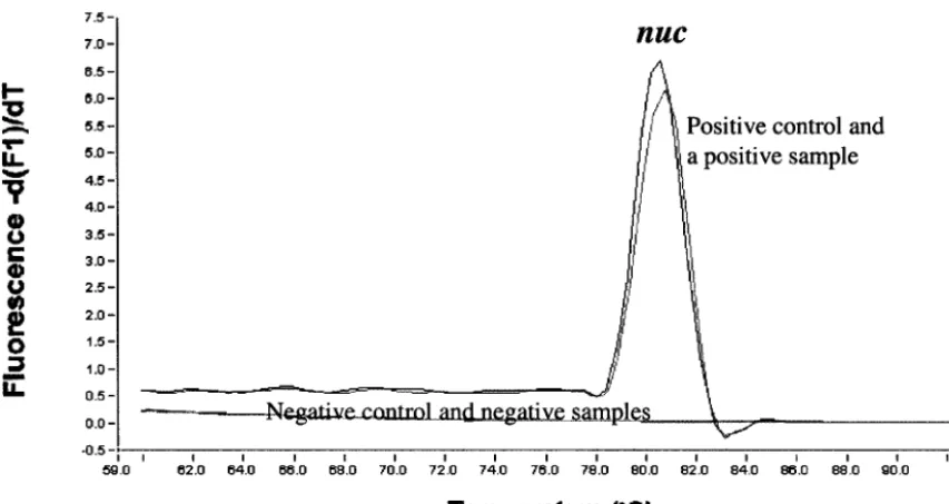
By use of the broth-PCR method, among the 304 clinical samples investigated, 44 samples (14.5%) were found to benuc
[image:2.603.48.540.81.316.2]positive after overnight cultivation in the MRSA broth (Fig. 1
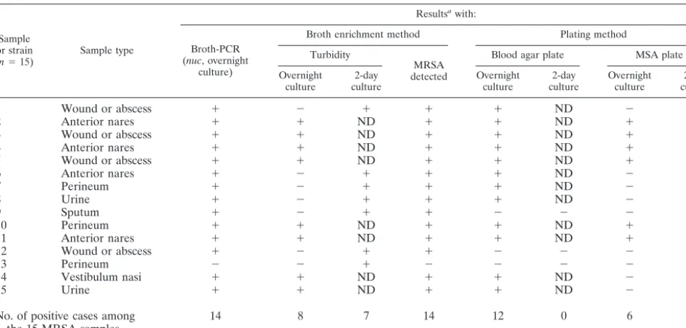
TABLE 1. The 15 MRSA strains detected by different screening methods and their sample types in the study
Sample or strain (n⫽15)
Sample type
Resultsawith:
Broth-PCR (nuc, overnight
culture)
Broth enrichment method Plating method
Turbidity
MRSA detected
Blood agar plate MSA plate
Overnight culture 2-day culture Overnight culture 2-day culture Overnight culture 2-day culture
1 Wound or abscess ⫹ ⫺ ⫹ ⫹ ⫹ ND ⫺ ⫺
2 Anterior nares ⫹ ⫹ ND ⫹ ⫹ ND ⫹ ND
3 Wound or abscess ⫹ ⫹ ND ⫹ ⫹ ND ⫹ ND
4 Anterior nares ⫹ ⫹ ND ⫹ ⫹ ND ⫹ ND
5 Wound or abscess ⫹ ⫹ ND ⫹ ⫹ ND ⫹ ND
6 Anterior nares ⫹ ⫺ ⫹ ⫹ ⫹ ND ⫺ ⫹
7 Perineum ⫹ ⫺ ⫹ ⫹ ⫹ ND ⫺ ⫹
8 Urine ⫹ ⫺ ⫹ ⫹ ⫹ ND ⫺ ⫹
9 Sputum ⫹ ⫺ ⫹ ⫹ ⫺ ⫺ ⫺ ⫹
10 Perineum ⫹ ⫹ ND ⫹ ⫹ ND ⫹ ND
11 Anterior nares ⫹ ⫹ ND ⫹ ⫹ ND ⫹ ND
12 Wound or abscess ⫹ ⫺ ⫹ ⫹ ⫺ ⫺ ⫺ ⫺
13 Perineum ⫺ ⫺ ⫹ ⫺ ⫺ ⫺ ⫺ ⫹
14 Vestibulum nasi ⫹ ⫹ ND ⫹ ⫹ ND ⫺ ⫹
15 Urine ⫹ ⫹ ND ⫹ ⫹ ND ⫺ ⫹
No. of positive cases among the 15 MRSA samples
14 8 7 14 12 0 6 7
a⫹
, positive;⫺, negative; ND, not determined.
VOL. 41, 2003 SCREENING AND IDENTIFICATION OF MRSA 2895
on May 15, 2020 by guest
http://jcm.asm.org/
and 2). Among the 260nuc-negative samples, one sample was proved to contain anucPCR inhibitor. Among the 15 MRSA strains detected in the study, 14 strains were derived from
nuc-positive samples and one was from anuc-negative sample (sample 13) in which nonucPCR inhibitor was detected. The strain (strain 13) missed in the broth-PCR assay was not de-tected by the broth enrichment method or the blood-agar plate method either, but it was found on the MSA plate, with only 2 colonies, after a 2-day cultivation (Table 1). The diagnostic values of the broth-PCR method were as follows: sensitivity, 93.3% (14 of 15); specificity, 89.6% (259 of 289); positive predictive value, 31.8% (14 of 44); and negative predictive value, 99.6% (259 of 260).
Among the 15 samples in which MRSA was found, turbid broth was observed in 8 samples after a 1-day incubation and in all 15 samples after incubation for 2 days (Table 1). How-ever, in one case (sample 13), MRSA was not isolated from the enriched broth; instead, a coagulase-negative staphylococcus
(CoNS) was isolated. The predominant bacteria isolated from the turbid broth in this study were CoNS (64% of the cases), while MRSA and MSSA accounted for only 11 and 4%, re-spectively. The diagnostic values of the broth-enrichment method, after 2 days of incubation, were as follows: sensitivity, 93.3% (14 of 15); specificity, 61.2% (177 of 289); positive predictive value, 11.1% (14 of 126); and negative predictive value, 99.4% (177 of 178).
By the plating method, when the two types of agar plates were taken together, 14 of the 15 MRSA strains were detected after incubation for 2 days, with a sensitivity (93.3%) as high as those of the other two methods, but the specificity was poor (10.1% [31 of 289]). MRSA was detected from sample 13, but not from sample 12 (Table 1), by this method.
The sensitivity assay showed that the detection limit of the broth-PCR fornucat the time of inoculation, time zero, was 104to 105 CFU/ml of inoculum in broth. After overnight
[image:3.603.79.506.68.294.2]in-cubation, the detection limit was improved to 100 to 101
FIG. 1. Tmcurves in the broth-PCR for detection of thenucgene (MRSA screening).
FIG. 2. Flowchart and results of the broth-PCR method in this study. a, the broth, when it turned turbid or after a maximal 5-day incubation, was spread to the agar plates and processed in a conventional way. b, one (sample 12) of the MRSA strains was not detected by the direct plating method. c, an MRSA strain was isolated from sample 13 on the MSA plate by the direct plating method.
on May 15, 2020 by guest
http://jcm.asm.org/
[image:3.603.77.500.556.695.2]CFU/ml of inoculum in broth (Fig. 3). After 3 and 6 days of incubation, the detection limits were 100to 101and 100to 102
CFU/ml of inoculum in broth, respectively.
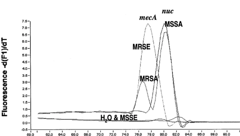
MRSA identification.All MRSA strains tested in the study
presented two peaks in the melting curve analysis; one peak was specific for themecAgene with aTmof 77.50 to 79.00°C,
and one was specific for thenucgene with a Tmof 79.90 to
80.60°C. MSSA strains had only anucpeak, MRSE strains had only amecApeak, and MSSE strains had no peak (Fig. 4). The
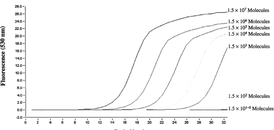
[image:4.603.110.472.69.345.2]mecAandnucgenes could be detected, by use of LightCycler real-time PCR, with an amount of DNA template as small as 1.5⫻102genomic molecules (Fig. 5).
FIG. 3. Sensitivity of the real-time PCR assay for detection of thenucgene from the overnight-cultured broth.
FIG. 4.Tmcurves for MRSA, MSSA, MRSE, and MSSE inmecAandnucduplex real-time PCR (MRSA identification).
VOL. 41, 2003 SCREENING AND IDENTIFICATION OF MRSA 2897
on May 15, 2020 by guest
http://jcm.asm.org/
[image:4.603.85.499.478.713.2]The specificity of the real-time PCR assay was further de-termined with a panel of 11 gram-negative and gram-positive standard strains of species other than S. aureus. No cross-reactivity was observed.
DISCUSSION
MRSA is now one of the most important nosocomial patho-gens worldwide. The prevalence of MRSA, however, varies markedly by country. The prevalence of MRSA in northern European countries is low; this is assumed to be due at least in part to the prompt implementation of aggressive infection con-trol measures (1, 6, 21, 22). Screening high-risk patients and health care workers for MRSA is one of the control measures. Several studies have found that such screening programs are cost-effective (3, 10, 12, 16).
The conventional culture methods are time- and labor-con-suming, and the diagnostic values are not as good as those of the new MRSA screening method. Especially because the number of samples for MRSA screening has increased dramat-ically in recent years, a more efficient method is needed to meet the clinical requirements. In the present study, we report a rapid and sensitive method, using MRSA-selective broth and real-time PCR, to screen for the presence of MRSA. This procedure started with enrichment of bacteria in a selective broth that favors the growth of MRSA, followed by detection of an S. aureus-specific gene (nuc) via real-time PCR. The
nuc-negative samples were further processed by a PCR inhib-itor assay to check fornucamplification-inhibitory substances.
nuc-negative samples without nuc PCR inhibitors were re-garded as MRSA negative. Compared to the broth enrichment method and the conventional plating method, which required incubation for 2 days, the overnight broth-PCR assay achieved the same sensitivity, 93.3%, and a higher specificity (89.6% versus 61.2 and 10.7%, respectively), positive predictive value
(31.8% versus 11.1 and 5.1%, respectively), and negative pre-dictive value (99.6% versus 99.4 and 96.9%, respectively).
The gene coding for methicillin resistance (mecA) was not targeted in this screening assay (broth-PCR), because the DNA template used at this step was directly extracted from the broth, which was a mixed culture. In these mixed cultures, CoNS were the predominant bacteria. It is known that meth-icillin resistance is frequent among CoNS on a global scale (8, 9, 17). At our hospital the incidence of methicillin resistance in CoNS is 45%. Therefore, the addition ofmecAin the screening method would not really increase the specificity and positive predictive value of the method, but it would give rise to false-positive results in cases such as those involving methicillin-resistant CoNS combined with MSSA.
The sensitivity assay showed that the nuc gene could be identified with an inoculum as low as 1 to 10 CFU/ml in the broth after overnight cultivation. Prolonged incubation did not produce higher sensitivity. The study indicated that overnight was a suitable interval for incubation before proceeding to the following PCR assay.
[image:5.603.49.528.64.292.2]Since the sample used fornucPCR was from the enriched broth, it is possible that PCR-inhibitory substances were present in the sample. To determine whethernucamplification inhibitors were present in the nuc-negative samples, a PCR internal inhibitor assay was performed as a complementary test to the broth-PCR method. The compositions of clinical sam-ples are usually complicated and display great variation, which could range from mixed culture to bacteria-free, so a possible way to check for PCR internal inhibitors is to add a minimal amount of positive DNA to the PCR system. In this study, the PCR inhibitor assay was aimed at determining ifnuc amplifi-cation was inhibited or disturbed in thosenuc-negative sam-ples, so the direct method was to add thenuc-positive DNA to the PCR system. One advantage of using the nuc-positive DNA was that all the other parameters in the PCR inhibitor
FIG. 5. Sensitivity of themecAandnucduplex real-time PCR assay, determined through serial dilutions of the template DNA extracted from MRSA strain CCUG 31966.
on May 15, 2020 by guest
http://jcm.asm.org/
assay, including the criteria in data analysis, were exactly the same as those in thenucPCR, so the inhibitory status of the
nuc PCR could be truly reflected through the PCR internal inhibitor assay. A negative result in the PCR inhibitor assay indicated thatnucamplification was inhibited or disturbed by the inhibitory substances in the sample DNA. Although only one sample was found to inhibitnucamplification in this study, and it was proved to be a non-MRSA sample (Fig. 2), we still think it necessary to include this assay in the broth-PCR method in order to reduce the number of falsenuc-negative cases.
With the application of this method, most of the negative samples (258 of 289 [89.3%]) can be identified the next morn-ing after samplmorn-ing, so the time to obtain the negative result can be reduced to 16 to 18 h. Meanwhile, as much as 84.9% (258 of 304) of the labor and cost spent on processingnuc-negative specimens on plates can be saved, which is especially cost-effective in countries such as Sweden, where the prevalence of MRSA remains low. This new MRSA-screening method has now been applied at our routine laboratory. The introduction of this new method is of significance in clinics. According to our policy, patients who are highly suspected to be colonized with MRSA, such as patients hospitalized abroad, are isolated in a single room until the cultures become MRSA negative. An earlier MRSA-negative report makes it possible for patients to go to open wards earlier, saving much expense.
One explanation for the missed MRSA strain in the broth-PCR assay and broth enrichment assay is that the inoculum of the bacteria in the broth was lower than the detection limits of the assays. PCR inhibitors were not detected from the ex-tracted DNA of this sample (sample 13), and noS. aureuswas isolated from the enriched broth.
Identification of MRSA by real-time PCR has been reported previously (7, 18, 20). In the study by Tan et al. (20), the detection of MRSA was performed with two separate PCRs, for an S. aureus-specific gene (Sa442) and the mecA gene, respectively, by using a cyanine 5-labeled probe and SYBR Green I. In the studies of Reischl et al. (18) and Grisold et al. (7), the PCR assay was carried out with two pairs of hybrid-ization probes labeled with different dyes. Here we report a real-time PCR assay which applies SYBR Green I as the only fluorescent agent for detectingmecAandnuc, simultaneously, in one PCR. By using the conditions described in the present study, bothmecAandnuccould be easily detected with a DNA template in a range of 1.5⫻102to 1.5⫻107genomic
mole-cules in the reaction mixture (Fig. 5). The DNA extracted from a single colony, by using our simple and rapid boiling proce-dure (described in Materials and Methods), usually has a con-centration of 1.5⫻106to 6.0⫻106molecules/l. Thus, the
duplex PCR with SYBR Green I described in this study is sensitive enough to be used as a routine diagnostic method. The specificity of this method has been tested with a panel of standard strains of species other thanS. aureus, and no cross-reactivity was observed. Furthermore, by the combination of the simple template DNA preparation with the rapid thermo-cycling of the LightCycler system, results are available within 1 h.
In conclusion, the broth-PCR method is an efficient MRSA-screening assay. The duplex real-time PCR for rapid
identifi-cation of MRSA is a valuable tool in a routine microbiological laboratory.
ACKNOWLEDGMENTS
We are grateful to Agneta Ahlin and Gerd Fesse´ for skillful tech-nical assistance.
REFERENCES
1. Boyce, J. M.2002. Understanding and controlling methicillin-resistant Staph-ylococcus aureusinfections. Infect. Control Hosp. Epidemiol.23:485–487. 2. Brakstad, O. G., K. Aasbakk, and J. A. Maeland.1992. Detection of
Staph-ylococcus aureusby polymerase chain reaction amplification of thenucgene. J. Clin. Microbiol.30:1654–1660.
3. Chaix, C., I. Durand-Zaleski, C. Alberti, and C. Brun-Buisson.1999. Control of endemic methicillin-resistantStaphylococcus aureus: a cost-benefit analy-sis in an intensive care unit. JAMA282:1745–1751.
4. Chambers, H. F.1997. Methicillin resistance in staphylococci: molecular and biochemical basis and clinical implications. Clin. Microbiol. Rev.10:781–791. 5. Enright, M. C., D. A. Robinson, G. Randle, E. J. Feil, H. Grundmann, and B. G. Spratt.2002. The evolutionary history of methicillin-resistant Staphy-lococcus aureus(MRSA). Proc. Natl. Acad. Sci. USA99:7687–7692. 6. Fluit, A. C., C. L. C. Wielders, J. Verhoef, and F.-J. Schmitz.2001.
Epide-miology and susceptibility of 3,051Staphylococcus aureusisolates from 25 university hospitals participating in the European SENTRY study. J. Clin. Microbiol.39:3727–3732.
7. Grisold, A. J., E. Leitner, G. Mu¨hlbauer, E. Marth, and H. H. Kessler.2002. Detection of methicillin-resistantStaphylococcus aureusand simultaneous confirmation by automated nucleic acid extraction and real-time PCR. J. Clin. Microbiol.40:2392–2397.
8. Jarlov, J. O.1999. Phenotypic characteristics of coagulase-negative staphy-lococci: typing and antibiotic susceptibility. APMIS91(Suppl.):1–42. 9. Jarlov, J. O., and N. Hoiby.1998. Coagulase-negative staphylococci in a
major Danish university hospital: diversity in antibiotic susceptibility be-tween wards. APMIS106:411–416.
10. Karchmer, T. B., L. J. Durbin, B. M. Simonton, and B. M. Farr.2002. Cost-effectiveness of active surveillance cultures and contact/droplet precau-tions for control of methicillin-resistantStaphylococcus aureus. J. Hosp. In-fect.51:126–132.
11. Kloos, W. E., and T. L. Bannerman.1999. Staphylococcus andMicrococcus, p. 264–282.InP. R. Murray, E. J. Baron, M. A. Pfaller, F. C. Tenover, and R. H. Yolken (ed.), Manual of clinical microbiology, 7th ed. American Society for Microbiology, Washington, D.C.
12. Kunori, T., B. Cookson, J. A. Roberts, S. Stone, and C. Kibbler.2002. Cost-effectiveness of different MRSA screening methods. J. Hosp. Infect.
51:189–200.
13. Livermore, D. M., and J. D. Williams.1996.-Lactams: mode of action and mechanisms of bacterial resistance, p. 502–578.InV. Lorian (ed.), Antibi-otics in laboratory medicine, 4th ed. Williams & Wilkins, Baltimore, Md. 14. Lowy, F. D.1998.Staphylococcus aureusinfections. N. Engl. J. Med.339:
520–532.
15. Oliveira, D. C., A. Tomasz, and H. de Lencastre.2002. Secrets of success of a human pathogen: molecular evolution of pandemic clones of methicillin-resistantStaphylococcus aureus. Lancet Infect. Dis.2:180–189.
16. Papia, G., M. Louie, A. Tralla, C. Johnson, V. Collins, and A. E. Simor.1999. Screening high-risk patients for methicillin-resistantStaphylococcus aureus
on admission to the hospital: is it cost effective? Infect. Control Hosp. Epidemiol.20:473–477.
17. Petinaki, E., F. Kontos, V. Miriagou, M. Maniati, F. Hatzi, A. N. Maniatis, and The Bacterial Resistance Study Group.2001. Survey of methicillin-resistant coagulase-negative staphylococci in the hospitals of central Greece. Int. J. Antimicrob. Agents18:563–566.
18. Reischl, U., H. J. Linde, M. Metz, B. Leppmeier, and N. Lehn.2000. Rapid identification of methicillin-resistantStaphylococcus aureus and simulta-neous species confirmation using real-time fluorescence PCR. J. Clin. Mi-crobiol.38:2429–2433.
19. Smyth, R. W., G. Kahlmeter, B. Olsson-Liljequist, and B. Hoffman.2001. Methods for identifying methicillin resistance inStaphylococcus aureus. J. Hosp. Infect.48:103–107.
20. Tan, T. Y., S. Corden, R. Barnes, and B. Cookson.2001. Rapid identification of methicillin-resistantStaphylococcus aureusfrom positive blood cultures by real-time fluorescence PCR. J. Clin. Microbiol.39:4529–4531.
21. Verhoef, J., D. Beaujean, H. Blok, A. Baars, A. Meyler, C. van der Werken, and A. Weersink.1999. A Dutch approach to methicillin-resistant Staphylo-coccus aureus. Eur. J. Clin. Microbiol. Infect. Dis.18:461–466.
22. Voss, A., D. Milatovic, C. Wallrauch-Schwarz, V. T. Rosdahl, and I. Braveny.
1994. Methicillin-resistantStaphylococcus aureus(MRSA) in Europe. Eur. J. Clin. Microbiol. Infect. Dis.13:50–55.
23. Waldvogel, F. A.2000. Gram-positive cocci, p. 2069–2092.InG. L. Mandell, J. E. Bennett, and R. Dolin (ed.), Principles and practice of infectious diseases, 5th ed. Churchill Livingstone, Philadelphia, Pa.
VOL. 41, 2003 SCREENING AND IDENTIFICATION OF MRSA 2899
on May 15, 2020 by guest
http://jcm.asm.org/
0095-1137/06/$08.00⫹0 doi:10.1128/JCM.44.2.675.2006
ERRATUM
Rapid Screening and Identification of Methicillin-Resistant
Staphylococcus aureus
from Clinical Samples by
Selective-Broth and Real-Time PCR Assay
Hong Fang and Go
¨ran Hedin
Department of Clinical Bacteriology, Huddinge University Hospital, SE-141 86 Stockholm, Sweden
Volume 41, no. 7, p. 2894–2899, 2003. Page 2894, column 2, lines 16 and 17 should read as follows: “. . . CCUG 31966 (a homogeneously, highly resistant strain) and CCUG 46147 (a heterogeneously, weakly resistant strain) . . .”