JOURNAL OFCLINICALMICROBIOLOGY, Apr. 2004, p. 1614–1619 Vol. 42, No. 4
0095-1137/04/$08.00⫹0 DOI: 10.1128/JCM.42.4.1614–1619.2004
Copyright © 2004, American Society for Microbiology. All Rights Reserved.
Simple Algorithm for Identification of
Bordetella pertussis
Pertactin
Gene Variants
Gae¨tan Muyldermans,
1Denis Pie´rard,
1* Nathalie Hoebrekx,
1Reza Advani,
2Shirley Van Amersfoorth,
3Iris De Schutter,
4Oriane Soetens,
1Leo Eeckhout,
1Anne Malfroot,
4and Sabine Lauwers
1Belgian Reference Laboratory for Pertussis, Department of Microbiology,1and Department of Pediatrics,4
Academisch Ziekenhuis Vrije Universiteit Brussel, Brussels, Belgium; Swedish Institute for Infectious
Disease Control, Solna, Sweden2; and Research Laboratory for Infectious Diseases,
National Institute for Public Health and Environment (RIVM),
Bilthoven, The Netherlands3
Received 18 July 2003/Returned for modification 5 September 2003/Accepted 12 January 2004
Studies performed in several countries have demonstrated the recent emergence and subsequent dominance of circulating Bordetella pertussis strains harboring pertactin and pertussis toxin variants not included in pertussis vaccines. Determination of the pertactin gene variants is commonly performed using a time-con-suming and expensive sequence analysis. We developed a simple and reliable pertactin typing algorithm suitable for large-scale screening. The assay correctly identified all pertactin alleles in representative strains. The typing of 231 clinical strains ofB. pertussisroutinely isolated in Belgium showed that this algorithm was adequate to identify less-frequentprntypes likeprn9andprn11.
Pertussis (whooping cough), mainly caused byBordetella
per-tussis, is still responsible for high morbidity and mortality
among children in many parts of the world. The reemergence of pertussis in several countries with high vaccine coverage (1, 3, 6) has led to renewed interest in whooping cough and in pertussis vaccine efficacy. Vaccine-induced immunity may se-lect among circulating strains for antigenic variants that are divergent from those of the vaccine strains (2, 5, 10, 12, 13). In a recent multilocus sequence typing study based on 15 genes coding for surface proteins, polymorphism was observed in the genes of pertactin (prn), pertussis toxin subunits S1 and S3
(ptxS1 and ptxS3), and the tracheal colonizing factor (tcfA)
(14).
Pertactin, a 69-kDa outer membrane protein, is a major virulence factor ofB. pertussisthat mediates adherence to host cells through an arginine-glycine-aspartic acid (RGD) se-quence (8). It is a component of many acellular subunit vac-cines. On the basis of polymorphism that occurs mainly in the repetitive region (11), 11prnalleles showing a variable number of GGXXP repeats (Fig. 1A) have been identified (EMBL GenBank database). As demonstrated in isolates collected in The Netherlands and Finland (12, 13), all isolates from the prevaccine period harbored the vaccine pertactin type prn1. However, these types were replaced during the 1990s by the nonvaccineprntypesprn2(72 and 27%, respectively, in these two countries),prn3 (12 and 63%, respectively), and prn4(0 and 9%, respectively). For the same period, high prevalences of nonvaccine strains were detected as well in other countries such as Italy (6%prn1, 41%prn2, 51%prn3, and 2%prn5), the United States (30%prn1and 70%prn2), and the United
King-dom (53% non-prn1) (2, 5, 10). The allelic variation in prn
typesprn1toprn4, which represent more than 98% of tested clinical isolates, is restricted to region 1. Typing of these alleles necessitates, therefore, the characterization of this region. However, some less frequentprntypes (prn5toprn11) showing variation in other regions have been identified. The determi-nation of the single nucleotide polymorphism T1595G, present
inprn6toprn8andprn10, provides a way to further
discrimi-nate these rare prn types against the predominantprn types
prn1toprn4.
After the introduction of the whole-cell vaccine in the 1950s, a progressive shift toward alleles not found in the vaccine strains has been observed. Since the currently circulating iso-lates differ from the vaccine strains (2, 4, 5, 10, 12, 13, 15), it is important to monitor the corresponding variations, particu-larly in view of the recent introduction of acellular vaccines in most national immunization programs. The composition of these vaccines is not uniform and differs in the number of antigens, which varies from 1 to 5, and in the antigenic sub-types included. The number of antigens or the inclusion of pertactin (only in vaccines with three or more components) may be responsible for differences in efficacy (7).
Monitoring of clinical isolates by simpler alternatives to se-quence analysis improves the turnaround time and expands the analytical capacity to additional strains. So far, the reference method for the determination of the pertactin type has been the sequencing of theprngene (11), a relatively time-consum-ing and expensive method. Recently, Ma¨kinen et al. (9) devel-oped a rapid identification method based on real-time PCR in combination with gel electrophoresis. Although the speed and simplicity of this approach make it an advantageous alternative to conventional sequencing of theprngene, this method needs an expensive real-time apparatus and does not differentiate new types such asprn6toprn11.
We developed a simple method for discriminating the most
* Corresponding author. Mailing address: Department of Microbi-ology, Academisch Ziekenhuis Vrije Universiteit Brussel, Laarbeek-laan 101, B-1090 Brussels, Belgium. Phone: 32 2 477 50 02. Fax: 32 2 477 50 15. E-mail: [email protected].
1614
on May 15, 2020 by guest
http://jcm.asm.org/
FIG. 1. (A) Polymorphism in theprngene region 1. The 11 known sequences are aligned. Dots represent identical bases; dashes represent deleted bases. The nucleotide numbers correspond to those ofprn1sequence accession number AJ011091 (GenBank database [http://www.ncbi .nlm.nih.gov/entrez]). (B) Primers prnC and prnE were designed to recognize a region of two constitutive GGAVP repeats and the overlap between GGAVP and GGFGP, respectively. They were used in two separate reactions with the prnD primer that hybridizes with a region downstream of region 1, as shown in panel C. (C) The amplification of isolates with primer pairs prnC-prnD and prnE-prnD allowed us to determine the total number of repeating units (GGAVP and GGFGP) and the number of GGFGP repeating units, respectively (as at right). n. a., no amplification.
on May 15, 2020 by guest
http://jcm.asm.org/
[image:2.603.53.523.284.643.2]frequent prn2 and prn3 types from the other less frequently occurring variants and to differentiateprn2andprn3from each other. Further identification of the few remaining isolates can then be performed by either sequencing or real-time PCR. A complete algorithm is proposed and successfully applied to 231
B. pertussisisolates, representing almost all the strains isolated
in Belgium from 1987 to 2001.
MATERIALS AND METHODS
Strains.The analysis was initially performed on a set of strains representing the differentprntypes kindly provided by J. Mertsola (University of Turku, Turku, Finland) and F. Mooi (National Institute for Public Health and the Environment, Bilthoven, The Netherlands). In addition, 231 clinicalB. pertussis isolates collected from 1987 through 2001 in our laboratory were used for this study.
Bacteria were grown on Regan-Lowe charcoal agar (Charcoal Agar; Oxoid Ltd., Basingstoke, England) containing 10% horse blood and cephalexin (Bor-detella Selective Supplement), incubated during 4 days at 35°C. For PCR, one isolated colony was resuspended in TE (10 mM Tris-HCl [pH 8], 1 mM EDTA) and boiled for 10 min.
Primers.Primers QJF3 and QJR1 were as designed by Ma¨kinen et al. (9). The others were designed on the basis of sequences available in the GenBank data-base (http://www.ncbi.nlm.nih.gov/entrez) and are shown in Table 1.
Sequence-specific PCR for typing the repetitive region 1.Two amplifications were performed for the determination of the polymorphism of theprnregion 1 using either the prnC-prnD or prnE-prnD primer pair (Fig. 1B and C). The PCR method was optimized for the following variables: Mg concentration, annealing temperature, standard or hot-start PCR, number of cycles, and input concentra-tion.
The 50-l reaction mixture contained 1⫻Reaction Buffer II (Applied Biosys-tems, Foster City, Calif.), 1.5 mM MgCl2, a 200M concentration of each deoxynucleoside triphosphate, a 1M concentration of each primer, and 1 U of Amplitaq Gold DNA polymerase (Applied Biosystems). Amplification was per-formed in a DNA Thermal Cycler 480 (Applied Biosystems) using the following program: 94°C for 10 min and then 40 cycles of 94°C for 1 min, 66°C for 1 min 30 s, and 72°C for 2 min.
The amplification product was further analyzed on 3% LSI MP agarose (Life Science International, Zellik, Belgium) gel, and the length of the DNA band was compared with those of control samples fromprn1toprn4.
Real-time PCR for the identification of the T1595G point mutation.The real-time PCR method was adapted from that of Ma¨kinen et al. (8) for SYBR Green I DNA detection. Briefly, two amplifications were performed for the determination of the polymorphism at position 1595. Primer pairs QJF3-QJR1 and QJF4-QJR1 preferentially amplify (i)prn1toprn5,prn9, andprn11and (ii) prn6toprn8andprn10, respectively. The PCR mixture was optimized for an I-cycler apparatus (Bio-Rad, Hercules, Calif.) using the qPCR Core Kit for SYBR Green I (Eurogentec, Seraing, Belgium) with the addition of 0.01M fluorescein and 1l of boiled sample. After an initial denaturation step at 94°C for 10 min, 50 cycles of denaturation at 94°C for 15 s, annealing at 62°C for 15 s, and extension at 72°C for 30 s were performed. Measurement of fluorescence at 495 nm at the end of each extension step allows the detection of the SYBR Green I bound to the amplified double-stranded DNA. The increase in fluores-cence signal during the PCR process correlates with PCR product accumulation. The parameter used to type the single nucleotide polymorphism was the cycle threshold (Ct). This is the cycle number at which the reaction begins to be exponential. It is determined by the intersection between the amplification curve and the Ct level, calculated as the 10-fold standard deviation of the observed fluorescence signal between cycles 5 and 20. QJF3 and QJF4 (Table 1) primers were designed to preferentially anneal and extend the alleles with T1595T and T1595G mutations, respectively. Using each of these primers in combination with the antisense QJR1 results in a difference in amplification efficiency. The PCR with the lowest Ct was considered to contain the primers with the most specific binding. After amplification, melting curve analysis of the PCR product was used to differentiate between specific and nonspecific amplification products and thereby confirming the previous amplification result. These curves were obtained by slowly changing the temperature of the reaction solution from 55 to 95°C while continuously collecting fluorescence data. This increase in tempera-ture induces the PCR products to denatempera-ture, which is accompanied by a decrease in the fluorescence from solutions containing the SYBR Green I dye. To improve the visualization of the melting temperature (Tm), melting peaks were derived from the initial melting curves (relative fluorescence units [RFU] versus tem-perature [T]) by plotting the negative derivative of fluorescence over tempera-ture versus temperatempera-ture [⫺d(RFU)/dTversusT].
Sequencing of the pertactin gene.Sequencing of theprngene from isolates Bord4, Bord49, and Bord68 was performed as described by Mooi et al. (13).
RESULTS
Development of the sequence-specific PCR for the identifi-cation ofprnregion 1 polymorphism.The polymorphism in the
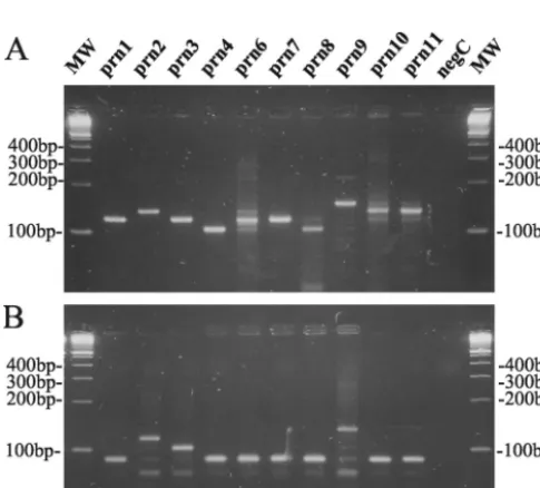
prngene is mainly confined to region 1 (13). The number of repetitive GGXXP units in region 1 was determined by two sequence-specific PCR tests as shown in Fig. 1. These reactions were performed on a representative isolate from eachprntype as described in Materials and Methods, except for prn5, for which no isolate was available. Using the primer pair prnC-prnD, amplification fragments were obtained with different lengths corresponding to the total number of repeat units GGAVP and GGFGP (Fig. 1C). This allowed us to discrimi-nate the prn1, prn3, prn6, andprn7 types (120-bp fragment) from theprn2,prn10, andprn11types (135-bp fragment), the
prn4 and prn8 types (105-bp fragment), and the prn9 type
[image:3.603.43.286.467.686.2]FIG. 2. Ethidium bromide-stained 3% LSI MP agarose (Life Sci-ence International) gel containingB. pertussis DNA amplified with primer pairs prnC-prnD (A) and prnE-prnD (B) from strains with knownprntypes.
TABLE 1. Primers used in this studya
Primer Sequence (5⬘to 3⬘) Position Reference
prnC GTGCGGTTCCCGGCGGTG 827–844 This study prnD GCTCCACGCTGGAGCCCG 946–929 This study prnE TGCGGTTCCCGGCGGCTTC 858–876 This study QJF3 GCTGGTGCAGACGCCAGT 1605–1622 9 QJF4 GCTGGTGCAGACGCCAGG 1605–1622 This study QJR1 CCGATATCGACCTTGCC 1676–1660 9
aThe position numbers indicate the position of bases relative to the first start codon ofprn1(accession number AJ011091). The differences between the prim-ers prnC and prnE as well as those between QJF3 and QJF4 are shown in boldface type.
1616 MUYLDERMANS ET AL. J. CLIN. MICROBIOL.
on May 15, 2020 by guest
http://jcm.asm.org/
(150-bp fragment) as shown in Fig. 2A. Due to the presence of point mutations in the prnC primer binding site (Fig. 1A and B), it was not possible to obtain single DNA bands for some
prntypes. Lowering the specificity of PCR conditions (increas-ing the Mg concentration or decreas(increas-ing the anneal(increas-ing temper-ature) increased the number and intensity of the secondary bands on agarose gel, while increasing the specificity of PCR conditions resulted in the disappearance of the appropriate fragments forprn6,prn8, andprn10(data not shown).
On the other hand, with the primer pair prnE-prnD, ampli-fication fragments with different sizes corresponding to the number of GGFGP repeat units (Fig. 1C) were obtained. It allowed us to discriminate theprntypesprn1,prn4,prn6,prn7,
prn8, prn10, and prn11 (89-bp fragment) from types prn3
(104-bp fragment),prn2(119-bp fragment), andprn9(134-bp fragment) as shown in Fig. 2B.
Compared to previous sequencing data (Fig. 1A), all type strains were correctly categorized by this PCR method. Com-bining the two sequence-specific amplification methods al-lowed us to differentiate the most frequentprntypes,prn2and
prn3, from the others as summarized in Fig. 3. In addition, this method allowed us to discriminate the 11 knownprntypes into the following seven groups: prn1, prn6, and prn7;prn2;prn3;
prn4andprn8;prn5;prn9; andprn10andprn11.
Real-time PCR for the identification of the T1595G point mutation. As the determination of polymorphism with the sequence-specific PCR developed in-house was unable to dis-criminate theprn1 type from the rare prn6orprn7 type, we performed a SYBR Green I real-time PCR adapted from a previously described method (9). This method could
discrimi-nateprn4fromprn8andprn10fromprn11as well.
Primers QJF3 and QJF4 differ by only one nucleotide at the
3⬘end (Table 1). Since the prntypesprn1 toprn5,prn9, and
prn11 contain a T at position 1595, these type strains are
preferentially recognized by the QJF3 primer, which contains a T at the 3⬘end. Preferential amplification is obtained with this sense primer in combination with the antisense QJR1 primer compared to the QJF4-QJR1 pair. On the other hand, asprn6
toprn8andprn10contain a G at position 1595, these alleles are
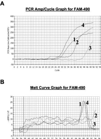
preferentially amplified by the QJF4-QJR1 primer pair. In Fig. 4A a comparison of the amplification profiles fromprn1 and
prn7are shown for the real-time amplification with the primer pairs QJF3-QJR1 and QJF4-QJR1. The lower value of the intersection between the curve and the Ct level with primer pair QJF3-QJR1 (30.7) compared to that for primer pair QJF4-QJR1 (37.4) confirms that T1595T is present in theprn1
[image:4.603.48.280.66.295.2]allele. In contrast, the Ct for the prn7using the primer pair QJF4-QJR1 is 15 cycles lower than that obtained with QJF3-QJR1. The melting curve analysis confirmed this interpreta-tion, as a specific melting peak was observed for theprn1strain amplified with the QJF3-QJR1 primer pair at 88.0°C and not for the QJF4-QJR1 primer pair (Fig. 4B). For theprn7strain, a specific melting peak (88.5°C) was observed only with the QJF4-QJR1 amplification. No specific amplification could be detected for the negative control and the melting curve anal-ysis did not show any peak (data not shown).
Table 2 summarizes the Ct for the representative strains and theirTms. The meanTmwas 88.0°C (range, 87.5 to 88.5°C).
Use of theprntyping algorithm for typing clinical isolates.
A total of 231 clinical isolates collected in Belgium from 1987 through 2001 were typed using our algorithm (Fig. 3): 22 (9.5%), 140 (60.6%), and 66 (28.6%) containedprn1,prn2, and
prn3, respectively. The sequence-specific PCR allowed us to discriminate three isolates (Bord4, Bord49, and Bord68) with
prntypes differing from the more-frequentprntypesprn1 to
prn3. Since Bord4 and Bord68 contained a 149-bp fragment in the amplification reaction mixture with primer pair prnC-prnD (Fig. 5A),prngenes from both isolates were classified asprn9. The presence of the 134-bp fragment in the amplification re-action mixture using the primer pair prnE-prnD (Fig. 5B) and the presence of the T1595T mutation confirmed this result (Table 2). On basis of the presence of the 134- and 89-bp fragments for the amplification reaction mixture using primer pairs prnC-prnD and prnE-prnD, respectively (Fig. 5), theprn
gene from isolate Bord49 was classified as aprn10 orprn11. The presence of the T1595T mutation as shown by the real-time PCR (Table 2) allowed us to further characterize this gene as aprn11. The type of these three rareprnvariants was subsequently confirmed by sequence analysis (GenBank acces-sion numbers AY382174, AY382175, and AY382176).
DISCUSSION
Sequencing theprngene in regions 1 and 2 is probably the most accurate method for typing the prngene. It was previ-ously demonstrated in numerous studies thatprn1,prn2, and
prn3types predominate in different parts of the world (2, 4, 5, 10, 12, 13, 15). Since these types differ only in region 1, it was proposed that region 1 in all isolates should be sequenced and when a novel variant of region 1 was found, that the complete
prngene should be sequenced (11). However, this method is time-consuming and expensive.
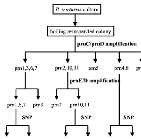
FIG. 3. Workflow for typingprnalleles. The sequence-specific PCR method using the primer pairs prnC-prnD and prnE-prnD allows the efficient identification of the major typesprn2 andprn3. A further identification of the minor typesprn1andprn4toprn11can then be performed by a single nucleotide polymorphism determination method (SNP) by either real-time PCR or sequencing.
on May 15, 2020 by guest
http://jcm.asm.org/
In this study, a simple algorithm was developed for typing the prngene from B. pertussis. The method proved to be ac-curate and has a short turnaround time of 1 day. It allows the differentiation of the commonprntypesprn1toprn3from each other. Althoughprn6andprn7could not be differentiated, the method allows the identification of less-frequent prn types. Furthermore, it is convenient for large-scale screening while rapidly providing an epidemiologic overview of the circulating strains. With these advantages, this method is a convenient alternative to the expensive and time-consuming sequence typ-ing method. An important characteristic of the algorithm is the possibility to detect novel types as shown by the characteriza-tion of the three isolates with rareprntypes.
The algorithm used in this work is based on two
amplifica-tion reacamplifica-tions using the primer pairs prnC-prnD and prnE-prnD. The former was mainly used for the differentiation of knownprntypes, with the latter providing additional discrim-ination and/or confirmation. Due to the presence of point mutations in the prnC primer binding site (Fig. 1A and B), it was not possible to fully optimize the PCR test. The presence of aspecific bands for some rare isolates demonstrates that the PCR conditions are highly critical. Whenever these aspecific bands are observed, performing the described real-time PCR or sequencing of the prn gene is necessary to confirm the observed types.
The primer pairs prnC-prnD and prnE-prnD were specifi-cally designed for the differentiation ofprn1toprn4, the only types that were known at the initiation of the study. Using both
FIG. 4. (A) Amplification profiles of real-time PCR with SYBR Green I. Curves 1 and 2, amplification profile for a representativeprn1strain with primer pairs QJF3-QJR1 and QJF4-QJR1, respectively; curves 3 and 4, amplification profile for a representativeprn7strain with primer pairs QJF3-QJR1 and QJF4-QJR1, respectively. (B) Melting curves from the specific amplification assays shown in panel A.
1618 MUYLDERMANS ET AL. J. CLIN. MICROBIOL.
on May 15, 2020 by guest
http://jcm.asm.org/
[image:5.603.132.460.69.517.2]amplifications, we succeeded in differentiating the 11 types known at present into seven groups. These groups were further differentiated by an additional real-time PCR method. Since the difference in the number of repeat units results in a size difference of only 15 bp, we designed a PCR in which short amplification products were obtained, allowing us to differen-tiate the types on basis of the differences in amplification fragments. In order to accurately estimate the size of the ob-tained fragments, a high-resolution agarose gel was needed, and the amplification products were always compared to those of the positive controls.
Our study demonstrated that 98% of the Belgian isolates
belong to theprntypesprn1toprn3. As much as 9% still belong to theprn1type, theprntype of the strains that are included in the conventional whole-cell vaccine. Since no isolates are avail-able for the prevaccination period in Belgium, no comparison could be performed for this period.
This method is convenient for large-scale screening of all available isolates in a short time rather than typing a fraction of the collected isolates when sequencing is performed. In the future, application of this method—which is faster and less laborious than sequencing—to other genetic loci such asptxS1,
ptxS3, and tcfA will improve the monitoring of the strains.
Finally, this method will also be a useful tool for the collection of typing data from the pre- and post-acellular vaccine eras.
ACKNOWLEDGMENT
This work was supported by OZR grant 624 (Vrije Universiteit Brussel, Brussels, Belgium).
REFERENCES
1. Andrews, R., A. Herceg, and C. Roberts.1997. Pertussis notifications in Australia, 1991 to 1997. Commun. Dis. Intell.21:145–148.
2. Cassiday, P., G. Sanden, K. Heuvelman, F. Mooi, K. M. Bisgard, and T. Popovic.2000. Polymorphism inBordetella pertussispertactin and pertussis toxin virulence factors in the United States, 1935–1999. J. Infect. Dis.182:
1402–1408.
3. De Melker, H. E., J. F. Schellekens, S. E. Neppelenbroek, F. R. Mooi, H. C. Rumke, and M. A. Conyn-Van Spaendonck.2000. Reemergence of pertussis in the highly vaccinated population of the Netherlands: observations on surveillance data. Emerg. Infect. Dis.6:348–357.
4. De Schutter, I., A. Malfroot, I. Dab, N. Hoebrekx, G. Muyldermans, D. Pie´rard, and S. Lauwers.2003. Molecular typing ofBordetella pertussis iso-lates recovered from Belgian children and their household members. Clin. Infect. Dis.36:1391–1396.
5. Fry, N. K., S. Neal, T. G. Harrison, E. Miller, R. Matthews, and R. C. George.2001. Genotypic variation in theBordetella pertussisvirulence factors pertactin and pertussis toxin in historical and recent clinical isolates in the United Kingdom. Infect. Immun.69:5520–5528.
6. Guris, D., P. M. Strebel, B. Bardenheier, M. Brennan, R. Tachdjian, E. Finch, M. Wharton, and J. R. Livengood.1999. Changing epidemiology of pertussis in the United States: increasing reported incidence among adoles-cents and adults, 1990–1996. Clin. Infect. Dis.28:1230–1237.
7. Jefferson, T., M. Rudin, and C. Di Pietrantonj.2003. Systematic review of the effects of pertussis vaccines in children. Vaccine21:2012–2023. 8. Leininger, E., C. A. Ewanowich, A. Bhargava, M. S. Peppler, J. G. Kenimer,
and M. J. Brennan.1992. Comparative roles of the Arg-Gly-Asp sequence present in theBordetella pertussisadhesins pertactin and filamentous hem-agglutinin. Infect. Immun.60:2380–2385.
9. Ma¨kinen, J., M. K. Viljanen, J. Mertsola, H. Arvilommi, and Q. He.2001. Rapid identification of Bordetella pertussispertactin gene variants using LightCycler real-time polymerase chain reaction combined with melting curve analysis and gel electrophoresis. Emerg. Infect. Dis.7:952–958. 10. Mastrantonio, P., P. Spigaglia, H. van Oirschot, H. G. van der Heide, K.
Heuvelman, P. Stefanelli, and F. R. Mooi.1999. Antigenic variants in Bor-detella pertussisstrains isolated from vaccinated and unvaccinated children. Microbiology145:2069–2075.
11. Mooi, F. R., H. Hallander, C. H. Wirsing von Konig, B. Hoet, and N. Guiso.
2000. Epidemiological typing ofBordetella pertussisisolates: recommenda-tions for a standard methodology. Eur. J. Clin. Microbiol. Infect. Dis.19:
174–181.
12. Mooi, F. R., Q. He, H. van Oirschot, and J. Mertsola.1999. Variation in the Bordetella pertussisvirulence factors pertussis toxin and pertactin in vaccine strains and clinical isolates in Finland. Infect. Immun.67:3133–3134. 13. Mooi, F. R., H. van Oirschot, K. Heuvelman, H. G. van der Heide, W.
Gaastra, and R. J. Willems.1998. Polymorphism in theBordetella pertussis virulence factors P.69/pertactin and pertussis toxin in The Netherlands: tem-poral trends and evidence for vaccine-driven evolution. Infect. Immun.66:
670–675.
14. van Loo, I. H., K. J. Heuvelman, A. J. King, and F. R. Mooi.2002. Multilocus sequence typing of Bordetella pertussis based on surface protein genes. J. Clin. Microbiol.40:1994–2001.
[image:6.603.48.279.462.683.2]15. Weber, C., C. Boursaux-Eude, G. Coralie, V. Caro, and N. Guiso.2001. Polymorphism ofBordetella pertussisisolates circulating for the last 10 years in France, where a single effective whole-cell vaccine has been used for more than 30 years. J. Clin. Microbiol.39:4396–4403.
TABLE 2. Ct values for representativeprnstrains and clinical isolates with rareprntypes for the QJF3-QJR1 and
QJF4-QJR1 amplifications
Strain typeprn Ct value (Tm
a[°C]) for:
Result QJF3-QJR1 QJF4-QJR1
prn1 30.7 (88.0) 37.4 (—) 1595T prn2 29.5 (88.0) 36.5 (—) 1595T prn3 29.6 (80.0) 36.6 (—) 1595T prn4 28.2 (87.5) 38.9 (—) 1595T prn6 41.7 (—) 28.9 (88.5) 1595G prn7 44.5 (—) 29.7 (88.5) 1595G prn8 40.8 (—) 27.3 (88.5) 1595G prn9 28.8 (88.0) 35.9 (—) 1595T prn10 40.0 (—) 27.9 (88.5) 1595G prn11 29.1 (88.0) 35.1 (—) 1595T
Bord4 prn9 34.1 (87.5) 40.2 (—) 1595T
Bord49 prn11 32.4 (88.5) 42.7 (—) 1595T Bord68 prn9 25.8 (87.5) 36.2 (—) 1595T
Negative control ⬎50 (—) 45.7 (—) Negative aT
mof the main peak. —, no specific melting peak observed.
FIG. 5. Ethidium bromide-stained 3% LSI MP agarose (Life Sci-ence International) gel containingB. pertussis DNA amplified with primer pairs prnC-prnD (A) and prnE-prnD (B).
![FIG. 1. (A) Polymorphism in the gene region 1. The 11 known sequences are aligned. Dots represent identical bases; dashes represent.nlm.nih.gov/entrez])](https://thumb-us.123doks.com/thumbv2/123dok_us/8250469.834187/2.603.53.523.284.643/polymorphism-region-sequences-aligned-represent-identical-represent-entrez.webp)