Progress and Challenges towards
Point-of-Care Diagnostic Development for
Dengue
Junxiong Pang,a,bPo Ying Chia,aDavid C. Lye,a,c,dYee Sin Leoa,b,c,d
Institute of Infectious Diseases and Epidemiology, Tan Tock Seng Hospital, Singaporea; Saw Swee Hock School of Public Health, National University of Singapore, Singaporeb; Lee Kong Chian School of Medicine, Nanyang Technological University, Singaporec; Yong Loo Lin School of Medicine, National University of Singapore, Singapored
ABSTRACT Dengue detection strategies involve viral RNA, antigen, and/or antibody detection. Each strategy has its advantages and disadvantages. Optimal, user-friendly, rapid diagnostic tests based on immunochromatographic assays are pragmatic point-of-care tests (POCTs) in regions where dengue is endemic where there are limited labora-tory capabilities and optimal storage conditions. Increasingly, there is a greater public health significance for a multiplexing assay that differentiates dengue from Zika or pathogens with similar clinical presentations. Although there have been many assay/ platform developments toward POCTs, independent validation and implementation re-main very limited. This review highlights the current key progress and challenges toward the development of a dengue POCT.
KEYWORDS dengue fever, biotechnology, clinical diagnosis, diagnostics tools, flavivirus, neutralizing antibodies, point-of-care test, serology, viral clearance
DENGUE EPIDEMIOLOGY AND CLINICAL SEVERITY
D
engue is the most prevalent arboviral disease in humans, with 3.6 billion peopleliving in areas with a significant risk of disease transmission and an estimated 390 million dengue virus infections and 96 million dengue cases annually (1). Dengue is endemic in the tropical and subtropical regions of the world due to the adaptability of Aedesmosquitoes in the human living environment. Due to global warming and climate change, a geographic expansion of the dengue epidemic beyond tropical regions has been observed (2). Dengue is caused by infection with the dengue virus
(DENV), which belongs to the family Flaviviridaeand genus flavivirus. There are four
antigenically distinct serotypes of dengue virus (DENV-1, DENV-2, DENV-3, and DENV-4), which may cocirculate in these regions where the dengue is hyperendemic. Rapid urbanization over the past decades has facilitated endemicity, as dengue is nantly found in semiurban and urban areas (3). Dengue virus infection was predomi-nantly a pediatric disease in Southeast Asia and western Pacific regions (4), where children of less than 15 years of age were at a higher risk of infection and dengue hemorrhagic fever (DHF) than adults (5). However, over the years, there has been a gradual increase in reports of dengue among older children and young adults age 15 years and above in Singapore (6–8), Vietnam (9), and Thailand (10–13).
Dengue fever (DF) causes a wide spectrum of presentations ranging from mild self-limiting illness to severe disease. Based on the 1997 WHO dengue classification, patients presenting with all four criteria of fever, hemorrhagic diathesis, thrombocyto-penia, and evidence of plasma leakage will be classified as having DHF, or dengue shock syndrome (DSS) if they present with symptoms of shock. The WHO modified the severity classification in 2009 into dengue, dengue with warning signs, and severe dengue (14). Seven warning signs were introduced to assist clinicians in the triage of
Accepted manuscript posted online13 September 2017
CitationPang J, Chia PY, Lye DC, Leo YS. 2017. Progress and challenges towards point-of-care diagnostic development for dengue. J Clin
Microbiol 55:3339 –3349.https://doi.org/10
.1128/JCM.00707-17.
EditorColleen Suzanne Kraft, Emory University
Copyright© 2017 American Society for
Microbiology.All Rights Reserved.
Address correspondence to Junxiong Pang, [email protected].
crossm
on May 16, 2020 by guest
http://jcm.asm.org/
patients who need inpatient care and/or closer monitoring. However, the diagnosis of dengue virus infections cannot rely solely on clinical manifestations, since many patients are either asymptomatic or present with a nonspecific fever requiring a differential diagnosis to distinguish exposure to DENVs from other febrile episode-inducing diseases. Therefore, rapid, accurate, relatively low-cost diagnostic tools for DENV are critical for the confirmation of suspected clinical cases, which in turn, is the key to effective disease management and control, especially in developing countries with limited and inaccessible health care resources. With more large outbreaks in the developing countries, the development of a reliable and accurate point-of-care test (POCT) for dengue detection remains an urgent task (15, 16).
As recommended by the WHO Special Programme for Research and Training in Tropical Diseases (TDR) (17), the specifications of an ideal dengue test are that it should (i) distinguish between dengue and other diseases with similar clinical presentations (such as malaria, chikungunya, and other flaviviruses), (ii) be highly sensitive, (iii) provide rapid results, (iv) be inexpensive, (v) be easy to use, and (vi) be stable at temperatures above 30°C for usage in the field and in primary health care settings, usually with very limited/no optimal storage options. Dengue nucleic acid amplification tests (NAAT), including PCR, serologic tests, including nonstructural protein (NS1) antigen tests, and antibody tests by enzyme-linked immunosorbent assay (ELISA) or immunochromatography are some of the recent advances in laboratory medicine. Some of these modalities have been developed as POCTs, allowing for rapid identifi-cation of patients with dengue virus infection in different settings. However, some technologies are still in the development phase in the laboratory setting. This review aims to highlight the current progress and challenges in dengue POCT development using key examples.
PROGRESS AND CHALLENGES IN THE DEVELOPMENT OF DENGUE VIRAL RNA DETECTION ASSAYS FOR POCT
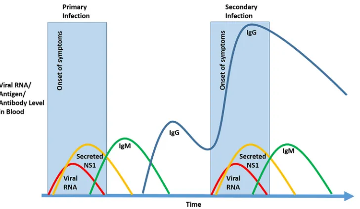
Dengue viral RNA can be detected using NAAT or PCR on tissues, whole blood, or sera taken from patients in the acute phase of the disease (Fig. 1), likely with undif-ferentiated fever. Various protocols have been developed that identify DENV using primers directed to serotype-specific regions of the genome (18). However, reverse transcriptase PCR (RT-PCR) is usually limited by a long processing time of about 2 h or more, the need for reasonable copies of viral load for first amplification (not suitable for patients presenting for more than 7 days), and multiple heat denaturation steps for the FIG 1Schematic profiles of dengue viremia, NS-1, and anti-dengue IgM and IgG in blood during primary and secondary dengue infections over time.
on May 16, 2020 by guest
http://jcm.asm.org/
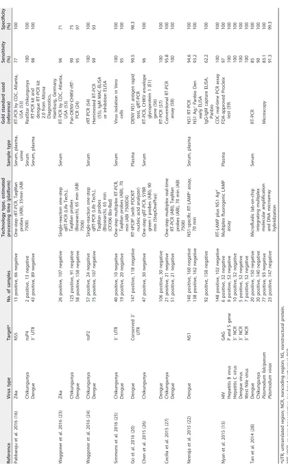
[image:2.585.43.407.69.281.2]cycling of DNA synthesis. Moreover, most RT-PCR detection assays rely heavily on a high precision thermocycler, which is expensive and bulky and not practical as a POCT. Recently, a novel, inexpensive, and user-friendly diagnostic assay based on a reverse transcription-insulated isothermal PCR (RT-iiPCR) method (19) was developed and validated to detect all four serotypes of DENV in clinical samples without an expensive thermocycler (20). This technology involves a fluorescent probe hydrolysis-based iiPCR for amplification and detection of nucleic acid. The iiPCR is highly sensitive and specific for the detection of both DNA and RNA. This assay could be performed with a single heating source with Rayleigh-Bénard convection driving the fluid cycling through temperature gradients. The three PCR steps, namely, denaturation, annealing, and extension, can be completed at different zones within the capillary tube. The integra-tion of an optical detecintegra-tion module in the device allows automatic detecintegra-tion and interpretation of iiPCR results. The diagnostic sensitivity and specificity of the pan-DENV RT-iiPCR with a portable POCKIT nucleic acid analyzer (GeneReach USA, Lexington, MA, USA) were 90.5% and 98.3%, respectively, compared to the results of a CDC multiplex DENV-1 to DENV-4 quantitative RT-PCR (qRT-PCR) assay as controls (Table 1). This new RT-iiPCR POCT, with lyophilized reagents, can provide a highly reliable, sensitive, and specific point-of-care diagnostic assay for the diagnosis of DENV in provincial clinics and hospitals in developing countries with no or limited accessibility to laboratory capabilities. However, a limited number of samples (approximately eight samples) can be processed at one time, which may not be cost- and time-effective during an outbreak setting. Furthermore, there is a lack of differentiation of dengue serotypes and other flaviviruses that may be endemic in the geographical areas of interest for surveillance purposes.
Another advanced technique, known as reverse transcription-loop-mediated iso-thermal amplification (RT-LAMP), has been developed (21) to combine the multiple processes of conventional PCR to detect RNA viruses into a one-step process for POCT development. The sample is mixed with the primers, reverse transcriptase, and DNA polymerase before the reaction takes place under a constant temperature in a simple heat block with a level of precision similar to that of conventional PCR. An NS1 serotype-specific RT-LAMP assay has been validated for the rapid detection and differ-entiation of dengue virus serotypes via the NS1 genomic region (22). This assay provides sensitivity and specificity of 94.6% and 100%, respectively (Table 1). To facilitate the field application of the RT-LAMP assay, monitoring of amplification can be done visually with an unaided eye after adding SYBR green I intercalating dye to the reaction mix. A positive reaction turns the reaction mixture green under white light, which fluoresces under UV irradiation. The reaction mix remains orange and nonfluo-rescent in the absence of amplification. This change of color is permanent and thus can be kept for record purposes. A similar assay using LAMP with additional multiplexing capability was also developed and validated, with close to 100% sensitivity and specificity for differentiating dengue virus from other common flaviviruses (15) (Table 1). However, it is not clear if the necessary reagents can be stored for a prolonged period of time in the field without a refrigerator.
With the increasing importance of differentiating dengue virus from other flavivi-ruses, there is an increasing number of assays developed and validated for detecting and differentiating dengue viral RNA from other diseases with similar clinical presen-tations, such as Zika (16, 23), chikungunya (16, 23–28), malaria (28), and those from other flaviviruses (15). Of significant interest to the recent Zika outbreak, a one-step multiplex RT-PCR assay using clinical samples from patients in Canada (16) and Nica-ragua (23) (Table 1) was highly sensitive and specific for differentiating dengue from Zika and chikungunya. Besides the common one-step multiplex real-time RT-PCR technology to identify and differentiate dengue from other common pathogens, a microfluidic lab-on-chip integrating multiplex molecular amplification with DNA mi-croarray hybridization was also developed and validated for the simultaneous detection and species differentiation of 26 globally important tropical pathogens (28) (Table 1). Its performance is comparable to those of the other one-step multiplex real-time PCR
on May 16, 2020 by guest
http://jcm.asm.org/
TABLE 1 Summary of recent key developments in dengue viral RNA detection assays Reference Virus type Target a No. of samples Technology type, estimated processing time (platform) Sample type Gold standard used (reference) Sensitivity (%) Specificity (%) Pabbaraju et al. 2016 (16 ) Zika NS5 13 positive, 66 negative One-step rRT-PCR, TaqMan probes (ABI), 35min (ABI 7500) Serum, plasma, urine RT-PCR by CDC, Atlanta, USA (53 ) 77 100 Chikungunya nsP4 2 positive, 13 negative Serum RealStar chikungunya RT-PCR kit and dengue RT-PCR kit 2.0 from Altona Diagnostics, Hamburg, Germany 100 100 Dengue 3 = UTR 43 positive, 89 negative Serum, plasma 98 100 Waggoner et al. 2016 (23 ) Zika 26 positive, 107 negative Single-reaction one-step qRT-PCR (Life Tech.), TaqMan probes (Biosearch), 65 min (ABI 7500) Serum RT-PCR by CDC, Atlanta, USA (53 ) 96 71 Chikungunya 125 positive, 91 negative Pan-DENV-CHIKV-rRT-PCR (24 ) 90 75 Dengue 58 positive, 158 negative 95 97 Waggoner et al. 2016 (24 ) Chikungunya nsP2 57 positive, 24 negative Single-reaction one-step qRT-PCR (Life Tech.), TaqMan probes (Biosearch), 65 min (CFX96 Bio-Rad) Serum rRT-PCR (54 ) 100 100 Dengue 75 positive, 107 negative Heminested RT-PCR (55 ), IgM MAC-ELISA or inhibition ELISA 99 93 Simmons et al. 2016 (25 ) Chikungunya 3 = UTR 40 positive, 10 negative One-step multiplex RT-PCR, TaqMan probes (ABI), 70 min (ABI 7500DX) Serum Virus isolation in Vero cells 100 100 Dengue 19 positive, 20 negative 95 100 Go et al. 2016 (20 ) Dengue Conserved 3 = UTR 147 positive, 118 negative RT-iiPCR b (with POCKIT nucleic acid analyzer) Plasma DENV NS1 antigen rapid test, qRT-PCR 90.5 98.3 Chen et al. 2015 (26 ) Chikungunya 47 positive, 30 negative One-step qRT-PCR, SYBR green I probes (ABI); 90 min (StepOnePlus) Serum RT-PCR, CHIKV envelope glycoprotein 1 (E1) gene (56 ) 96 100 Dengue 106 positive, 30 negative RT-PCR (57 ) 100 100 Cecilia et al. 2015 (27 ) Chikungunya 21 positive, 21 negative One-step multiplex real-time RT-PCR (ABI), TaqMan probes (ABI), 70 min (ABI 7500) Conventional RT-PCR assay (58 ) 95.8 100 Dengue 51 positive, 21 negative 100 100 Neeraja et al. 2015 (22 ) Dengue NS1 140 positive, 160 negative NS1-specific RT-LAMP cassay, 70 min Serum, plasma NS1 RT-PCR 94.6 100 138 positive, 162 negative NS1 Ag, cPanbio Den early ELISA 93.2 100 92 positive, 158 negative IgG-IgM capture ELISA, Panbio 62.2 100 148 positive, 102 negative RT-LAMP plus NS1 Ag d CDC real-time PCR assay 100 100 Nyan et al. 2015 (15 ) HIV GAG 6 positive, 52 negative Multiplex fluorogenic LAMP assay Plasma FDA-approved Procleix test (59 ) 97 100 Hepatitis B virus P and S gene 9 positive, 52 negative 100 100 Hepatitis C virus 5 = NCR 10 positive, 52 negative 100 100 Dengue virus 3 = NCR 5 positive, 52 negative 100 100 West Nile virus 5 = NCR 7 positive, 52 negative 100 100 Tan et al. 2014 (28 ) Dengue 20 positive, 150 negative Microfluidic lab-on-chip integrating multiplex molecular amplification and DNA microarray hybridization Serum RT-PCR 85 100 Chikungunya 30 positive, 140 negative 90 100 Plasmodium falciparum 77 positive, 93 negative Microscopy 83.1 100 Plasmodium vivax 23 positive, 147 negative 91.3 99.3 aUTR, untranslated region; NCR, noncoding region; NS, nonstructural protein. bRT-iiPCR, reverse transcription-insulated isothermal PCR. cRT-LAMP, reverse transcription-loop-mediated isothermal amplification. dAg, antigen.
on May 16, 2020 by guest
http://jcm.asm.org/
[image:4.585.54.474.61.739.2]assays, and at the same time, provides higher confidence for clinical diagnosis by eliminating many other pathogens with similar clinical presentations, albeit at an operating cost that is likely much higher.
There are two main challenges in the application of dengue viral RNA POCTs, namely, sample processing and nuclei acid extraction. A number of studies have been reported for plasma separation based on the mechanisms of size exclusion, hydrody-namic forces, or microchannel geometry. Although these approaches can extract plasma from unprocessed whole blood without using a centrifuge, they still require the use of an external instrument (either a pump or a vacuum), and involve low-volume blood processing, hemolysis, dilution of blood, or cell contamination (29). Increasingly, paper-based platforms for sample pretreatment were developed. These include pro-cesses for sample collection and storage, separation, extraction, and concentration. Briefly, a piece of polyethersulfone (PES) filter paper is attached to a cellulose absorbent pad for capillary force movement of the sample. For nucleic acid extraction, specimens are lysed and then added to the PES filter paper. RNA is isolated on the PES filter paper and subsequently purified by rinsing with ethanol. This extraction method is easy and rapid, providing a centrifugation-free method for nucleic acid extraction outside labo-ratory settings, which, as demonstrated, can be coupled with loop-mediated isothermal amplification (LAMP) and lateral flow strips for amplification and detection, respectively (30). In addition, due to the significantly low viral genomic concentrations at the later stages of infection (usually 7 days postinfection), viral POCTs may be limiting, and serology with antigen-specific tests will be necessary to achieve accurate dengue diagnosis.
PROGRESS AND CHALLENGES IN THE DEVELOPMENT OF DENGUE ANTIGEN DETECTION ASSAYS FOR POCT
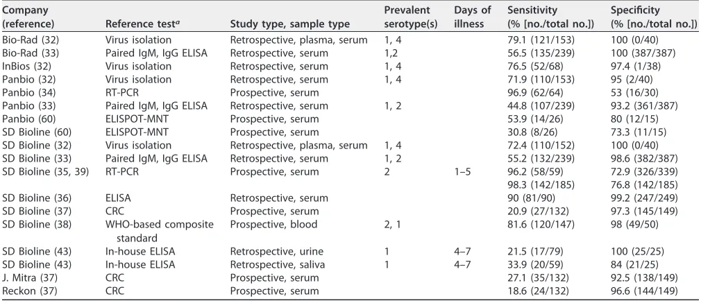
Nonstructural (NS) 1 protein is a highly conserved 46- to 55-kDa glycoprotein that is critical to form the dengue virus particles. NS1 is eventually secreted as a soluble hexamer from DENV-infected cells and circulates in the bloodstream of infected patients. During the viremia phase of dengue virus infection, NS1 antigen is produced concomitantly during the virus replication process and is likely to be present in the bloodstream 2 to 3 days longer after the viremia phase. In addition, the fact that NS1 is more stable than viral RNA, makes NS1 antigen a more common target for detection during outbreak in the field (Fig. 1). The detection of NS1 antigen has been used to rapidly diagnose acute dengue (31). A number of commercially available NS1 ELISAs and lateral flow rapid tests are available, and these tests required approval by the FDA or CE before they can be used for diagnostic purposes. There has been various studies comparing these POCT kits with different reference tests, with sensitivities ranging widely from 18.6 to 96.9% and specificities ranging from 53 to 100% (32–39) (Table 2). One study in Malaysia reported that the SD dengue NS1 Ag ELISA kit produced by Standard Diagnostics, Inc., South Korea, has a higher sensitivity of 77% and a specificity of 98% (39) compared to another study which reported that the SD Bioline dengue Duo
(IgG/IgM⫹NS1 Ag) test has a sensitivity of 30.8% and a specificity of 73.3%. However,
it should be emphasized that ELISAs use serum samples that would still require a laboratory to process. As a result, this would take a much longer time for diagnosis than the SD Bioline dengue Duo test to guide clinical management in an outbreak.
One of the challenges of antigen-based assays is the fluctuation of NS1 antigenemia levels throughout the course of illness. A decrease in NS1 levels occurs earlier in secondary infections (40), leading to an overall lower antigen-based test sensitivity when used in isolation. The sensitivities of these POCTs also vary according to sero-types, with DENV-1 being the most sensitive and DENV-4 being the least (41). A false-positive rate of 8.1% for NS1 POCT has been reported in a study that involved 148 samples (33). In that study, false-positive results were observed in patients with other viral and bacterial infections, as well as in healthy donors. False-positive results for DENV NS1 have also been reported in patients with Zika virus infection (42). Concurrent flavivirus infections are more likely to result in false positives as the IgG antibodies used
on May 16, 2020 by guest
http://jcm.asm.org/
to detect the dengue NS1 protein can cross-react with NS1 proteins of other viruses. This may result in an overestimation of the burden of dengue. As such, in areas where flaviviruses are endemic, it would be a good practice to perform PCR for DENV confirmation after patients are identified by screening with the NS1 protein detection POCT.
Of some interest is the use of urine and saliva samples for immunochromatographic POCT kits to detect dengue NS1 protein (43). Although the reported sensitivity is low (15 to 20%) in both urine and saliva samples to detect NS1 protein using POCT, it is as good as the in-house ELISAs with a kappa coefficient of 0.88. The sensitivity for both urine and saliva samples is higher from samples collected at approximately day 4 to 7 postfever than in those collected the first 3 days postfever. The utility of urine and saliva samples for POCT may not be applicable for early dengue diagnosis, but these samples will be very useful in situations when blood cannot be drawn readily for surveillance and outbreak monitoring purposes (44).
With increasing importance for multiplexing to accurately diagnose diseases that present common symptoms, such as the symptoms of malaria and dengue fever, three-dimensional, paper-based microfluidic devices, combining the advantages of lateral flow with vertical flow of fluids between multiple layers, have the potential to fulfill this need. Recently, a multiplex, patterned paper immunoassay for the detection of biomarkers (antigens) of malaria and dengue fever, namely, malaria HRP2, malaria pLDH, and dengue NS1 type 2, was developed but has yet to be clinically validated (45). It showed comparable performance between the singleplex and multiplex platform-based immunoassays. Additionally, it demonstrated high specificity in clinical blood samples. These suggest multiplex paper-based devices can be an essential component of diagnostic assays used at the point-of-care in the near future, particularly in areas where malaria and dengue fever are endemic, to determine the cause of undifferen-tiated febrile illness.
PROGRESS AND CHALLENGES IN THE DEVELOPMENT OF ANTI-DENGUE ANTIBODY DETECTION ASSAYS FOR POCT
Serological testing is widely adopted for the diagnosis of dengue, particularly in developing countries. This is largely due to the ease of use compared with techniques such as cell culture or RNA detection. During a primary dengue virus infection, the IgM response is typically of a higher titer and more specific than during secondary
infec-TABLE 2Summary of recent key developments in dengue antigen detection assays
Company
(reference) Reference testa Study type, sample type
Prevalent serotype(s)
Days of illness
Sensitivity (% [no./total no.])
Specificity (% [no./total no.])
Bio-Rad (32) Virus isolation Retrospective, plasma, serum 1, 4 79.1 (121/153) 100 (0/40) Bio-Rad (33) Paired IgM, IgG ELISA Retrospective, serum 1,2 56.5 (135/239) 100 (387/387)
InBios (32) Virus isolation Retrospective, serum 1, 4 76.5 (52/68) 97.4 (1/38)
Panbio (32) Virus isolation Retrospective, serum 1, 4 71.9 (110/153) 95 (2/40)
Panbio (34) RT-PCR Prospective, serum 96.9 (62/64) 53 (16/30)
Panbio (33) Paired IgM, IgG ELISA Retrospective, serum 1, 2 44.8 (107/239) 93.2 (361/387)
Panbio (60) ELISPOT-MNT Prospective, serum 53.9 (14/26) 80 (12/15)
SD Bioline (60) ELISPOT-MNT Prospective, serum 30.8 (8/26) 73.3 (11/15)
SD Bioline (32) Virus isolation Retrospective, plasma, serum 1, 4 72.4 (110/152) 100 (0/40) SD Bioline (33) Paired IgM, IgG ELISA Retrospective, serum 1, 2 55.2 (132/239) 98.6 (382/387)
SD Bioline (35, 39) RT-PCR Prospective, serum 2 1–5 96.2 (58/59) 72.9 (326/339)
98.3 (142/185) 76.8 (142/185)
SD Bioline (36) ELISA Retrospective, serum 90 (81/90) 99.2 (247/249)
SD Bioline (37) CRC Prospective, serum 20.9 (27/132) 97.3 (145/149)
SD Bioline (38) WHO-based composite standard
Prospective, blood 2, 1 81.6 (120/147) 98 (49/50)
SD Bioline (43) In-house ELISA Retrospective, urine 1 4–7 21.5 (17/79) 100 (25/25)
SD Bioline (43) In-house ELISA Retrospective, saliva 1 4–7 33.9 (20/59) 84 (21/25)
J. Mitra (37) CRC Prospective, serum 27.1 (35/132) 92.5 (138/149)
Reckon (37) CRC Prospective, serum 18.6 (24/132) 96.6 (144/149)
aRT-PCR, real-time PCR; CRC, composite reference criteria consisting of clinical and laboratory features and exclusion of other etiologies that present similarly (37).
on May 16, 2020 by guest
http://jcm.asm.org/
[image:6.585.40.545.83.300.2]tions. The titer of the IgG response is usually higher after the second week of illness than the first week of illness. During secondary infection, the IgG response usually increases more rapidly in the first week of illness than during primary infection (Fig. 1). The IgG antibodies are also more cross-reactive against other flaviviruses, known as the neutralizing heterotypic antibodies, which confer some level of cross-protection over a short period of time compared with the neutralizing homotypic antibodies, which confer lifelong protection against the specific dengue virus serotype (46). In contrast, cross-reactive but nonneutralizing antibodies may lead to increased disease severity due to the phenomenon of antibody-dependent enhancement (47).
Traditionally, hemagglutination inhibition (HAI) assays and neutralization tests (NTs) have been used for dengue diagnosis down to the serotype level. However, these methods are time consuming, labor intensive with low throughput, and are not cost-effective. Several commercially available laboratory-based IgM and IgG ELISA kits are also available and have sensitivities ranging from 21% to 99% and from 8% to 89%, respectively, and specificities ranging from 52% to 100% for IgM and 63.5% to 100% for IgG, compared with gold standard laboratory-based ELISAs (18). With advances in technology development, the ELISA, especially M antibody capture (MAC)-ELISA, sig-nifies the most important advance for IgM detection for routine dengue diagnosis. The MAC-ELISA is based on detecting IgM in serum using human-specific IgM that is bound to the solid phase. MAC-ELISAs are frequently run as a nonquantitative single dilution test, and positive results are commonly reported as a “recent flavivirus infection.” However, because IgM circulates for up to 3 months or longer, its presence might not be diagnostic of a current illness. To diagnose a current dengue virus infection, the demonstration of a seroconversion or at least 4-fold changes in antibody titers in paired sera is required. In areas where dengue is not endemic, IgM-based assays can be used in clinical surveillance for viral illness or for random population-based serological surveys, with the likelihood that any positive results detected indicate recent infections (within the past 2 to 3 months).
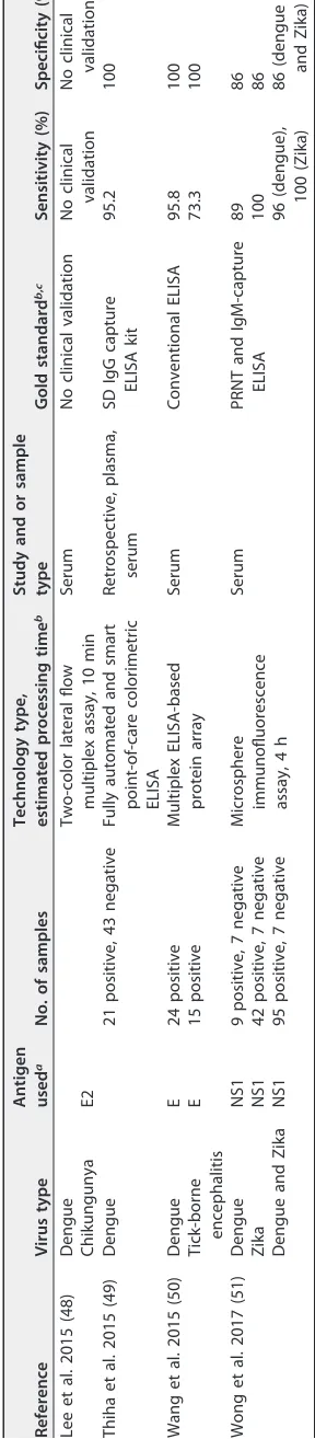
Several rapid IgM-based dengue diagnostic tests have also been developed as a quick and easy method for use at the point of care and exist in different formats, including particle agglutination and lateral-flow immunochromatographic strips, with or without plastic cassettes. Most of these tests use recombinant antigens from all four dengue virus serotypes, and the results are available within 15 to 90 min. Several studies have evaluated these commercially available rapid IgM- and IgG-based kits and have been reviewed recently (18). The IgM- and IgG-based kits have sensitivities ranging from 53% to 82% and from 62% to 89%, respectively, and specificities ranging from 75% to 100% and 67% to 95%, respectively, compared with gold standard laboratory-based ELISAs. Even though the rapid tests’ performances are not as sensitive and specific as those of ELISAs, the performance is still acceptable, with results available in a short time frame without laboratory requirement. Recently, a novel lateral-flow assay scheme that uses two-color latex labels for rapid multiplex detection of IgG/IgM antibodies to DENV and chikungunya virus in 10 min was reported by Lee et al. (48) (Table 3). With further clinical validation performed, this assay has significant potential as a POCT for the differential diagnosis of numerous pathogens of interest analyzed quantitatively in an automated point-of-care setting. Another novel integrated device was reported to detect and interpret the ELISA results on a portable lab-on-compact-disc (LOCD) platform by Thiha et al. (49) (Table 3). The system applies absorption spectrophotometry to measure the absorbance (optical density) of the sample using a monochromatic light source and an optical sensor. The device allows automated analysis of the results in a quantitative manner, with 95% sensitivity and 100% specificity (Table 3) in dengue virus detection compared with gold standard commer-cial ELISA microplate readers.
The biggest challenge for the application of IgM- and IgG-based assays is in areas where there are other flaviviruses circulating throughout the year. The IgM- and IgG-based assays may not be able to diagnose dengue accurately because of the cross-reactivity of antibodies against flaviviruses. Recently, an immunoassay was
on May 16, 2020 by guest
http://jcm.asm.org/
TABLE
3
Summary
of
recent
key
developments
in
anti-dengue
antibody
detection
platforms
Reference
Virus
type
Antigen used
a
No.
of
samples
Technology
type,
estimated
processing
time
b
Study
and
or
sample
type
Gold
standard
b
,
c
Sensitivity
(%)
Specificity
(%)
Lee
et
al.
2015
(48
)
Dengue
Two-color
lateral
flow
multiplex
assay,
10
min
Serum
No
clinical
validation
No
clinical
validation
No
clinical
validation
Chikungunya
E2
Thiha
et
al.
2015
(49
)
Dengue
21
positive,
43
negative
Fully
automated
and
smart
point-of-care
colorimetric
ELISA
Retrospective,
plasma,
serum
SD
IgG
capture
ELISA
kit
95.2
100
Wang
et
al.
2015
(50
)
Dengue
E
24
positive
Multiplex
ELISA-based
protein
array
Serum
Conventional
ELISA
95.8
100
Tick-borne
encephalitis
E
15
positive
73.3
100
Wong
et
al.
2017
(51
)
Dengue
NS1
9
positive,
7
negative
Microsphere
immunofluorescence assay,
4
h
Serum
PRNT
and
IgM-capture
ELISA
89
86
Zika
NS1
42
positive,
7
negative
100
86
Dengue
and
Zika
NS1
95
positive,
7
negative
96
(dengue), 100
(Zika)
86
(dengue and
Zika)
aE,
envelope
protein;
NS,
nonstructural
protein.
bELISA,
enzyme-linked
immunosorbent
assay.
cPRNT,
plaque
reduction
neutralization
test.
on May 16, 2020 by guest
http://jcm.asm.org/
[image:8.585.125.269.82.739.2]veloped and validated by Wang et al. (50) (Table 3) that simultaneously measures multiple antigen–antibody reactions. This ELISA-based microarray is emerging as a strong candidate platform for multiplex protein analysis due to its high-throughput potential, assay sensitivity and stringency, ease of handling, and low sample volume demand compared with conventional ELISA, and it provides significant clinical value. The platform was based on an indirect ELISA, and 15 antigens were constructed for
specific antibody detection against five Flaviviridae viruses (Japanese B, tick-borne
encephalitis, West Nile, dengue, and yellow fever viruses) and four serotypes of dengue virus. Dengue virus was detected with a sensitivity and specificity of 95.8% and 100%, respectively (Table 3). Additionally, a multiplex microsphere immunoassay (MIA) that captures the diagnostic power of detecting the viral envelope protein (that elicits robust yet cross-reactive IgG antibodies to other flaviviruses) and the differential power of detecting viral nonstructural proteins NS1 and NS5 (that induce more virus-type-specific IgG antibodies) was developed and validated by Wong et al. for differentiating dengue and Zika viruses (51) (Table 3). The sensitivity and specificity for detecting dengue virus are 89% and 86%, respectively, while those for detecting Zika virus are 100% and 86%, respectively (Table 3). In addition, it is capable of detecting coinfection with a sensitivity and specificity of 96% and 86%, respectively. This technology may be further enhanced to include other flaviviruses that may be endemic with dengue virus in different parts of the world.
THE FUTURE OF DENGUE POINT-OF-CARE DIAGNOSTIC ASSAYS
Ideally, dengue should be diagnosed at the primary level of care and during the early undifferentiated febrile phase. Regardless of the detection strategies, multiplexing technology should be the ultimate way forward for dengue POCT, especially in tropical countries where pathogens, especially flaviviruses, resulting in similar clinical presen-tations are prevalent. Multiplexing capability provides a higher confidence level of diagnosis when other pathogens of concern can be ruled out. It is likely to be more cost-effective to achieve the final diagnosis in the shortest time possible for prompt clinical management. However, it is always a challenge to balance between maximizing sensitivity/specificity of each pathogen and the multiplexing capabilities. Moreover, for serological assays, dengue diagnosis can be confounded by the cross-reactivity of IgG antibodies if other flaviviruses are endemic. Future effort should be focused on iden-tifying specific epitopes minimizing cross-reactivity, particularly in a setting where patients usually present late and the viremia is not detectable for a viral genomic assay. Ideally, multiplex POC NAAT-IgM test combinations using whole blood, in a minimally powered system with high connectivity, will enhance patient management in a primary health care setting.
There is a critical need to have a set of well-characterized dengue biological samples, which can include blood, urine, plasma and saliva, for a systematic comparison among the different POCTs for evaluation, auditing, or licensing purposes. In addition, with the differences in environmental factors and users’ performances in different settings, the performance of a newly developed POCT should be validated with an independent set of clinical samples to ensure the optimal accuracy of dengue diagno-ses and compared with a set of dengue-positive control samples.
There is no doubt of the need for specific, inexpensive dengue POC diagnostic tests that can be used for clinical management, surveillance, and outbreak investigations and that permit early intervention to treat patients and prevent or control epidemics, particularly in developing countries. However, there is a significant gap between the development and the implementation of these POCTs in developing countries. This is largely due to the fact that market incentives are usually small, especially for small and medium enterprises pursuing this area of research and development. As a result, there is a lack of awareness on the availability of these POCTs, which is critical for imple-mentation in the primary health care setting (52). There is a critical need for more evaluation studies to be conducted in a well-coordinated multicountry trial, with the establishment of a strong laboratory network and reference center, based on
on May 16, 2020 by guest
http://jcm.asm.org/
mendations from WHO (17), to address the generalizability and cost-effectiveness of using the commercial POCTs of interest. In addition, a stronger private–public partner-ship has to be fostered in order to materialize these evaluations to generate sufficient evidence for future development of POCT application guidelines for dengue diagnosis.
ACKNOWLEDGMENTS
This research was supported by a National Medical Research Council grant in Singapore (NMRC/CG/03/2013). The funders had no role in the study design, data collection or analysis, the decision to publish, or preparation of the manuscript.
All authors declare no conflict of interest.
REFERENCES
1. Bhatt S, Gething PW, Brady OJ, Messina JP, Farlow AW, Moyes CL, Drake JM, Brownstein JS, Hoen AG, Sankoh O, Myers MF, George DB, Jaenisch T, Wint GR, Simmons CP, Scott TW, Farrar JJ, Hay SI. 2013. The global distribution and burden of dengue. Nature 496:504 –507.https://doi.org/
10.1038/nature12060.
2. Junxiong P, Yee-Sin L. 2015. Clustering, climate and dengue transmis-sion. Expert Rev Anti Infect Ther 13:731–740.https://doi.org/10.1586/
14787210.2015.1028364.
3. Simmons CP, Farrar JJ, Nguyen vV, Wills B. 2012. Dengue. N Engl J Med 366:1423–1432.https://doi.org/10.1056/NEJMra1110265.
4. World Health Organization. 1997. Dengue haemorrhagic fever: diagno-sis, treatment, prevention and control, 2nd ed. World Health Organiza-tion, Geneva, Switzerland.
5. Guzmán MG, Kouri G, Bravo J, Valdes L, Vazquez S, Halstead SB. 2002. Effect of age on outcome of secondary dengue 2 infections. Int J Infect Dis 6:118 –124.https://doi.org/10.1016/S1201-9712(02)90072-X. 6. Goh KT. 1995. Changing epidemiology of dengue in Singapore. Lancet
346:1098.https://doi.org/10.1016/S0140-6736(95)91771-3.
7. Ooi EE, Goh KT, Chee Wang DN. 2003. Effect of increasing age on the trend of dengue and dengue hemorrhagic fever in Singapore. Int J Infect Dis 7:231–232.https://doi.org/10.1016/S1201-9712(03)90057-9. 8. Ooi EE, Goh KT, Gubler DJ. 2006. Dengue prevention and 35 years of
vector control in Singapore. Emerg Infect Dis 12:887– 893.https://doi
.org/10.3201/eid1206.051210.
9. Anders KL, Nguyet NM, Chau NV, Hung NT, Thuy TT, Lien le B, Farrar J, Wills B, Hien TT, Simmons CP. 2011. Epidemiological factors associated with dengue shock syndrome and mortality in hospitalized dengue patients in Ho Chi Minh City, Vietnam. Am J Trop Med Hyg 84:127–134.
https://doi.org/10.4269/ajtmh.2011.10-0476.
10. Chareonsook O, Foy HM, Teeraratkul A, Silarug N. 1999. Changing epidemiology of dengue hemorrhagic fever in Thailand. Epidemiol In-fect 122:161–166.https://doi.org/10.1017/S0950268898001617. 11. Tantawichien T. 2012. Dengue fever and dengue haemorrhagic fever in
adolescents and adults. Paediatr Int Child Health 32(Suppl 1):S22–S27.
https://doi.org/10.1179/2046904712Z.00000000049.
12. Wichmann O, Hongsiriwon S, Bowonwatanuwong C, Chotivanich K, Sukthana Y, Pukrittayakamee S. 2004. Risk factors and clinical features associated with severe dengue infection in adults and children dur-ing the 2001 epidemic in Chonburi, Thailand. Trop Med Int Health 9:1022–1029.https://doi.org/10.1111/j.1365-3156.2004.01295.x. 13. Pongsumpun P, Yoksan S, Tan IM. 2002. A comparison of the age
distributions in the dengue hemorrhagic fever epidemics in Santiago de Cuba (1997) and Thailand (1998). Southeast Asian J Trop Med Public Health 33:255–258.
14. World Health Organization. 2009. Dengue: guidelines for diagnosis, treatment, prevention and control. World Health Organization, Geneva, Switzerland.
15. Nyan DC, Swinson KL. 2015. A novel multiplex isothermal amplification method for rapid detection and identification of viruses. Sci Rep 5:17925.
https://doi.org/10.1038/srep17925.
16. Pabbaraju K, Wong S, Gill K, Fonseca K, Tipples GA, Tellier R. 2016. Simultaneous detection of Zika, chikungunya and dengue viruses by a multiplex real-time RT-PCR assay. J Clin Virol 83:66 –71.https://doi.org/
10.1016/j.jcv.2016.09.001.
17. Peeling RW, Artsob H, Pelegrino JL, Buchy P, Cardosa MJ, Devi S, Enria DA, Farrar J, Gubler DJ, Guzman MG, Halstead SB, Hunsperger E, Kliks S, Margolis HS, Nathanson CM, Nguyen VC, Rizzo N, Vazquez S, Yoksan S.
2010. Evaluation of diagnostic tests: dengue. Nat Rev Microbiol 8:S30 –S38.https://doi.org/10.1038/nrmicro2459.
18. Fatima A, Wang J. 2015. Review: progress in the diagnosis of dengue virus infections and importance of point of care test: a review. Pak J Pharm Sci 28:271–280.
19. Tsai YL, Wang HT, Chang HF, Tsai CF, Lin CK, Teng PH, Su C, Jeng CC, Lee PY. 2012. Development of TaqMan probe-based insulated isothermal PCR (iiPCR) for sensitive and specific on-site pathogen detection. PLoS One 7:e45278.https://doi.org/10.1371/journal.pone.0045278.
20. Go YY, Rajapakse RP, Kularatne SA, Lee PY, Ku KB, Nam S, Chou PH, Tsai YL, Liu YL, Chang HF, Wang HT, Balasuriya UB. 2016. A pan-dengue virus reverse transcription-insulated isothermal PCR assay intended for point-of-need diagnosis of dengue virus infection by use of the POCKIT nucleic acid analyzer. J Clin Microbiol 54:1528 –1535.https://doi.org/10.1128/
JCM.00225-16.
21. Notomi T, Okayama H, Masubuchi H, Yonekawa T, Watanabe K, Amino N, Hase T. 2000. Loop-mediated isothermal amplification of DNA. Nucleic Acids Res 28:E63.https://doi.org/10.1093/nar/28.12.e63.
22. Neeraja M, Lakshmi V, Lavanya V, Priyanka EN, Parida MM, Dash PK, Sharma S, Rao PV, Reddy G. 2015. Rapid detection and differentiation of dengue virus serotypes by NS1 specific reverse transcription loop-mediated isothermal amplification (RT-LAMP) assay in patients present-ing to a tertiary care hospital in Hyderabad, India. J Virol Methods 211:22–31.https://doi.org/10.1016/j.jviromet.2014.10.005.
23. Waggoner JJ, Gresh L, Mohamed-Hadley A, Ballesteros G, Davila MJ, Tellez Y, Sahoo MK, Balmaseda A, Harris E, Pinsky BA. 2016. Single-reaction multiplex reverse transcription PCR for detection of Zika, chi-kungunya, and dengue viruses. Emerg Infect Dis 22:1295–1297.https://
doi.org/10.3201/eid2207.160326.
24. Waggoner JJ, Ballesteros G, Gresh L, Mohamed-Hadley A, Tellez Y, Sahoo MK, Abeynayake J, Balmaseda A, Harris E, Pinsky BA. 2016. Clinical evaluation of a single-reaction real-time RT-PCR for pan-dengue and chikungunya virus detection. J Clin Virol 78:57– 61.https://doi.org/10
.1016/j.jcv.2016.01.007.
25. Simmons M, Myers T, Guevara C, Jungkind D, Williams M, Houng HS. 2016. Development and validation of a quantitative, one-step, multiplex, real-time reverse transcriptase PCR assay for detection of dengue and chikungunya viruses. J Clin Microbiol 54:1766 –1773.https://doi.org/10
.1128/JCM.00299-16.
26. Chen H, Parimelalagan M, Lai YL, Lee KS, Koay ES, Hapuarachchi HC, Ng LC, Ho PS, Chu JJ. 2015. Development and evaluation of a SYBR green-based real-time multiplex RT-PCR assay for simultaneous detection and serotyping of dengue and chikungunya viruses. J Mol Diagn 17:722–728.
https://doi.org/10.1016/j.jmoldx.2015.06.008.
27. Cecilia D, Kakade M, Alagarasu K, Patil J, Salunke A, Parashar D, Shah PS. 2015. Development of a multiplex real-time RT-PCR assay for simulta-neous detection of dengue and chikungunya viruses. Arch Virol 160: 323–327.https://doi.org/10.1007/s00705-014-2217-x.
28. Tan JJ, Capozzoli M, Sato M, Watthanaworawit W, Ling CL, Mauduit M, Malleret B, Gruner AC, Tan R, Nosten FH, Snounou G, Renia L, Ng LF. 2014. An integrated lab-on-chip for rapid identification and simultane-ous differentiation of tropical pathogens. PLoS Negl Trop Dis 8:e3043.
https://doi.org/10.1371/journal.pntd.0003043.
29. Wang S, Lifson MA, Inci F, Liang LG, Sheng YF, Demirci U. 2016. Advances in addressing technical challenges of point-of-care diagnostics in resource-limited settings. Expert Rev Mol Diagn 16:449 – 459.https://doi
.org/10.1586/14737159.2016.1142877.
on May 16, 2020 by guest
http://jcm.asm.org/
30. Tang RH, Yang H, Choi JR, Gong Y, Feng SS, Pingguan-Murphy B, Huang QS, Shi JL, Mei QB, Xu F. 2017. Advances in paper-based sample pre-treatment for point-of-care testing. Crit Rev Biotechnol 37:411– 428.
https://doi.org/10.3109/07388551.2016.1164664.
31. Shu PY, Yang CF, Kao JF, Su CL, Chang SF, Lin CC, Yang WC, Shih H, Yang SY, Wu PF, Wu HS, Huang JH. 2009. Application of the dengue virus NS1 antigen rapid test for on-site detection of imported dengue cases at airports. Clin Vaccine Immunol 16:589 –591.https://doi.org/10.1128/CVI
.00475-08.
32. Pal S, Dauner AL, Mitra I, Forshey BM, Garcia P, Morrison AC, Halsey ES, Kochel TJ, Wu SJ. 2014. Evaluation of dengue NS1 antigen rapid tests and ELISA kits using clinical samples. PLoS One 9:e113411.https://doi
.org/10.1371/journal.pone.0113411.
33. Blacksell SD, Jarman RG, Gibbons RV, Tanganuchitcharnchai A, Mammen MP, Jr, Nisalak A, Kalayanarooj S, Bailey MS, Premaratna R, de Silva HJ, Day NP, Lalloo DG. 2012. Comparison of seven commercial antigen and antibody enzyme-linked immunosorbent assays for detection of acute dengue infection. Clin Vaccine Immunol 19:804 – 810.https://doi.org/10
.1128/CVI.05717-11.
34. Anand AM, Sistla S, Dhodapkar R, Hamide A, Biswal N, Srinivasan B. 2016. Evaluation of NS1 antigen detection for early diagnosis of dengue in a tertiary hospital in southern India. J Clin Diagn Res 10:DC01–DC04.
https://doi.org/10.7860/JCDR/2016/15758.7562.
35. Shih HI, Hsu HC, Wu CJ, Lin CH, Chang CM, Tu YF, Hsieh CC, Chi CH, Sung TC. 2016. Applications of a rapid and sensitive dengue DUO rapid immunochromatographic test kit as a diagnostic strategy during a dengue type 2 epidemic in an urban city. PLoS One 11:e0158437.
https://doi.org/10.1371/journal.pone.0158437.
36. Vickers IE, Harvey KM, Brown MG, Nelson K, DuCasse MB, Lindo JF. 2015. The performance of the SD BIOLINE dengue DUO(R) rapid immunochro-matographic test kit for the detection of NS1 antigen, IgM and IgG antibodies during a dengue type 1 epidemic in Jamaica. J Biomed Sci 22:55.https://doi.org/10.1186/s12929-015-0164-9.
37. Mitra S, Choudhari R, Nori H, Abhilash KP, Jayaseelan V, Abraham AM, Cherian AO, Prakash JA, Muliyil J. 2016. Comparative evaluation of validity and cost-benefit analysis of rapid diagnostic test (RDT) kits in diagnosis of dengue infection using composite reference criteria: a cross-sectional study from south India. J Vector Borne Dis 53:30 –36. 38. Gan VC, Tan LK, Lye DC, Pok KY, Mok SQ, Chua RC, Leo YS, Ng LC. 2014.
Diagnosing dengue at the point-of-care: utility of a rapid combined diagnostic kit in Singapore. PLoS One 9:e90037.https://doi.org/10.1371/
journal.pone.0090037.
39. Wang SM, Sekaran SD. 2010. Evaluation of a commercial SD dengue virus NS1 antigen capture enzyme-linked immunosorbent assay kit for early diagnosis of dengue virus infection. J Clin Microbiol 48:2793–2797.
https://doi.org/10.1128/JCM.02142-09.
40. Duyen HT, Ngoc TV, Ha do T, Hang VT, Kieu NT, Young PR, Farrar JJ, Simmons CP, Wolbers M, Wills BA. 2011. Kinetics of plasma viremia and soluble nonstructural protein 1 concentrations in dengue: differential effects according to serotype and immune status. J Infect Dis 203: 1292–1300.https://doi.org/10.1093/infdis/jir014.
41. Huang C-H, Kuo L-L, Yang KD, Lin P-S, Lu P-L, Lin C-C, Chang K, Chen T-C, Lin W-R, Lin C-Y, Chen Y-H, Wu H-S. 2014. Laboratory diagnostics of dengue fever: an emphasis on the role of commercial dengue virus nonstructural protein 1 antigen rapid test. J Microbiol Immunol Infect 46:358 –365.https://doi.org/10.1016/j.jmii.2012.07.011.
42. Gyurech D, Schilling J, Schmidt-Chanasit J, Cassinotti P, Kaeppeli F, Dobec M. 2016. False-positive dengue NS1 antigen test in a traveller with an acute Zika virus infection imported into Switzerland. Swiss Med Wkly 146:w14296.https://doi.org/10.4414/smw.2016.14296.
43. Andries A-C, Duong V, Ong S, Ros S, Sakuntabhai A, Horwood P, Dussart P, Buchy P. 2016. Evaluation of the performances of six commercial kits designed for dengue NS1 and anti-dengue IgM, IgG and IgA detection in urine and saliva clinical specimens. BMC Infect Dis 16:201.https://doi
.org/10.1186/s12879-016-1551-x.
44. Andries A-C, Duong V, Ly S, Cappelle J, Kim KS, Lorn Try P, Ros S, Ong S, Huy R, Horwood P, Flamand M, Sakuntabhai A, Tarantola A, Buchy P. 2015. Value of routine dengue diagnostic tests in urine and saliva specimens. PLoS Negl Trop Dis 9:e0004100. https://doi.org/10.1371/
journal.pntd.0004100.
45. Deraney RN, Mace CR, Rolland JP, Schonhorn JE. 2016. Multiplexed, patterned-paper immunoassay for detection of malaria and dengue fever. Anal Chem 88:6161– 6165.https://doi.org/10.1021/acs.analchem
.6b00854.
46. Vaughn DW, Green S, Kalayanarooj S, Innis BL, Nimmannitya S, Suntaya-korn S, Endy TP, Raengsakulrach B, Rothman AL, Ennis FA, Nisalak A. 2000. Dengue viremia titer, antibody response pattern, and virus sero-type correlate with disease severity. J Infect Dis 181:2–9.https://doi.org/
10.1086/315215.
47. Dejnirattisai W, Jumnainsong A, Onsirisakul N, Fitton P, Vasanawathana S, Limpitikul W, Puttikhunt C, Edwards C, Duangchinda T, Supasa S, Chawansuntati K, Malasit P, Mongkolsapaya J, Screaton G. 2010. Cross-reacting antibodies enhance dengue virus infection in humans. Science 328:745–748.https://doi.org/10.1126/science.1185181.
48. Lee S, Mehta S, Erickson D. 2016. Two-color lateral flow assay for multiplex detection of causative agents behind acute febrile ill-nesses. Anal Chem 88:8359 –8363.https://doi.org/10.1021/acs.analchem
.6b01828.
49. Thiha A, Ibrahim F. 2015. A colorimetric enzyme-linked immunosor-bent assay (ELISA) detection platform for a point-of-care dengue detection system on a lab-on-compact-disc. Sensors (Basel) 15: 11431–11441.https://doi.org/10.3390/s150511431.
50. Wang D, Zheng Y, Kang X, Zhang X, Hao H, Chen W, Liu L, Li X, Li L, Yuan Q, Chen F, Yang Y, Jiang Y, Jiang H. 2015. A multiplex ELISA-based protein array for screening diagnostic antigens and diagnosis of Flavi-viridaeinfection. Eur J Clin Microbiol Infect Dis 34:1327–1336.https://
doi.org/10.1007/s10096-015-2353-6.
51. Wong SJ, Furuya A, Zou J, Xie X, Dupuis AP, II, Kramer LD, Shi PY. 2017. A multiplex microsphere immunoassay for Zika virus diagnosis. EBio-Medicine 16:136 –140.https://doi.org/10.1016/j.ebiom.2017.01.008. 52. Pang J, Hildon ZJ, Thein TL, Jin J, Leo YS. 2017. Assessing changes in
knowledge, attitude and practices on dengue diagnosis and manage-ment among primary care physicians after the largest dengue epidemic in Singapore. BMC Infect Dis 17:428.https://doi.org/10.1186/s12879-017
-2525-3.
53. Lanciotti RS, Kosoy OL, Laven JJ, Velez JO, Lambert AJ, Johnson AJ, Stanfield SM, Duffy MR. 2008. Genetic and serologic properties of Zika virus associated with an epidemic, Yap State, Micronesia, 2007. Emerg Infect Dis 14:1232–1239.https://doi.org/10.3201/eid1408.080287. 54. Lanciotti RS, Kosoy OL, Laven JJ, Panella AJ, Velez JO, Lambert AJ,
Campbell GL. 2007. Chikungunya virus in US travelers returning from India, 2006. Emerg Infect Dis 13:764 –767. https://doi.org/10.3201/
eid1305.070015.
55. Lanciotti RS, Calisher CH, Gubler DJ, Chang GJ, Vorndam AV. 1992. Rapid detection and typing of dengue viruses from clinical samples by using reverse transcriptase-polymerase chain reaction. J Clin Microbiol 30: 545–551.
56. Pastorino B, Bessaud M, Grandadam M, Murri S, Tolou HJ, Peyrefitte CN. 2005. Development of a TaqMan RT-PCR assay without RNA extraction step for the detection and quantification of African chikungunya viruses. J Virol Methods 124:65–71. https://doi.org/10.1016/j.jviromet.2004.11
.002.
57. Lai YL, Chung YK, Tan HC, Yap HF, Yap G, Ooi EE, Ng LC. 2007. Cost-effective real-time reverse transcriptase PCR (RT-PCR) to screen for den-gue virus followed by rapid single-tube multiplex RT-PCR for serotyping of the virus. J Clin Microbiol 45:935–941.https://doi.org/10.1128/JCM
.01258-06.
58. Klungthong C, Gibbons RV, Thaisomboonsuk B, Nisalak A, Kalayanarooj S, Thirawuth V, Nutkumhang N, Mammen MP, Jr, Jarman RG. 2007. Dengue virus detection using whole blood for reverse transcriptase PCR and virus isolation. J Clin Microbiol 45:2480 –2485.https://doi.org/10
.1128/JCM.00305-07.
59. Ziermann R, Sanchez-Guerrero SA. 2008. PROCLEIX West Nile virus assay based on transcription-mediated amplification. Expert Rev Mol Diagn 8:239 –245.https://doi.org/10.1586/14737159.8.3.239.
60. Piedrahita LD, Agudelo IY, Trujillo AI, Ramirez RE, Osorio JE, Restrepo BN. 2016. Evaluation of commercially available assays for diagnosis of acute dengue in schoolchildren during an epidemic period in Medellin, Co-lombia. Am J Trop Med Hyg 95:315–321.https://doi.org/10.4269/ajtmh
.15-0492.