Copyright © 1997, American Society for Microbiology
Analysis of an Outbreak of Non-Phage-Typeable
Methicillin-Resistant Staphylococcus aureus by Using a Randomly
Amplified Polymorphic DNA Assay
A. TAMBIC, E. G. M. POWER,* H. TALSANIA, R. M. ANTHONY,
ANDG. L. FRENCH
Department of Microbiology, UMDS of Guy’s and St. Thomas’s Hospitals, London SE1 7EH, United Kingdom
Received 24 March 1997/Returned for modification 20 May 1997/Accepted 29 July 1997
A cluster of methicillin-resistant Staphylococcus aureus (MRSA) infections among patients on an intensive
care unit (ICU) was detected by routine infection control surveillance. In the period from 5 January to 22 June
1995, 10 patients on the ICU and a further 6 patients (5 on one ward that had received colonized patients
transferred from the ICU) were affected by MRSA strains with the same antibiotic susceptibility patterns.
Seven (44%) of these 16 colonized patients developed MRSA bacteremia. MRSA isolates with the same
characteristics were also found on the hands of one member of the ICU staff. The isolates were untypeable by
phage typing, but 15 of 17 outbreak strains analyzed genetically had identical randomly amplified polymorphic
DNA (RAPD) and pulsed-field gel electrophoresis (PFGE) profiles. A single strain of MRSA that was
non-typeable by phage typing and that was isolated on the ICU on 1 January and six nonnon-typeable and
epidemio-logically unrelated MRSA isolates all had RAPD profiles distinct from that of the outbreak strain.
Implemen-tation of strict infection control measures stopped the further spread of MRSA on the ICU, the affected general
ward, and seven other wards that received MRSA carriers from the ICU. Although nontypeable by phage typing
and not previously recognized as an epidemic strain, this strain of MRSA was readily transmissible and highly
virulent. RAPD typing was found to be a simple, rapid, and effective method for the epidemiological
investi-gation of this outbreak, and performance of typing by this method was simpler and less time-consuming than
that of typing by PFGE. RAPD typing may have more general application for the study of S. aureus infections
in hospitals.
Until the mid-1970s, methicillin-resistant Staphylococcus
au-reus (MRSA) strains usually occurred sporadically, were
usu-ally resistant only to beta-lactam antibiotics, and were not a
serious clinical problem. Today, MRSA strains have become
resistant to a wide range of other antibiotics and are a major
cause of nosocomial infections throughout the world (3, 5). A
variety of typing techniques are available to help determine the
source and transmission routes of MRSA strains within a
hos-pital (16, 27), with the most common technique being
bacte-riophage typing (17). However, some MRSA strains may be
nontypeable by this method, and the limitations of phenotypic
typing have stimulated the development of DNA-based
tech-niques. Plasmid profile analysis was the first genotypic method
used in epidemiological studies of S. aureus (11, 13, 27).
Chro-mosomal DNA has been analyzed by a variety of techniques,
including conventional restriction enzyme analysis, restriction
fragment length polymorphism analysis, ribotyping,
pulsed-field gel electrophoresis (PFGE), and PCR-based methods.
Randomly amplified polymorphic DNA (RAPD) assays use
short primers with an arbitrary sequence to amplify genomic
DNA in a low-stringency PCR. These primers randomly
hy-bridize with chromosomal sequences that vary among different
strains and that produce different amplification products.
These products can be separated by gel electrophoresis to
produce fingerprints or patterns characteristic of different
ep-idemiological types. The method is attractive because it is
simple to perform and, theoretically, can be applied to any
organism (18). In this paper we describe the use of RAPD
analysis to investigate a cluster of MRSA infections detected
by routine infection control surveillance on the intensive care
unit (ICU) at St. Thomas’s Hospital.
MATERIALS AND METHODS
MRSA surveillance and control measures.Surveys for MRSA isolates were
performed daily through the pathology laboratory computer system. The index patient (patient 1) was admitted to the ICU on 2 January 1995, and he was found to be MRSA positive on 5 January (Table 1; Fig. 1). Another patient (patient A) colonized with an MRSA strain with a susceptibility pattern different from that of the strain colonizing patient 1 had been on the ICU on 3 January, and an MRSA strain with a pattern similar to that of the strain colonizing patient 1 was detected on 30 January (patient 2). Patients in the ICU were screened for MRSA carriage by taking swabs from the anterior nares, throat, axilla, groin, and per-ineum. MRSA strains were isolated from wound swabs, blood cultures, sputum, and MRSA screen swabs. All the staff in the ICU were screened on two occasions by taking swabs of their hands, anterior nares, and any cutaneous lesions. By 21 March another nine patients on the same ward had been infected or colonized with MRSA strains with the same antibiotic susceptibility pattern as the index strain (resistance to penicillin, gentamicin, amoxicillin-clavulanate, erythromy-cin, methicillin, clindamyerythromy-cin, rifampin, and neomycin and susceptibility to tetra-cycline, co-trimoxazole, fusidic acid, mupirocin, and vancomycin). By 22 June 1995, another five patients (patients 10, 12, 15, 16, and 17) on another ward (general surgical ward P) had been affected by MRSA strains with the same antibiotic susceptibility patterns as the index strain. In addition, a similar strain was isolated from a patient (patient 6) on cardiothoracic surgical ward D on 15 February.
Once the outbreak was recognized, hospital wards were visited by members of the infection control team. Initially, efforts were concentrated on the ICU, but later other wards receiving patient transfers from the ICU were targeted. Al-though it was hospital policy to implement the control measures for MRSA described in the guidelines produced by the Working Party of the British Society for Antimicrobial Chemotherapy and the Hospital Infection Society (20), it was obvious that these guidelines were not always being followed. The policy was circulated and explained to staff and was strictly applied. Patients and staff on wards with MRSA were screened for carriage; carriers were isolated or sent home and were treated with mupirocin nasal ointment and chlorhexidine baths. Carriers remained in isolation or were excluded until three screens showed that
* Corresponding author. Mailing address: Department of
Microbi-ology, UMDS of Guy’s and St. Thomas’s Hospitals, London SE1 7EH,
United Kingdom. Phone: 44 171 922 8385. Fax: 44 171 928 0730.
E-mail: [email protected].
3092
on May 15, 2020 by guest
http://jcm.asm.org/
they were clear of MRSA. Hand washing with chlorhexidine disinfectants by staff after caring for each patient was strictly enforced. Regular visits to the wards ensured that the policy was being followed, and laboratory surveillance for MRSA isolates continued. No further MRSA strains with the same susceptibility pattern as the index strain were isolated from any other patient from 18 June to 1 August 1995.
Strain identification.All S. aureus isolates were identified by routine
labora-tory procedures. Gram-positive, catalase-positive cocci were tested for mannitol fermentation on salt mannitol agar plates, clumping factor was detected by using the Staphaurex kit (Wellcome Diagnostics), and organisms were confirmed to be S. aureus by the tube coagulase test.
Antibiotic susceptibility testing. Antibiotic susceptibility testing was
per-formed by the disk diffusion method for methicillin, penicillin, gentamicin, amoxicillin-clavulanate, erythromycin, clindamycin, rifampin, tetracycline, co-trimoxazole, fusidic acid, mupirocin, neomycin, and vancomycin according to British Society for Antimicrobial Chemotherapy guidelines (30).
Bacteriophage typing. Bacteriophage susceptibility testing was kindly
per-formed by the Central Public Health Laboratory, Colindale, London, United Kingdom, with the international set of S. aureus typing phages.
DNA isolation.DNA was isolated as described previously (1), except that the
pelleted bacterial cells were first treated with 50ml of lysostaphin (50mg/ml) and 100ml of lysozyme (2 mg/ml) (Sigma Chemical Co.) for 1 h at 37°C.
Typing by RAPD analysis.We used four primers that in pilot experiments had
shown a good ability to discriminate S. aureus strains for typing purposes. The nucleotide sequences of the primers are listed in Table 1. For each sample we used 200mM (each) nucleotide (Sigma), 5ml of 103reaction buffer, 500 nM primer, 50 ng of template DNA, and 2 U of Taq XL DNA polymerase (Northum-bria Biochemicals Ltd., Cramlington, United Kingdom), and the mixture was made up to 50ml with molecular biology-grade water. This mixture was overlaid with 50ml of mineral oil. RAPD cycling parameters were 94°C for 5 s, 34°C for 30 s, and 72°C for 1 min for 35 cycles, with a final extension of 72°C for 5 min. Reactions were carried out in a Hybaid Thermal Reactor (Hybaid, Teddington, United Kingdom). The products were electrophoresed on 2% agarose gels in 0.53TBE (Tris, borate, EDTA) buffer. Synthetic PCR molecular size markers (Cambridge Bioscience Ltd., Cambridge, United Kingdom) were included in each gel. The gels were run for 5 to 6 h at 60 V to allow for the complete separation of the bands. DNA profiles were visualized with UV light after ethidium bromide staining and were photographed with a red filter and Polaroid
667 film. Analyses were performed with the two individual primers EP017 and EP007 alone and with the combinations EP0151KAY1 and EP0071KAY1 (Table 2).
Computer analysis of RAPD fingerprints.Polaroid photographs of the gels
were scanned and saved as 256-color TIF files. These images were normalized, a similarity matrix was produced by the Dice coefficient, and a dendrogram was constructed from the resulting data by the unweighted pair group method with arithmetic means; these analyses were performed by the GelCompar computer program (Applied Maths, Kortrijk, Belgium) (26).
PFGE typing.The isolates were also subjected to typing by PFGE. Briefly, 5 to
10 colonies of an overnight growth from a Columbia blood agar plate incubated at 37°C were harvested into 100ml of 1 M NaCl–10 mM EDTA (pH 7.5), and 3 ml of 1 mg of lysostaphin per ml was added. The preparation was mixed with an equal volume of 1% low-melting-point agarose at 50°C, allowed to set in a mold, and incubated at 37°C for 4 h in 1 ml of 1 M NaCl–10 mM EDTA (pH 7.5). The liquid was removed and replaced with 1 ml of 0.5 M EDTA (pH 7.5) containing 1 mg of proteinase K (Boehringer Mannheim) and 1% N-laurylsarcosine (Sig-ma), and then the mixture was incubated at 50°C for 18 h. The plugs were then washed and digested with the restriction enzyme SmaI as described previously (14). Pulsed-field gel electrophoresis was carried out on a Bio-Rad Dr II system on a 1% agarose gel in 0.53TBE at 14°C for 24 h with a ramped pulse time of from 19 to 100 s. DNA profiles were visualized as described above.
RESULTS
Description of the outbreak.
Between 5 January and 21
March 1995, MRSA strains with the same antibiotic
suscepti-bility patterns were isolated from 10 patients on the ICU. Six
of these patients (60%) developed MRSA bacteremia. Among
the ICU staff, MRSA strains with the same susceptibility
pat-terns were found on the hands of one nurse during the first
screening only.
[image:2.612.54.547.82.349.2]The patient colonized with the index strain was admitted
from the community with a history of having spent some hours
in the Accident and Emergency Unit of another hospital 2 days
TABLE 1. Characteristics of patients and MRSA isolates
Patient Ward isolationSite of
Date of isolation of first MRSA isolate (day.mo.yr)
Susceptibility patterna RAPD
type
A
ICU
Wound
03.01.1995
P,G,A,E,M,N,TMP
A
1
ICU
Blood
05.01.1995
P,G,A,E,M,C,R,N
B
2
ICU
Wound
30.01.1995
P,G,A,E,M,C,R,N
B
3
ICU
Blood
02.02.1995
P,G,A,E,M,C,R,N
B
4
ICU
Sputum
08.02.1995
P,G,A,E,M,C,R,N
ND
b5
ICU
Wound
09.02.1995
P,G,A,E,M,C,R,N
B
6
Ward D
Wound
15.02.1995
P,G,A,E,M,C,R,N
B
7
ICU
Blood
19.02.1995
P,G,A,E,M,C,R,N
B
8
cICU
Screen
19.02.1995
P,G,A,E,M,C,R,N
B
9
ICU
Blood
07.03.1995
P,G,A,E,M,C,R,N
B
10
Ward P
Wound
15.03.1995
P,G,A,E,M,C,R,N
B
11
ICU
Blood
17.03.1995
P,G,A,E,M,C,R,N
B
12
Ward P
Wound
19.03.1995
P,G,A,E,M,C,R,N
B
13
ICU
Wound
20.03.1995
P,G,A,E,M,C,R,N
B
14
ICU
Blood
21.03.1995
P,G,A,E,M,C,R,N
B
15
Ward P
Wound
10.05.1995
P,G,A,E,M,C,R,N
ND
16
Ward P
Wound
12.05.1995
P,G,A,E,M,C,R,N
B
17
Ward P
Blood
22.06.1995
P,G,A,E,M,C,R,N
B
18
Unrelated
dP,G,A,E,M,C,R,N,Tc
C
19
Unrelated
P,G,A,E,M,C,R,N,Tc
D
20
Unrelated
P,G,A,E,M,C,R,N,Tc
E
21
Unrelated
P,G,A,E,M,N,Mu
F
22
Unrelated
P,G,A,E,M,N,Tc,FA
G
23
Unrelated
P,G,A,E,M,C,R,N,Tc
H
aResistant to penicillin (P), gentamicin (G), amoxicillin-clavulanate (A), erythromycin (E), methicillin (M), clindamycin (C), rifampin (R), neomycin (N),
trimethoprim (TMP), mupirocin (Mu), fusidic acid (FA), or tetracycline (Tc).
bND, not done.
cPatient 8 was an ICU nurse.
dUnrelated isolates were obtained from other hospitals.
on May 15, 2020 by guest
http://jcm.asm.org/
previously. No MRSA strain with the same or similar antibiotic
susceptibility pattern had been isolated on the ICU during the
previous 2 months. The most recent previous isolate of MRSA
(from patient A; Table 1) on the ICU had an antibiotic
sus-ceptibility pattern quite different from that of the index strain,
but a strain (from patient 6; Table 1) with the same
suscepti-bility pattern as the strains involved in the ICU outbreak was
isolated concurrently on another ward (ward D) on 15
Febru-ary 1995. No MRSA-positive patients from the ICU had
re-cently been transferred to that ward.
All the ICU patients involved in the outbreak had
overlap-ping stays on that ward (Fig. 1). Two of them died while in the
ICU; one was transferred to two other wards before returning
to the ICU, where he died; the other seven patients were
transferred to seven different wards. Two patients were
trans-ferred to general surgical ward P and were still found to be
MRSA carriers after transfer. Another five patients on ward P
were subsequently colonized or infected with an MRSA strain
with the same susceptibility pattern as the outbreak strain (Fig.
1; Table 1). One of these patients developed MRSA
bactere-mia.
Following strict implementation of the British guidelines for
the control of MRSA (20) on the ICU and the other affected
wards, the outbreak was brought under control, and no further
isolates of the outbreak strain were found.
Strain characteristics.
None of the 17 strains involved in the
outbreak could be typed by phage typing (Table 1). They all
had the same antibiotic susceptibility pattern. One of the 11
outbreak strains isolated from ICU patients and staff and 1 of
5 outbreak strains isolated from ward P patients were lost
before RAPD and PFGE analyses could be performed. Fifteen
of the remaining 16 isolates were indistinguishable by RAPD
analysis when they were tested with the four different primers
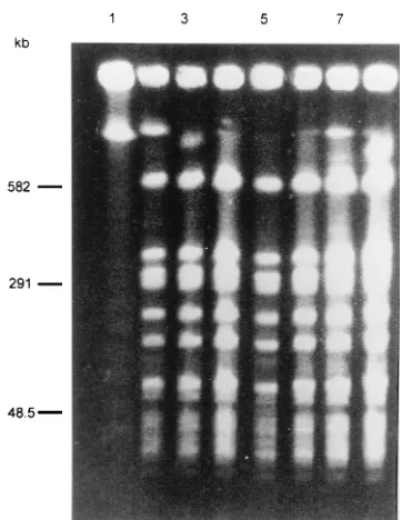
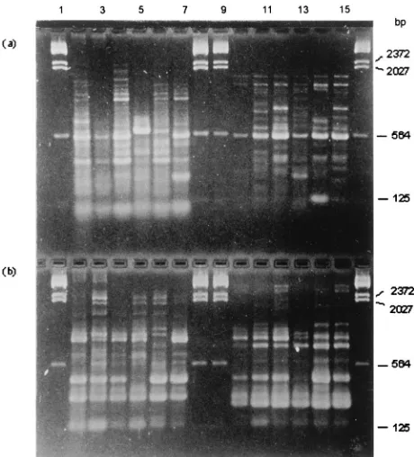
or primer combinations and by PFGE (Fig. 2 and 3),
respec-tively. Strain A, which was nontypeable by phage typing and
which had an antibiogram different from those of the outbreak
strains, was isolated on the ICU on 1 January, before the
outbreak, and had a RAPD pattern different from those of the
outbreak strains. Six other strains of MRSA nontypeable by
phage typing and epidemiologically unrelated to the outbreak
described here also had RAPD profiles different from those of
the outbreak strains (Fig. 4).
Cluster analysis of RAPD results.
The results of cluster
analysis of the RAPD results with GelCompar software are
presented in Fig. 5. The 15 outbreak isolates included on the
dendrogram fell into a single cluster at a similarity level of
76%. The isolate from patient A from the ICU was not part of
the outbreak and did not cluster with any of the other isolates
analyzed. The six epidemiologically unrelated isolates (isolates
from patients 18 to 23) did not cluster with the outbreak
strains. The bacteriophage lambda markers formed a cluster at
a similarity level of 84%. The variation among the outbreak
strains and the lambda markers detected by this analysis
prob-ably does not represent genuine differences but probprob-ably
rep-resents artifacts due to variations in the PCR, electrophoresis
conditions, and subsequent manipulation of the images.
DISCUSSION
[image:3.612.57.285.68.422.2]As a result of a routine infection control surveillance, a
cluster of MRSA isolates with the same antibiotic susceptibility
patterns was noticed on an ICU. Since the antibiogram may be
poorly discriminatory (2, 22), the MRSA strains were phage
typed, which is still a mainstay in the epidemiological analysis
of S. aureus infections (7, 28). In this case, however, all the
MRSA isolates involved in the outbreak were nontypeable by
phage typing. They were therefore typed by RAPD analysis
and PFGE and were found to have identical profiles by these
two methods. Another MRSA strain isolated from an ICU
patient just before this outbreak was also nontypeable by
phage typing but had an antibiotic susceptibility pattern
differ-ent from those of the outbreak isolates and a distinct RAPD
fingerprint. Eleven other epidemiologically unrelated isolates
of MRSA nontypeable by phage typing showed six distinct
RAPD and PFGE patterns. On the basis of these DNA typing
results, we concluded that 13 MRSA strains nontypeable by
phage typing and 2 epidemiologically related strains that were
also nontypeable by phage typing but that were lost before
RAPD analysis could be performed were all part of a single
hospital outbreak.
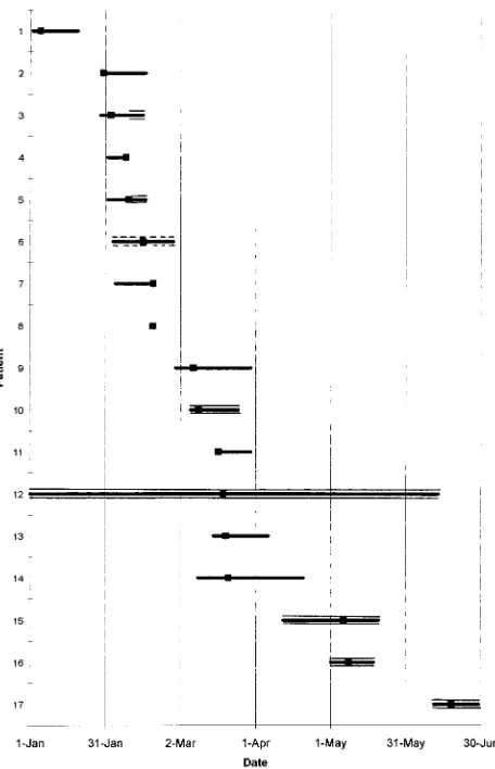
FIG. 1. Lengths of stay on the ward for patients infected or colonized with the epidemic strain of MRSA, as indicated by the groups of lines. The squares indicate the time of isolation of the first isolate of MRSA. The stay on the ICU is indicated by a single solid line, the stay on ward P is indicated by triple solid lines, and the stay on ward D is indicated by two dashed lines and one solid line.
TABLE 2. Arbitrary primers used for RAPD typing
Primer Sequence Source or referenceEP017
5
9
-
TACACCCGTCAACATTGAGG-
3
9
This study
EP015
5
9
-
ACAACTGCTC-
3
9
This study
KAY1
5
9
-
AGCAGCCTGC-
3
9
29
EP007
5
9
-
AGCACGCTGTCAATCATGTA-
3
9
This study
on May 15, 2020 by guest
http://jcm.asm.org/
[image:3.612.50.290.668.726.2]The patient colonized with the index strain was admitted
from the community with a history of a brief stay in another
hospital 2 days prior to admission to St. Thomas’s Hospital,
and the other hospital may have been the source of the
out-break strain. However, an MRSA strain with the same
antibi-otic susceptibility pattern and RAPD profile as the index strain
was isolated on another ward (ward D) on 15 February, but
that ward had no history of a recent patient transfer from the
ICU. It is thus possible that the outbreak strain was introduced
into the ICU from ward D via a transfer of an unrecognized
MRSA carrier prior to identification of the index strain. Coello
et al. (6) showed that a substantial number of asymptomatic
MRSA carriers can be missed if screening is not done.
[image:4.612.64.537.69.338.2]In the ICU outbreak reported here we found that the stays
of infected or colonized patients in the ICU overlapped, and
cross-infection presumably occurred by cross-infection from
patient to patient. The fact that MRSA was found on the hands
of one staff nurse supports the widely held view that the hands
of personnel are the principal route of MRSA transmission (4).
It is clear that ICUs play a significant role in transferring
MRSA isolates to other wards. It is more difficult to control the
spread of MRSA in ICUs because patients are frequently
sub-jected to invasive procedures and cohort nursing is not always
possible. At the time of the outbreak, standard British
infec-tion control procedures (30) for MRSA were hospital policy
but were not being strictly followed in all the wards. There was
no further spread of infection on seven of eight wards which
received MRSA carriers from the ICU, but on ward P, five
patients acquired MRSA following the transfer from the ICU
of two patients who were carriers of the outbreak strain.
Fol-lowing confirmation of an outbreak strain by phenotypic and
genotypic characteristics, all wards were visited by members of
the infection control team, and the infection control policies
were explained and reinforced. Patient isolation, strict hand
disinfection, use of barrier precautions, and nasal treatment of
MRSA carriers with mupirocin ointment and chlorhexidine
baths prevented the further spread of MRSA. Wards were
FIG. 2. RAPD fingerprints of MRSA isolates produced by primer EP007 (a), primers EP0071KAY1 (b), primers EP0151KAY1 (c), and primer EP017 (d). Lanes 1 and 18, bacteriophage lambda HindIII molecular size markers; lanes 2 to 4, patient isolates 1 to 3, respectively; lanes 5 to 11, patient isolates 5 to 11, respectively; lane 12, patient A isolate; lanes 13 to 15, patient isolates 12 to 14, respectively; lanes 16 and 17, patient isolates 16 and 17, respectively. Patient isolate numbers correspond to the patient numbers in Table 1.FIG. 3. PFGE fingerprints of MRSA isolates. Lane 1, bacteriophage lambda concatemers; lanes 2 to 4, patient isolates 1 to 3, respectively; lanes 5 to 8, patient isolates 5 to 8, respectively. Patient isolate numbers correspond to the patient numbers in Table 1.
on May 15, 2020 by guest
http://jcm.asm.org/
[image:4.612.79.260.456.690.2]subsequently visited at least twice weekly, and laboratory
sur-veillance of MRSA isolates continued. Up to 1 August 1995, no
further cases of infection with the outbreak strain were
de-tected. Since then, only sporadic cases of the outbreak strain
have been observed. The standard infection control
proce-dures described above have been implemented, and we have
experienced no more outbreaks with this strain.
It is apparent from this outbreak that this strain of MRSA
nontypeable by phage typing had a high degree of
transmissi-bility. It was also highly virulent, causing bacteremias in 7 of
the 15 affected patients. Although six of these cases of
bacte-remia occurred in ICU patients and host factors were
undoubt-edly involved, we do not normally see such a high incidence of
invasive S. aureus infection in our ICU. We believe that this
strain of MRSA is an epidemic type which is not easily
recog-nized because it is nontypeable by phage typing.
Strict microbiological monitoring and epidemiological
inves-tigation are essential for controlling MRSA in hospitals (4, 5).
The antibiogram provides useful information for routine
sur-veillance (2, 4, 16, 22, 27), but in outbreaks suspected of being
caused by MRSA, additional typing should be performed.
Plasmid analysis was the first DNA-based method to be
applied to S. aureus, and in some studies good discrimination
was achieved by restriction endonuclease enzyme analysis of
plasmid DNA (8, 9). However, the plasmid profiles of isolates
are not necessarily consistent over time, because strains can
spontaneously loose plasmids or acquire new ones (23, 27).
Analysis of restriction fragment length polymorphisms
(includ-ing ribotyp(includ-ing) of chromosomal DNA has frequently been used
to type S. aureus strains (12, 15). The discriminatory power of
this method depends on the restriction enzymes used (21).
PFGE of chromosomal digests with infrequently cutting
en-zymes has been proposed as the method of choice for typing
MRSA (19, 25), being a highly reproducible technique with
good discriminatory power (7, 16), and we applied this
tech-nique to our isolates. RAPD analysis and PFGE gave similar
results, but PFGE was both more time-consuming and
techni-cally demanding.
The RAPD assay, also called arbitrarily primed PCR, is
rapid and technically simple. Different studies comparing the
discriminatory power of RAPD analysis with those of other
techniques have come to different conclusions. Van Belkum et
al. (28) showed that RAPD analysis could discriminate 23
genotypes in a collection of 48 MRSA isolates, whereas only 13
different phage types could be distinguished. However, this
high discriminatory power was achieved by using the combined
DNA fingerprints obtained with several primers or primer
pairs (four PCR tests). The same investigators found that some
S. aureus strains had constant PFGE types but variable RAPD
types, and vice versa, suggesting that additional resolution
might be achieved by combined analyses (29). Saulnier et al.
(24) reported that RAPD analysis was less discriminatory than
PFGE in typing MRSA, but Hojo et al. (10) found that RAPD
typing results correlated well with the results of PFGE. The
variable RAPD typing results achieved by different workers
may be due to their use of different primers.
In this study we analyzed MRSA isolates by four PCR tests
using primers or primer pairs which had shown good
discrim-inatory power in a pilot study. The method is relatively easy to
perform, and even multiple tests with different primers are not
too cumbersome. The fact that this technique can be used to
type many other bacteria and yeasts (18, 31) makes it especially
attractive for use in clinical laboratories. The major problem
with RAPD analysis, as with all the other electrophoretic
tech-niques, is the lack of standards for interpreting the results
between laboratories.
Nevertheless, RAPD typing has proven to be valuable for
determining the epidemiology of MRSA isolates. It is
partic-FIG. 4. RAPD fingerprints of unrelated MRSA isolates. (a) Lanes 1, 8, 9, [image:5.612.54.284.69.323.2]and 16, bacteriophage lambda HindIII molecular size marker; lanes 2 to 7, patient isolates 18 to 23, respectively, typed with primer EP017; lanes 10 to 15, patient isolates 18 to 23 typed with primer combination EP015 and KAY1. (b) Lanes 1, 8, 9, and 16, bacteriophage lambda HindIII molecular size markers; lanes 2 to 7, patient isolates 18 to 23, respectively, typed with primer combination EP0071KAY1; lanes 10 to 15, patient isolates 18 to 23 typed with primer EP007. Patient isolate numbers correspond to the patient numbers in Table 1.
FIG. 5. GelCompar software analysis of epidemiologically related and unre-lated MRSA isolates nontypeable by phage typing. Clustering was done with the unweighted pair group method with arithmetic averages (UPGMA) algorithm by using fine correlation on gel tracks. For interpretation of the results, see the text.
on May 15, 2020 by guest
http://jcm.asm.org/
ularly useful, as in the outbreak described here, for typing
isolates nontypeable by phage typing, because an apparent
outbreak may in fact be due to different strains. The technique
is easier and less time-consuming than PFGE and does not
require pulsed-field equipment. The use of RAPD typing in
the investigation of MRSA outbreaks should be further
ex-plored.
ACKNOWLEDGMENTS
This work was supported by the South East Thames Regional
Health Authority, London, United Kingdom, and by the British
Coun-cil.
Many thanks go to Ian Phillips and Vanya Gant for helpful
com-ments during the preparation of the manuscript and arranging A.
Tambic’s visit.
REFERENCES
1. Ausubel, F. M., R. Brent, R. E. Kingston, D. D. Moore, J. G. Seidman, J. A.
Smith, and K. Struhl.1990. Current protocols in molecular biology, vol. 1.
John Wiley & Sons, Chichester, United Kingdom.
2. Blanc, D. S., C. Lugeon, A. Wenger, H. H. Siegrist, and P. Francioli. 1994. Quantitative antibiogram typing using inhibition zone diameters compared with ribotyping for epidemiological typing of methicillin-resistant Staphylo-coccus aureus. J. Clin. Microbiol. 32:2505–2509.
3. Boyce, J. M. 1994. Methicillin-resistant Staphylococcus aureus: a continuing infection control challenge. Eur. J. Clin. Microbiol. Infect. Dis. 13:45–49. 4. Boyce, J. M., M. M. Jackson, G. Pugliese, M. D. Batt, D. Fleming, J. S.
Garrer, A. I. Hartstein, C. A. Kauffman, M. Simmons, and R. Weinstein.
1994. Methicillin-resistant Staphylococcus aureus (MRSA): a briefing for acute care hospitals and nursing facilities. Infect. Control Hosp. Epidemiol.
15:105–115.
5. Brumfitt, W., and J. Hamilton-Miller. 1989. Methicillin-resistant Staphylo-coccus aureus. N. Engl. J. Med. 320:1188–1196.
6. Coello, R., J. Jimenez, M. Garcia, P. Arroyo, D. Minguez, C. Fernandez, F.
Cruzet, and C. Gaspar.1994. Prospective study on infection, colonization
and carriage of methicillin-resistant Staphylococcus aureus in an outbreak affecting 990 patients. Eur. J. Clin. Microbiol. Infect. Dis. 13:74–81. 7. Couto, I., J. Melo-Cristino, M. L. T. Fernandes, N. Garcia, N. Serrano, M. J.
Salgado, A. Torres-Pereira, I. S. Sanches, and H. de Lencastre.1995.
Un-usually large number of methicillin-resistant Staphylococcus aureus clones in a Portuguese hospital. J. Clin. Microbiol. 33:2032–2035.
8. Hartstein, A. I., M. A. Denny, V. H. Morthland, A. M. LeMonte, and M. A.
Pfaller. 1995. Control of methicillin-resistant Staphylococcus aureus in a
hospital and an intensive care unit. Infect. Control Hosp. Epidemiol. 16:405– 411.
9. Hartstein, A. I., C. L. Phelps, R. Y. Kwok, and M. E. Mulligan. 1995. In vivo stability and discriminatory power of methicillin-resistant Staphylococcus aureus typing by restriction endonuclease enzyme analysis of plasmid DNA compared with those of other molecular methods. J. Clin. Microbiol. 33: 2022–2026.
10. Hojo, S., J. Fujita, K. Negayama, T. Ohnishi, G. Xu, Y. Yamaji, H. Ohada,
and J. Takahara.1995. DNA fingerprinting by arbitrary primed polymerase
chain reaction (AP-PCR) for methicillin-resistant Staphylococcus aureus. J. Jpn. Assoc. Infect. Dis. 69:506–510.
11. Locksley, R. M., M. L. Cohen, T. C. Quinn, L. S. Tompkins, M. B. Coyle,
J. M. Kirishra, and G. W. Counts.1982. Multiply antibiotic-resistant
Staph-ylococcus aureus: introduction, transmission and evolution of nosocomial infection. Ann. Intern. Med. 97:317–324.
12. Lugeon, C., S. Blans, A. Wenger, and P. Francioli. 1995. Molecular epide-miology of methicillin-resistant Staphylococcus aureus at a low incidence hospital over a 4-year period. Infect. Control Hosp. Epidemiol. 16:260–267. 13. McGowan, J. E., Jr., P. M. Terry, T. Huang, C. L. Houk, and J. Davies. 1979.
Nosocomial infections with gentamicin-resistant Staphylococcus aureus: plas-mid analysis as an epidemiologic tool. J. Infect. Dis. 140:864–872. 14. Mitsuda, T., K. Arai, S. Fujita, and S. Yokota. 1995. Epidemiological analysis
of strains of methicillin resistant Staphylococcus aureus (MRSA) infection in the nursery; prognosis of MRSA carrier infants. J. Hosp. Infect. 31:123–134. 15. Montserrat, I., F. March, M. Simon, T. Lloret, M. D. Ferrer, P. Coll, and G.
Prats.1994. Application of molecular epidemiology techniques in the study
of food poisoning caused by Staphylococcus aureus. Med. Clin. 103:361–365. 16. Mulligan, M. E., and R. D. Arbeit. 1991. Epidemiologic and clinical utility of typing systems for differentiating among strains of methicillin-resistant Staphylococcus aureus. Infect. Control Hosp. Epidemiol. 12:20–28. 17. Pitt, T. L. 1994. Bacterial typing systems: the way ahead. J. Med. Microbiol.
40:1–2.
18. Power, E. G. M. 1996. RAPD typing in microbiology—a technical review. J. Hosp. Infect. 34:247–265.
19. Prevost, G., B. Jaulhac, and Y. Piemont. 1992. DNA fingerprinting by pulsed-field gel electrophoresis is more effective than ribotyping in distin-guishing among methicillin-resistant Staphylococcus aureus isolates. J. Clin. Microbiol. 30:967–973.
20. Report of a Combined Working Party of the Hospital Infection Society and
the British Society for Antimicrobial Chemotherapy.1990. Revised
guide-lines for the control of epidemic methicillin-resistant Staphylococcus aureus. J. Hosp. Infect. 16:351–377.
21. Richardson, J. F., P. Aparicio, R. R. Marples, and B. D. Cookson. 1994. Ribotyping of Staphylococcus aureus: an assessment using well-defined strains. Epidemiol. Infect. 112:93–101.
22. Rossney, A. S., D. C. Coleman, and C. T. Keane. 1994. Evaluation of an antibiogram-resistogram typing scheme for methicillin-resistant Staphylococ-cus aureus. J. Med. Microbiol. 41:441–447.
23. Sabria-Leal, M., V. H. Morthland, M. L. Pedro-Botet, N. Sopena, M.
Gime-nez-Perez, M. L. Branchini, and M. A. Pfaller.1994. Molecular epidemiology
for local outbreaks of methicillin-resistant Staphylococcus aureus (MRSA). The need for several methods. Eur. J. Epidemiol. 10:325–330.
24. Saulnier, P., C. Bourneix, G. Prevost, and A. Andremont. 1993. Random amplified polymorphic DNA assay is less discriminant than pulsed-field gel electrophoresis for typing strain of methicillin-resistant Staphylococcus au-reus. J. Clin. Microbiol. 31:982–985.
25. Schlichting, C., C. Branger, J. M. Fournier, W. Witte, A. Boutonnier, C.
Wolz, P. Goullet, and G. Doring.1993. Typing of Staphylococcus aureus by
pulsed-field gel electrophoresis, zymotyping, capsular typing, and phage typ-ing: resolution of clonal relationships. J. Clin. Microbiol. 31:227–232. 26. Seward, R. J., B. Ehrenstein, H. J. Grundmann, and K. J. Towner. 1997.
Direct comparison of two commercially available computer programs for analysing DNA fingerprinting gels. J. Med. Microbiol. 46:314–320. 27. Tenover, F. C., R. Arbeit, G. Archer, J. Biddle, S. Byrne, R. Goering, G.
Hancock, G. A. Hebert, B. Hill, R. Hollis, W. R. Jarvis, B. Kreiswirth, W. Eisner, J. Maslow, L. K. MacDougal, J. M. Miller, M. Mulligan, and M. A.
Pfaller.1994. Comparison of traditional and molecular methods of typing
isolates of Staphylococcus aureus. J. Clin. Microbiol. 32:407–415. 28. van Belkum, A., R. Bax, P. Peerbooms, W. H. F. Goessens, N. Leeuwen, and
W. G. V. Quint.1993. Comparison of phage typing and DNA fingerprinting
by polymerase chain reaction for discrimination of methicillin-resistant Staphylococcus aureus strains. J. Clin. Microbiol. 31:798–803.
29. van Belkum, A., J. Kluytmans, W. van Leeuwen, R. Bax, W. Quint, E. Peters,
A. Fluit, C. Vandenbroucke-Grauls, A. van den Brule, H. Koeleman, W. Melchers, J. Meis, A. Elaichouni, M. Vaneechoutte, F. Moonens, N. Maes,
M. Struelens, F. Tenover, and H. Verbrugh.1995. Multicenter evaluation of
arbitrarily primed PCR for typing of Staphylococcus aureus strains. J. Clin. Microbiol. 33:1537–1547.
30. Working Party of the British Society for Antimicrobial Chemotherapy. 1991. A guide to sensitivity testing. J. Antimicrob. Chemother. 27(Suppl. D):1–50. 31. Young, K. A., E. G. M. Power, M. S. Dryden, and I. Phillips. 1994. RAPD typing of clinical isolates of Staphylococcus haemolyticus. Lett. Appl. Micro-biol. 18:86–89.