The cloning of animals from adult cells has demonstrated that the developmental state of adult cells can be reprogrammed into that of embryonic cells by uncharacterized factors within the oocyte. More recently, transcription factors have been identified that can induce pluripotency in somatic cells without the use of oocytes, generating induced pluripotent stem (iPS) cells. iPS cells provide a unique platform to dissect the molecular mechanisms that underlie epigenetic reprogramming.
Moreover, iPS cells can teach us about principles of normal development and disease, and might ultimately facilitate the treatment of patients by custom-tailored cell therapy.
Introduction
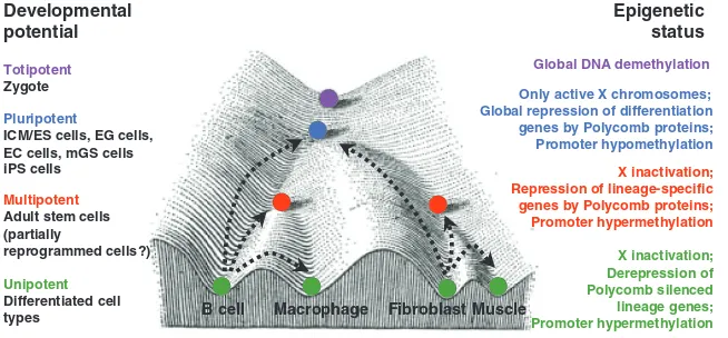
Mammalian development is a unidirectional process during which there is a progressive loss of developmental potential. It begins with the formation of a unicellular zygote and ends with the establishment of the 220 specialized cell types of the mammalian body. According to their decreasing differentiation potential, specific terms have been assigned to the individual cell populations that arise during development, including totipotency, pluripotency, multipotency and unipotency (Fig. 1, see also Glossary in Box 1). Each cell population is thought to have a characteristic epigenetic pattern that correlates with its differentiation potential (Fig. 1). As shown in Fig. 1 (which is adapted from C. H. Waddington’s ‘epigenetic landscape’ model) (Waddington, 1957), a marble rolling down a hill into one of several valleys illustrates the declining developmental potential of individual cell populations. At each bifurcation point, the potential of the marble (cell) to choose different routes (cell fates) diminishes.
Under certain experimental conditions, differentiated cells can revert into a less differentiated state, a process termed ‘nuclear reprogramming’ (Box 2). Examples include the generation of pluripotent embryonic stem (ES) cells from unipotent B lymphocytes or neurons by somatic cell nuclear transfer (SCNT) (Eggan et al., 2004; Hochedlinger and Jaenisch, 2002a; Li et al., 2004), or the derivation of pluripotent embryonic germ (EG) cells from unipotent primordial germ cells (PGCs) upon cell explantation (Matsui et al., 1992; Resnick et al., 1992). Reprogramming also describes the conversion of one differentiated cell type into another, for instance of a B lymphocyte into a macrophage (Xie et al., 2004), or a fibroblast into a muscle cell (Davis et al., 1987), following the expression of a single transcription factor (Fig. 1). Because these two examples of cell fate change may not involve a gain in differentiation potential, the term ‘lineage conversion’ or ‘transdifferentiation’ is currently used to describe them (Box 2). In Fig. 1, such cell fate changes are illustrated by the marble moving uphill Development 136, 509-523 (2009) doi:10.1242/dev.020867
Epigenetic reprogramming and induced pluripotency
Konrad Hochedlinger1and Kathrin Plath2
1Massachusetts General Hospital Cancer Center and Center for Regenerative
Medicine, Department of Stem Cell and Regenerative Biology, Harvard Stem Cell Institute, Harvard University, 185 Cambridge Street, Boston, MA 02114, USA.
2University of California Los Angeles, David Geffen School of Medicine, Department
of Biological Chemistry, Jonsson Comprehensive Cancer Center, Molecular Biology Institute, Eli and Edythe Broad Center of Regenerative Medicine and Stem Cell Research, 615 Charles E. Young Drive South, BSRB 390D, Los Angeles, CA 90024, USA.
e-mails: [email protected]; [email protected]
Box 1. Glossary of terms
Totipotency
Ability of a cell to give rise to all cells of an organism, including embryonic and extraembryonic tissues. Zygotes are totipotent.
Pluripotency
Ability of a cell to give rise to all cells of the embryo. Cells of the inner cell mass (ICM; see below) and its derivative, embryonic stem (ES) cells, are pluripotent.
Multipotency
Ability of a cell to give rise to different cell types of a given cell lineage. These cells include most adult stem cells, such as gut stem cells, skin stem cells, hematopoietic stem cells and neural stem cells.
Unipotency
Capacity of a cell to sustain only one cell type or cell lineage. Examples are terminally differentiated cells, certain adult stem cells (testis stem cells) and committed progenitors (erythroblasts).
Inner cell mass (ICM)
Cells of the blastocyst embryo that appear transiently during development and give rise to the three germ layers of the developing embryo.
Embryonic stem (ES) cells
Pluripotent cell line derived from the ICM upon explantation in culture, which can differentiate in vitro into many different lineages and cell types, and, upon injection into blastocysts, can give rise to all tissues including the germline.
Primordial germ cells (PGCs)
PGCs give rise to oocytes and sperm in vivo and to embryonic germ (EG) cells when explanted in vitro.
Embryonic germ (EG) cells
Pluripotent cell line derived from explanted PGCs. In contrast to pluripotent ICM and ES cells, PGCs are unipotent but become pluripotent upon explantation in culture.
Embryonic carcinoma (EC) cells
Pluripotent cell line originating from transformed PGCs. EC cells are derived from teratocarcinomas.
Germline stem (GS) cells
Unipotent cell line derived from mouse testes, which reconstitutes spermatogenesis when transplanted into sterile recipients.
Multipotent germline stem (mGS) cells
Pluripotent stem cell line derived from GS cells. mGS cells cannot reconstitute spermatogenesis, but have gained the potential to produce teratomas and chimeric animals.
Induced pluripotent stem (iPS) cells
Cells generated by the overexpression of specific transcription factors in mouse or human somatic cells, which are molecularly and functionally highly similar to ES cell counterparts.
Insertional mutagenesis
Insertion of a viral genome near endogenous genes, resulting in gene activation or silencing. Retrovirus-mediated insertional mutagenesis in hematopoietic cells can enhance self-renewal in vitro and cause cancer in vivo.
D
E
V
E
LO
P
M
E
N
(dedifferentiation) or across valleys (lineage conversion). In addition to experimentally induced changes of cell fate, the term reprogramming is also associated with the molecular changes that occur during normal germ cell development (Surani et al., 2007), which are not accompanied by a change in cell fate (Box 2).
Studies on the different forms of reprogramming have yielded important insights into the molecular mechanisms of normal development and disease, such as olfactory receptor choice (Eggan et al., 2004; Li et al., 2004) and cancer (Blelloch et al., 2004; Hochedlinger et al., 2004; Li et al., 2003), and have recently led to the identification of a defined set of transcription factors that can convert a mature cell into a pluripotent state in the culture dish, creating induced pluripotent stem (iPS) cells (Takahashi and Yamanaka, 2006). Reprogramming to pluripotency with defined factors enables the efficient derivation of patient-specific, autologous stem cells that have considerable potential in the study and treatment of human diseases. Indeed, skin cells reprogrammed to pluripotency with the ‘Yamanaka’ transcription factors have
recently been shown to alleviate the symptoms of Parkinson’s disease (Wernig et al., 2008c) and sickle cell anemia (Hanna et al., 2007) in mouse models. In addition to cellular therapy, patient-specific iPS cells may enable the establishment of in vitro models of complex genetic diseases, which are not yet well understood at the molecular level. To this end, patient-specific iPS cell lines have been derived from individuals with a variety of diseases, including diabetes, Parkinson’s disease and Amyotrophic Lateral Sclerosis (ALS) (Dimos et al., 2008; Park et al., 2008a); these cell lines are expected to facilitate the in vitro identification of novel drugs for the treatment of these disorders.
In this manuscript, we set recent advances in the induction of pluripotency in somatic cells with transcription factors into historical context and discuss the mechanisms that underlie this process in relation to alternative routes to reprogramming cell fate, including routes that are induced experimentally or that occur during normal development.
Scientific milestones leading to reprogramming with defined factors
Nuclear transfer (NT) was developed to assess whether the nuclei of differentiated cells remain equivalent to the nuclei of embryonic cells, and constituted the first attempts to reprogram an adult cell into a pluripotent embryonic state. Seminal experiments (see Box 3 for milestones in nuclear reprogramming) in amphibians in the 1950s and 1960s (Briggs and King, 1952; Gurdon, 1962), and later in mammals (Wilmut et al., 1997), demonstrated that the genomes of individual adult cells, and even those of terminally differentiated cells (Eggan et al., 2004; Hochedlinger and Jaenisch, 2002a; Inoue et al., 2005; Li et al., 2004), remain able to generate viable cloned animals, indicating that the developmental restrictions imposed on the genome during differentiation must be due to reversible epigenetic modifications, rather than to permanent genetic changes (Hochedlinger and Jaenisch, 2002b). These studies also indicated that the oocyte must contain factors that mediate the reprogramming of adult cells into an embryonic state; factors that should be identifiable and that could be used to induce pluripotency when expressed in somatic cells.
The derivation and stable maintenance of pluripotent cell lines was also instrumental for the reprogramming of somatic cells to pluripotency in vitro (see Box 3). Specifically, the study of teratocarcinomas in the 1950s and 1960s (Stevens, 1967; Stevens and Little, 1954) led to the isolation of pluripotent embryonal carcinoma (EC) cell lines from teratocarcinomas (Finch and Ephrussi, 1967; Kahan and Ephrussi, 1970; Kleinsmith and Pierce, 1964), and subsequently to the derivation of ES cells from
Totipotent
Zygote
Unipotent
Differentiated cell types
Developmental potential
Epigenetic status
muscle B cell Macrophage Fibroblast
X inactivation; Repression of lineage-specific genes by Polycomb proteins; Promoter hypermethylation
Muscle
Pluripotent
ICM/ES cells, EG cells, EC cells, mGS cells iPS cells
Only active X chromosomes; Global repression of differentiation genes by Polycomb proteins; Promoter hypomethylation Global DNA demethylation
X inactivation; Derepression of Polycomb silenced lineage genes; Promoter hypermethylation
Multipotent
Adult stem cells (partially
[image:2.612.50.377.61.213.2]reprogrammed cells?)
Fig. 1. The developmental potential and epigenetic states of cells at different stages of development.A modification of C. H.
Waddington’s epigenetic landscape model, showing cell populations with different developmental potentials (left) and their respective epigenetic states (right). Developmental restrictions can be illustrated as marbles rolling down a landscape into one of several valleys (cell fates). Colored marbles
correspond to different differentiation states (purple, totipotent; blue, pluripotent; red, multipotent; green, unipotent). Examples of reprogramming processes are shown by dashed arrows. Adapted, with permission, from Waddington (Waddington, 1957).
Box 2. Definitions of nuclear reprogramming
The term nuclear reprogramming is used to describe either functional or molecular changes to cells undergoing fate changes. When used as a functional term, reprogramming describes experimentally induced, stable changes in cell fate. It is most often used in the context of the reprogramming of adult cells into pluripotent cells, which can be achieved in various ways; for example, by somatic cell nuclear transfer (SCNT), by the fusion of somatic cells with pluripotent cells, by explanting germline cells (see Glossary, Box 1), or by the expression of a defined set of transcription factors in somatic cells (for a review, see Jaenisch and Young, 2008). Functional reprogramming also includes the stable conversion of one differentiated cell type into another by transcription factors; for example, the conversion of B cells into macrophages, fibroblasts into muscle cells or pancreatic acinar cells into β cells. The terms ‘transdifferentiation’ and ‘lineage conversion’ are used to describe this latter type of reprogramming because it is unclear if it involves the de-differentiation of cells into a less-differentiated progenitor cell, as it occurs during the reprogramming of adult cells into pluripotent cells. When used as a molecular term, reprogramming describes the molecular changes that cells undergo as their fate changes. For example, during the epigenetic reprogramming of cells, the promoter regions of pluripotency genes undergo demethylation following either SCNT or induced pluripotency. Epigenetic reprogramming has also been used to describe certain molecular changes that occur during development, irrespective of changes to the differentiation state of cells, such as the DNA and histone methylation changes that occur during germ cell maturation.
D
E
V
E
LO
P
M
E
N
blastocysts (Evans and Kaufman, 1981; Martin, 1981) and of embryonic germ (EG) cells from primordial germ cells (PGCs) (Matsui et al., 1992; Resnick et al., 1992) (Box 1). While these cell lines remain undifferentiated and immortal in culture, they undergo differentiation into all cell types when reintroduced into blastocysts (Bradley et al., 1984; Brinster, 1974; Matsui et al., 1992; Mintz and Illmensee, 1975).
Another crucial observation for reprogramming studies was that EC, ES and EG cells can reprogram other somatic cells when fused with them, generating pluripotent tetraploid hybrid cells (Cowan et al., 2005; Miller and Ruddle, 1976; Tada et al., 1997; Tada et al., 2001). These experiments indicated that pluripotent cells also harbor reprogramming activity and that the pluripotent state is dominant over the differentiated state (Box 3). Further investigations indicated that nuclear factors are responsible for reprogramming by cell fusion (Do and Scholer, 2004), as well as by NT (Egli et al., 2007), and hence suggested that transcription factors might be involved. Indeed, more recent work has indicated that the overexpression of the transcription factor Nanog, which is required for the establishment of ES cells (Chambers et al., 2003; Mitsui et al., 2003), enhances the formation of reprogrammed cell hybrids by up to 200-fold (Silva et al., 2006). However, the underlying mechanisms remain elusive.
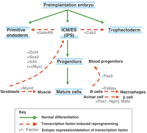
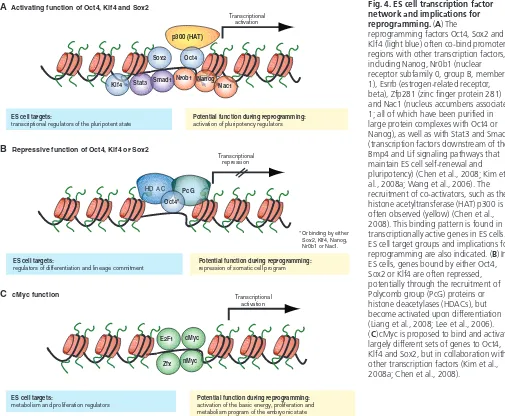
Previous work had already indicated that individual transcription factors, when overexpressed or deleted, could induce cell fate changes in somatic cells. For example, experiments by Harold Weintraub and colleagues showed that the overexpression of the myogenic transcription factor Myod was sufficient to convert fibroblasts into myogenic cells (Davis et al., 1987) (Fig. 2). Similarly, the elimination of Pax5from mouse B cells results in their dedifferentiation into progenitors that can give rise to multiple hematopoietic lineages (Nutt et al., 1999), and the overexpression of the transcription factor Cebpα (CCAAT enhancer binding protein alpha) can reprogram mouse B and T cells into macrophages (Laiosa et al., 2006; Xie et al., 2004) (Fig. 2). Similar to the transforming effects of transcription factors in adult cell lineages, which result in lineage conversion, the perturbation of embryonic transcription factors can induce major
cell fate changes in embryonic cells (Fig. 2). For instance, the ectopic expression of the transcription factor Cdx2 in ES cells results in the formation of trophectodermal stem cells from ES cells (Niwa et al., 2005). Likewise, the ectopic expression of Gata factors induces the formation of primitive endoderm (Fujikura et al., 2002; Shimosato et al., 2007). Together, these data indicated that the overexpression of individual transcription factors in closely related cells could induce stable cell fate changes. These observations provided the rationale for subsequent attempts to reprogram cell types beyond the boundaries of their cell lineage and differentiation state, including attempts to induce pluripotency in differentiated cells through the overexpression of transcription factors.
Generation of iPS cells from mouse and human somatic cells
Kazutoshi Takahashi and Shinya Yamanaka extended the observations that ES cells contain dominant reprogramming activity and that transcription factors are potent inducers of cell fate changes by identifying four transcription factors, Oct4, Sox2, Klf4 and cMyc, from 24 predominantly ES cell-specific genes, that were sufficient to reprogram adult mouse cells (fibroblasts) into ES-like iPS cells when expressed retrovirally (Fig. 3) (Takahashi et al., 2007). Initially, reprogrammed cells were identified based on drug selection for the expression of the ES cell-specific, but non-essential, gene Fbx15. These first-generation iPS cells were similar, but not identical, to ES cells (Fig. 3). For example, their transcriptional and epigenetic patterns appeared to be only partially reset from the fibroblast to the ES cell state. Moreover, these cells did not support the development of viable chimeric mice upon injection into blastocysts, which is indicative of a partially reprogrammed state.
ICM/ES (iPS)
Progenitors
Mature cells Preimplantation embryo
Trophectoderm Primitive
endoderm
B cells Fibroblasts
Blood progenitors
+Myod +Cebp
–Pax5 +Cdx2 +Gata4/6
+Oct4 +Sox2 +Klf4 (+cMyc)
Macrophages Muscle
Normal differentiation
Transcription factor-induced reprogramming +/– Factor Ectopic expression/deletion of transcription factor
Acinar cell cell +Pdx1, Ngn3, Mafa
[image:3.612.315.560.62.283.2]Key
Fig. 2. Examples of transcription factor-mediated
reprogramming.Hierarchy of cell populations (blue shading) that appear during normal development and their relationship to each other (green lines). Dashed red lines illustrate examples of transcription factor-induced reprogramming. The bracketed cMycgene indicates that this factor is dispensable for reprogramming. ICM, inner cell mass; ES, embryonic stem cell; iPS, induced pluripotent stem cell.
Box 3. Key milestones that led to the reprogramming of somatic cells into iPS cells by transcription factors 1952 First nuclear transfer experiments with frogs (Briggs and King,
1952).
1962 Cloned tadpoles generated from frog intestinal cells (Gurdon, 1962).
1964 Demonstration that single, teratoma-derived embryonic carcinoma (EC) cells are pluripotent (Kleinsmith and Pierce, 1964).
1976 Demonstration that EC cells can reprogram somatic cells in hybrids (Miller and Ruddle, 1976).
1981 Isolation of mouse embryonic stem (ES) cells from blastocysts (Evans and Kaufman, 1981; Martin, 1981).
1987 Reprogramming of fibroblasts into muscle cells by Myod (Davis et al., 1987).
1992 Isolation of mouse embryonic germ (EG) cells from fetal germ cells (Resnick et al., 1992; Matsui et al., 1992).
1997 First animal cloned from an adult cell (Dolly) (Wilmut et al., 1997).
1998 Derivation of human ES cells (Thomson et al., 1998).
2004 Reprogramming of B cells into macrophages by Cebpα(Xie et al., 2004).
2006 First induced pluripotent stem (iPS) cells generated from adult mouse fibroblasts (Takahashi and Yamanaka, 2006).
D
E
V
E
LO
P
M
E
N
Subsequent studies, however, showed that the identification of iPS cells based on drug selection using the promoters of the essential pluripotency genes Oct4or Nanog(Maherali et al., 2007; Okita et al., 2007; Wernig et al., 2007) gives rise to cells that are more similar to ES cells. The Oct4and Nanoggenes may become reactivated more selectively in cells undergoing faithful reprogramming, whereas Fbx15may be reactivated more broadly in treated cells, thus enriching for partially reprogrammed cells. Another important observation was that a delay in drug selection yields more faithfully reprogrammed iPS colonies (Maherali et al., 2007; Okita et al., 2007; Wernig et al., 2007). This led to experiments showing that morphological criteria alone are sufficient to obtain iPS cells (Blelloch et al., 2007; Maherali et al., 2007; Meissner et al., 2007). At the molecular level, completely reprogrammed iPS cells show transcriptional patterns that are highly similar to those in ES cells, as well as DNA demethylation of the promoter regions of Oct4and
Nanogand, in female cells, the reactivation of the somatically silent X chromosome. Moreover, iPS cells exhibit global patterns of histone methylation, including histones H3 lysine 4 (K4) and lysine 27 (K27) trimethylation, that are virtually indistinguishable from those in ES cells (Maherali et al., 2007; Mikkelsen et al., 2008; Okita et al., 2007; Wernig et al., 2007) (Fig. 3). At the functional level, completely reprogrammed iPS cells produce viable chimeric mice and contribute to the germline, and even support the development of embryos that are derived entirely from iPS cells (Hanna et al., 2008; Kim et al., 2008b; Wernig et al., 2007).
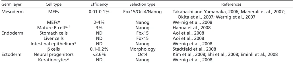
Since the 2006 landmark paper by Kazutoshi Takahashi and Shinya Yamanaka, iPS cell have been generated from the cells of multiple tissues, including blood (Hanna et al., 2008), liver (Aoi et al., 2008), stomach (Aoi et al., 2008), pancreas (Stadtfeld et al., 2008a), brain (Eminli et al., 2008; Kim et al., 2008b; Shi et al., 2008b), intestine and adrenals (Wernig et al., 2008a) (Table 1). Moreover, human fibroblasts (Lowry et al., 2008; Park et al., 2008b; Takahashi et al., 2007) and keratinocytes (Aasen et al., 2008; Maherali et al., 2008) have been converted into iPS cells using the same, and also using a different combination of factors, including OCT4, SOX2, LIN28 and NANOG (Yu et al., 2007). These results suggest that reprogramming to pluripotency with defined transcription factors is a process that can be induced in many cell types derived from all three germ layers.
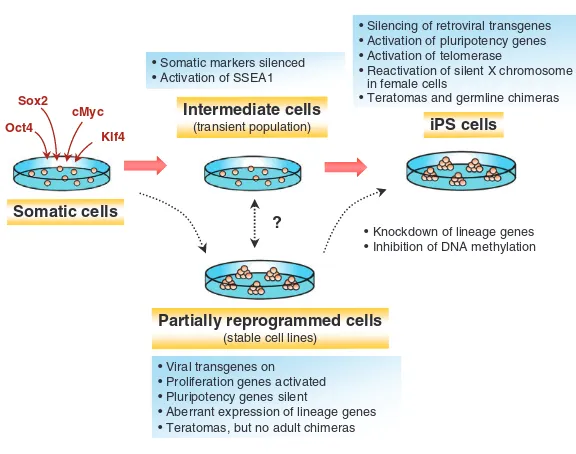
Intermediate stages of reprogramming
Transcription factor-induced reprogramming to pluripotency is a gradual process; it takes 1-2 weeks to generate iPS cells from mouse fibroblasts (Brambrink et al., 2008; Stadtfeld et al., 2008b). To dissect the mechanism of reprogramming, it has been informative to study partially reprogrammed cells (Fig. 3), such as cells generated by the method of Fbx15 selection that was initially used to identify iPS cells (Takahashi and Yamanaka, 2006). Partially reprogrammed cells are also frequently obtained when morphological criteria alone are employed to isolate iPS cells (Mikkelsen et al., 2008). In partially reprogrammed cells lines, the retroviral transgenes that are generally used to deliver the various reprogramming factors are not silenced, and the endogenous pluripotency genes show incomplete demethylation and reactivation (Mikkelsen et al., 2008; Takahashi and Yamanaka, 2006) (Fig. 3). Genome-wide expression analyses have shown that partially reprogrammed cell lines derived from B cells and fibroblasts are more similar to each other than to their cells of origin, suggesting that there could be one or several common intermediate states in which somatic cells get trapped in the culture dish, irrespective of the cell of origin (see Fig. 1).
Interestingly, partially reprogrammed cell lines show the activation of lineage-specific genes that are not normally expressed in the starting cell population or in pluripotent cells, such as Gata6,
Sox9and Pax7(Mikkelsen et al., 2008). Consistent with the notion that the ectopic expression of these lineage-specific transcription factors might prevent a cell from being converted into a pluripotent state, the knockdown of any of these genes resulted in a more efficient transition from the partially to a fully reprogrammed state (Mikkelsen et al., 2008) (Fig. 3). In agreement with the finding that the inhibition of differentiation-associated pathways is important for inducing pluripotency, the treatment of partially reprogrammed iPS cells with inhibitors of the extracellular signal-related kinase (ERK) and glycogen synthase kinase 3 (GSK3)/Wnt signaling cascades (Ying et al., 2008) facilitates their efficient conversion into fully reprogrammed iPS cells (Silva et al., 2008). Interestingly, the inhibition of differentiation seems to be important also during normal germ cell development (Surani et al., 2007). In mammals, for example, the repression of the differentiation-associated Hox genes by the SET domain protein Blimp1 is essential for the
Intermediate cells
(transient population)
Somatic cells
Partially reprogrammed cells
(stable cell lines)
• Knockdown of lineage genes • Inhibition of DNA methylation
iPS cells
?
• Somatic markers silenced • Activation of SSEA1
• Silencing of retroviral transgenes • Activation of pluripotency genes • Activation of telomerase
• Reactivation of silent X chromosome in female cells
• Teratomas and germline chimeras
• Viral transgenes on • Proliferation genes activated • Pluripotency genes silent
• Aberrant expression of lineage genes • Teratomas, but no adult chimeras Oct4
Sox2 cMyc
[image:4.612.51.339.55.282.2]Klf4
Fig. 3. Steps involved in direct reprogramming to pluripotency.The starting, intermediate and end stages of reprogramming to pluripotency that can be identified during the generation of iPS cells are shown.
‘Intermediate cells’ appear only transiently before converting into iPS cells, whereas ‘partially reprogrammed cells’ can be stably propagated and converted into iPS cells upon treatment with DNA demethylating agents and knockdown of lineage-specific genes. Although not proven, it is assumed that partially reprogrammed cells originate from transient intermediate cells. The defining molecular and cellular characteristics are shown above and below each cell population.
D
E
V
E
LO
P
M
E
N
specification of PGCs in vivo (Ohinata et al., 2005), indicating similar principles between the maintenance of germ cell fate and the induction of pluripotency.
While the analysis of partially reprogrammed cell states has been informative for understanding molecular barriers to reprogramming, a more detailed analysis of the earlier and later stages of reprogramming is crucial for establishing the sequence of transcriptional and epigenetic events that lead to a pluripotent state. In attempts to define such early intermediates, two studies have shown that the reprogramming of murine fibroblasts into iPS cells follows a defined sequence of molecular events that begins with the downregulation of somatic markers, such as Thy1 and collagens, followed by the reactivation of the embryonic marker stage-specific embryonic antigen 1 (SSEA1; Fig. 3) (Brambrink et al., 2008; Stadtfeld et al., 2008b). SSEA1-positive cells then gradually reactivate other markers associated with pluripotency, including
Oct4, Sox2, Nanog, telomerase (tert), and the silent X chromosome in female fibroblasts. The reactivation of these late markers correlates with the time window when cells become independent of retroviral transgene expression and enter a self-sustaining pluripotent state. It is possible that the partially reprogrammed cell lines described above are the trapped equivalent of the transient SSEA1-expressing cell population, although direct evidence for this relationship is lacking (Fig. 3). The observation that the somatic markers of a cell become downregulated before it progresses to a pluripotent state supports the notion that the silencing of its differentiation program is an important initial step towards re-establishing pluripotency. It further suggests that the differentiation state of the cell of origin for iPS cells might affect the efficiency and kinetics of the reprogramming process.
The differentiation status of the starting cell and reprogramming efficiency
NT is a very inefficient process (~1-3% of cloned blastocysts develop into live newborns) (Hochedlinger and Jaenisch, 2006) and the derivation of iPS cells is even less efficient, ranging from 0.01% to 0.1% (Table 2). The low efficiency of both processes has been argued to depend on the presence of rare stem cells within the starting population. For example, adult stem cells are present in many tissues at about the same frequency as the success rate of reprogramming. Some adult stem cells share transcriptional regulators with ES cells, such as the Zinc finger protein X-linked (Zfx) (Galan-Caridad et al., 2007) and Sox2 (Ellis et al., 2004), and may require less epigenetic reprogramming than terminally differentiated cells. While NT experiments have clearly demonstrated that fully differentiated lymphocytes (Hochedlinger
and Jaenisch, 2002a; Inoue et al., 2005) and neurons (Eggan et al., 2004; Li et al., 2004) can be reprogrammed into pluripotent ES cells, these experiments did not exclude the possibility that adult stem cells were the selective donors in most successful cloning experiments. In agreement with this idea, neural stem cells (NSCs) and keratinocyte stem cells give rise to cloned mice with greater efficiency than do mature fibroblasts, epidermal transit amplifying cells, or neurons (Blelloch et al., 2006; Eggan et al., 2004; Li et al., 2007; Li et al., 2004; Wakayama and Yanagimachi, 1999). By contrast, experiments in the hematopoietic system suggest that differentiated granulocytes are more efficient donors for NT than are hematopoietic stem cells (Inoue et al., 2006; Sung et al., 2006), although these experiments have recently been challenged (Hochedlinger and Jaenisch, 2007).
Consistent with results from NT experiments is the finding that murine NSCs can give rise to iPS cells up to 50 times more efficiently than can mouse fibroblasts (Kim et al., 2008b). Moreover, human keratinocytes undergo faster reprogramming than do human fibroblasts (Aasen et al., 2008; Maherali et al., 2008), and one study even observed an increased efficiency over fibroblasts (Aasen et al., 2008), suggesting that keratinocytes have a transcriptional state that is more amenable to reprogramming than that of fibroblasts or, alternatively, that keratinocyte cultures contain more progenitors than fibroblast cultures. It will undoubtedly be interesting to revisit the question of whether the differentiation state of a cell affects its reprogramming efficiency into iPS cells in a more defined system, such as the hematopoietic lineage.
[image:5.612.47.562.69.181.2]The identity of the starting cells that give rise to iPS cells remains controversial. Two recent experiments addressed the cell-of-origin question in different cellular systems and came to different conclusions. In the first set of experiments, Hanna et al. attempted to reprogram B lymphocytes into iPS cells to evaluate whether terminally differentiated cells can give rise to iPS cells (Hanna et al., 2008). B cells carry differentiation-associated DNA rearrangements, which serve as unambiguous genetic markers of their differentiation state (Hochedlinger and Jaenisch, 2002a). Interestingly, the ectopic expression of Oct4, Sox2, cMycand Klf4alone was insufficient to reprogram B lymphocytes into iPS cells, even when employing a ‘secondary’ system, in which most, if not all, cells express the four factors homogeneously (Wernig et al., 2008a) (Table 1). The authors had to either overexpress the transcription factor Cebpαor knock down the transcription factor Pax5, in addition to overexpressing the four factors, to generate iPS cells. The elimination of Pax5 has previously been shown to endow B cells with multipotency (Nutt et al., 1999) and the ectopic expression of Cebpα leads to the downregulation of Pax5 and thus to the reprogramming of B cells
Table 1. Reprogramming of different mouse cell types into iPS cells
Germ layer Cell type Efficiency Selection type References
Mesoderm MEFs 0.01-0.1% Fbx15/Oct4/Nanog Takahashi and Yamanaka, 2006; Maherali et al., 2007;
Okita et al., 2007; Wernig et al., 2007
MEFs* 2-4% Nanog Wernig et al., 2008
Mature B cell*,† 3% Nanog Hanna et al., 2008
Endoderm Stomach cells ND Fbx15 Aoi et al., 2008
Liver cells ND Fbx15 Aoi et al., 2008
Intestinal epithelium* ND Nanog Wernig et al., 2008
βcells 0.1-0.2% Morphology Stadtfeld et al., 2008
Ectoderm Neural progenitors <3.6% Oct4 Kim et al., 2008; Shi et al., 2008; Eminli et al., 2008
Keratinocytes* ND Nanog Wernig et al., 2008
*Secondary cells. Differentiated cells derived from iPS cells carrying the four viral transgenes under the control of a doxycycline-inducible promoter.
†Note that mature B cells required ectopic expression of Cebpαin addition to the four reprogramming factors to produce iPS cells.
MEFs, murine embryonic fibroblasts. ND, not determined.
D
E
V
E
LO
P
M
E
N
into macrophages (Xie et al., 2004). By contrast, progenitor B (pro-B) cells were permissive to being reprogrammed by the four factors alone, consistent with the notion that the differentiation state of the starting cell might affect reprogramming efficiency.
In another set of experiments, Stadtfeld et al. used genetically marked, terminally differentiated pancreatic β cells for reprogramming into iPS cells (Stadtfeld et al., 2008a). βcells gave rise to iPS cells at a frequency comparable to that of fibroblasts (0.1-0.2%; Table 1), demonstrating that this terminally differentiated cell type can be reprogrammed into iPS cells by just four factors and that adult stem cells are unlikely to be the selective cell type in successful reprogramming experiments. There are several explanations for the different outcomes of the reprogramming of B cells and βcells. First, B lymphocytes belong to the mesodermal lineage, whereas β cells are derived from endoderm; liver and stomach cells, which are also endodermal derivatives, have recently been suggested to be more amenable to reprogramming than fibroblasts (which are mesodermal in origin) (Aoi et al., 2008). Alternatively, βcells could be more easily reprogrammed than lymphocytes because the pancreas is not organized into a hierarchical structure, as the hematopoietic system is (which contains stem, progenitor and differentiated cells) (Orkin and Zon, 2008), but rather reproduces its
βcell pool by self duplication (Dor et al., 2004).
Technical limitations to reprogramming
Another explanation for the low efficiency of reprogramming could be that insertional mutagenesis, which can be caused by the retroviruses typically used to deliver the reprogramming factors, is potentially required for the nucleus of the starting cell to undergo reprogramming. Previous data have shown that retroviruses can activate endogenous genes in explanted hematopoietic stem cells, which promoted their turnover and survival (Kustikova et al., 2005). Similarly, one or several of the viral copies present in iPS cells might integrate into, and activate, a gene(s) that facilitates the reacquisition of a pluripotent, self-renewing state. However, the sequencing of viral insertion sites in iPS cells derived from fibroblasts (Varas et al., 2008), liver and stomach cells (Aoi et al., 2008) did not reveal any common integration sites, suggesting that insertional mutagenesis does not play an essential role in the induction of pluripotency. The possibility that retroviral insertion is required for the generation of iPS cells was finally excluded by two recent independent studies that
produced mouse iPS cells by transiently introducing the four reprogramming factors into somatic cells using either non-integrating adenoviruses (Stadtfeld et al., 2008c) or transient plasmid transfection (Okita et al., 2008). The efficiency of producing iPS cells with these transient expression methods was one to two orders of magnitude lower than the rates achieved using retroviral or lentiviral delivery methods, and will thus require further optimization to be useful in research or for future therapeutic purposes.
When using retroviruses, fibroblast-derived iPS cells carry ~10-20 proviral transgenes that express Oct4, Sox2, Klf4 and cMyc
[image:6.612.49.557.69.240.2](Maherali et al., 2007; Takahashi and Yamanaka, 2006; Wernig et al., 2007), which are found at different copy numbers per clone, suggesting that precise relative amounts of the individual transcription factors are important for reprogramming. This is consistent with observations that Oct4 and Sox2 levels in ES cells are crucial for maintaining a self-renewing pluripotent state (Kopp et al., 2008; Niwa et al., 2000). In further support of this idea, reprogramming of NSCs into iPS cells in the absence of exogenous Sox2 expression increases the overall efficiency by roughly fourfold (Eminli et al., 2008). It is conceivable that the frequency at which a single somatic cell receives the four viral transgenes at the appropriate stoichiometry is extremely low, resulting in the low overall efficiency. If viral infection is indeed the rate-limiting step, one would predict that cells that can reactivate all four factors at the correct stoichiometry should give rise to iPS cells at an efficiency close to 100%. The use of iPS cells in which the four transgenes can be reactivated with a doxycycline-inducible system (‘secondary system’) has allowed this question to be addressed. When mouse or human fibroblasts derived from such iPS cells were treated with doxycycline, between 3-5% of the cells gave rise to iPS cells (Hockemeyer et al., 2008; Maherali et al., 2008; Wernig et al., 2008a) (Table 1). This is a 30- to 100-fold increase in efficiency over primary cells infected directly with viruses, suggesting that viral infection and expression are parameters that affect reprogramming efficiency. However, these experiments also suggest that the expression of the four factors alone is insufficient in itself to reprogram adult cells to pluripotency and that additional rare events must affect the overall efficiency of reprogramming. As we discuss next, these events probably involve stochastic epigenetic events.
Table 2. Comparing efficiencies between reprogramming cells to pluripotency and lineage conversion
Starting cell type(s) Product cell type Treatment Efficiency References
Blastomere (2-cell) ES cell Explantation 50-69% Wakayama et al., 2007
Blastomere (4-cell) ES cell Explantation 22-40% Wakayama et al., 2007
Blastomere (8-cell) ES cell Explantation 10-16% Wakayama et al., 2007; Chung et al., 2006
ICM cell ES cell Explantation 12% Brook and Gardner, 1997
PGC EG cell Explantation 1-3% Durcova-Hills et al., 2006
GS cell mGS cell Explantation 0.001% Kanatsu-Shinohara et al., 2004
ES/NSC Hybrid cell Fusion <0.0006% Silva et al., 2006
ES/NSC (+Nanog) Hybrid cell Fusion 3-4% Silva et al., 2006
ES/ES Hybrid cell Fusion 3-4% Silva et al., 2006
Somatic cell* iPS cell Oct4, Sox2, Klf4, cMyc 3-5% Hanna et al., 2008; Wernig et al., 2008;
Maherali et al., 2008; Hockemeyer et al., 2008
Pro-B cells Macrophages Cebpα 65% Xie et al., 2004
Mature B cells Macrophages Cebpα 35% Xie et al., 2004
Pre-T cells Macrophages Cebpα 60% Laiosa et al., 2006
Fibroblasts Myogenic cell Myod 25-50% Davis et al., 1987
Acinar cell βcells Pdx1, Ngn3, Mafa 20% Zhou et al., 2008
*Secondary cells. Differentiated cells derived from iPS cells carrying the four viral transgenes under the control of a doxycycline promoter.
ICM, inner cell mass; PGC, primordial germ cell; GS cell, germline stem cell; ES cell, embronic stem cell; NSC, neural stem cell; EG cell, embryonic germ cell; mGS, multipotent germline stem cell.
D
E
V
E
LO
P
M
E
N
Stochastic epigenetic events: their impact on reprogramming
Several lines of evidence support the notion that stochastic epigenetic events contribute to the low efficiency of reprogramming. Indirect evidence for the involvement of such events comes from the observations that reprogramming is a gradual process that takes several weeks, and that the expression of the four factors alone is insufficient to efficiently convert somatic cells into pluripotent cells (see above). Moreover, a study that used reporter gene expression has shown that the transcriptional status of genetically identical cells can be very different. Some clonally derived daughter cells obtained from early appearing iPS colonies carrying an Oct4-GFP reporter reactivate GFP early, while others reactive it late, and some do not express it at all, despite carrying identical proviral integrations (Meissner et al., 2007). Thus, even though the cells are genetically identical, their transcriptional pattern, and therefore their epigenetic state, is different. Indeed, genome-wide analyses have confirmed that striking differences exist in the transcriptional and epigenetic signatures of partially and completely reprogrammed sibling clones (Mikkelsen et al., 2008). Importantly, the treatment of somatic cells or cells undergoing reprogramming with compounds that affect chromatin modifications, including DNA and histone methylation inhibitors (Huangfu et al., 2008a; Huangfu et al., 2008b; Mikkelsen et al., 2008; Shi et al., 2008b), enhances the efficiency of reprogramming significantly and facilitates the complete conversion of partially reprogrammed cells that would otherwise fail to reprogram into a pluripotent state (Fig. 3).
The requirement for stochastic epigenetic events to occur during the formation of iPS cell lines might be common to other approaches that aim to derive pluripotent cell lines from unipotent cells, given the uniformly low efficiencies of their generation. For example, the derivation of EG cells from PGCs (Durcova-Hills et al., 2006) and that of multipotent germline stem (mGS) cells from germ line stem (GS) cells (Conrad et al., 2008; Guan et al., 2006; Kanatsu-Shinohara et al., 2004; Seandel et al., 2007) is thought to be even less efficient, ranging from 1-3% and around 0.001%, respectively, even though both PGCs and GS cells express many pluripotency genes, including Oct4and Sox2(Table 2) (Imamura et al., 2006; Surani et al., 2007). Similarly, the reprogramming efficiency of somatic cells by ES cells in hybrids is less than 0.0006%, increasing to 3-4% when Nanog is overexpressed from the ES cell genome (Silva et al., 2006) (Table 2). These low efficiencies of reprogramming somatic cells into pluripotent cells are in contrast to the frequencies at which ES cell lines are generated from pluripotent blastomeres or ICM cells. Specifically, the efficiencies of deriving ES cell lines from single blastomeres of cleavage-stage embryos range from 50-69% for two-cell blastomeres, 22-40% for four-two-cell blastomeres and 10-16% for eight-cell blastomeres (Chung et al., 2006; Wakayama et al., 2007) (Table 2). Moreover, it has been estimated that, on average, three out of the ~25 ICM cells found in a blastocyst (12%) give rise to ES cell lines (Brook and Gardner, 1997). Together, these results suggest that undifferentiated blastomeres and ICM cells require fewer epigenetic changes to convert into ES cell lines than do the more differentiated PGCs and GSCs, which require more changes and thus convert less efficiently into pluripotent cell lines. Interestingly, the requirement for stochastic epigenetic events might not be limited to the derivation of pluripotent cell lines in vitro; they appear to also play a role in normal development (Box 4).
Assuming that the stochastic events crucial for the derivation of ES cells from ICM cells are also important for the establishment of EG, mGS and iPS cells, one might predict that the same genes and compounds that enhance ES cell derivation
should also facilitate the derivation of pluripotent cell types from other sources. Indeed, the treatment of partially reprogrammed iPS cells with inhibitors of the ERK kinase and GSK3 signalling cascades, both of which are crucial for the derivation of ES cells (Ying et al., 2008), results in the efficient conversion of partially reprogrammed cell lines into fully reprogrammed iPS cells (Silva et al., 2008). Moreover, Wnt pathway activation has beneficial effects not only on the growth of ES cells (Sato et al., 2004), but also for the reprogramming of somatic cells into pluripotent cells by transcription factors (Marson et al., 2008) and for cell fusion between somatic cells and ES cells (Lluis et al., 2008). Likewise, loss of the tumor suppressor protein p53, which normally inhibits the immortal growth of primary fibroblasts, enhances the transformation of PGCs into embryonal carcinomas (Lam and Nadeau, 2003) and increases the number of mGS cells derived from GS cells (Kanatsu-Shinohara et al., 2004), as well as the number of iPS colonies derived from fibroblasts (Zhao et al., 2008), possibly by conferring immortality and/or by de-repressing Nanog (Lin et al., 2005).
Reprogramming to pluripotency versus lineage conversion
How does the efficiency of reprogramming adult cells into a pluripotent state compare with the efficiency with which one differentiated cell type converts into another? Interestingly, the direct conversion of B cells and pre-T cells into macrophages by the retroviral expression of Cebpα(Laiosa et al., 2006; Xie et al., 2004), that of fibroblasts into myogenic cells by the retroviral expression of Myod (Davis et al., 1987), and that of acinar cells into βcells by adenoviral delivery in vivo of Pdx1, Ngn3 and Mafa (Zhou et al., 2008) does not appear to be restrained by major epigenetic barriers, based on the high efficiency of lineage switching (Table 2). For example, 25-50% of fibroblasts that express Myod convert into myogenic colonies (Davis et al., 1987). Pro- and pre-B cells that ectopically express Cebpα transform into macrophages at a frequency of ~65% and mature B cells at a frequency of ~35% (Xie et al., 2004). Similarly, ~60% of pre-T cells convert into
Box 4. Transcriptional fluctuations occurring during normal development
During normal development, stochastic fluctuations in gene expression are thought to influence cell fate decisions. For instance, subpopulations of clonally derived hematopoietic progenitor cells have been found to exhibit metastable states (which persist over multiple cell divisions), together with global changes in gene expression and different tendencies to give rise to the erythroid or myeloid lineages (Chang et al., 2008). The stochastic expression of Nanog and Cdx2 during mouse preimplantation development has also been observed and is assumed to play roles in the allocation of the ICM and trophectoderm (Dietrich and Hiiragi, 2007). Similarly, ES cells show fluctuations in Nanog (Chambers et al., 2007), Zinc finger protein 42 (Zfp42 or Rex1) (Toyooka et al., 2008) and Stella (Hayashi et al., 2008) expression, which has been correlated with their ability to maintain pluripotency or to differentiate, respectively. Specifically, it is assumed that ES cells that express the transcription factor maintain pluripotency, whereas those that lose expression become permissive for differentiation. Whether cells losing individual pluripotency markers are poised to differentiate into the same or different lineages remains to be determined. It is also unclear whether the mechanisms that underlie stochastic transcriptional changes during normal development are identical to those that occur during the derivation of pluripotent cell lines.
D
E
V
E
LO
P
M
E
N
macrophages upon overexpression of Cebpα(Laiosa et al., 2006). Pancreatic acinar cells infected with adenoviruses that express Pdx1, Ngn3 and Mafa convert into insulin-expressing β cells at a frequency of 20% (Zhou et al., 2008). It is conceivable, therefore, that changing cell fates within closely related cell types requires less epigenetic remodeling, leading to the high efficiency of conversion. Consistent with this idea, the expression of Cebpαand PU.1 in more distantly related fibroblasts gave rise to cells that were similar, but not identical, to macrophages that had been obtained from B cells, and their phenotype was not maintained upon silencing of the viral transgenes (Feng et al., 2008).
These observations raise the general question of whether transcription factor-induced conversion across cell lineages and germ layers can ever generate epigenetically stable cell fates that closely mirror cell types found in vivo. It is possible that such dramatic switches in cell identity require more extensive changes in the epigenetic signature or involve very stable chromatin marks that can only be reset after going through a pluripotent ground state, i.e. they require de-differentiation and subsequent re-differentiation. A related question, which was initially raised in NT experiments, is whether iPS cells retain an ‘epigenetic memory’ of their cell of origin (see Box 5).
Possible mechanisms of transcription factor-induced reprogramming
Differential requirement for the reprogramming factors An open question is how the reprogramming factors induce the epigenetic changes that are associated with reprogramming to pluripotency. Experiments in which several combinations and orthologs of the four transcription factors were tested demonstrated that not all four factors are required for reprogramming (Nakagawa et al., 2008; Wernig et al., 2008b; Yu et al., 2007). The fact that KLF4 and cMYC can be replaced by NANOG and by the RNA-binding protein LIN28 in human fibroblast reprogramming experiments suggests that different molecular pathways can lead to reprogramming or, alternatively, that these factors perform highly similar functions during this process. In support of the latter, LIN28 was recently found to function as a negative regulator of microRNA processing in ES cells, specifically of members of the let-7 family (Viswanathan et al., 2008). cMYC represses the transcription of similar miRNAs, suggesting that LIN28 and cMYC could perturb the same regulatory mechanisms that contribute to reprogramming (Chang et al., 2008).
iPS colonies can be generated from mouse or human fibroblasts in the absence of cMyc altogether, albeit at lower frequency and with delayed kinetics (Nakagawa et al., 2008; Wernig et al., 2008b). The effect of cMyc can be partially compensated by treating cells with either the histone deacetylase (HDAC) inhibitor valproic acid (VPA) or a ligand of the β-catenin pathway, Wnt3a (Huangfu et al., 2008a; Marson et al., 2008). Moreover, VPA can replace the function of both cMYC and KLF4 in human cell reprogramming, such that only the expression of OCT4 and SOX2 are required to generate iPS cells (Huangfu et al., 2008b).
The starting cell also has an effect on the requirement for reprogramming factors. As mentioned before, NSCs, which already express high levels of endogenous Sox2, require only the ectopic expression of Oct4 with either Klf4 or cMyc to produce iPS cells (Eminli et al., 2008; Kim et al., 2008b; Shi et al., 2008b), and hepatocytes are as efficiently reprogrammed in the absence of cMyc as in its presence (Aoi et al., 2008). Because cMyc, Klf4 and Sox2 (but not Oct4) are expressed in multiple adult tissues
and can be replaced by other orthologs during reprogramming into iPS cells (Nakagawa et al., 2008), Oct4 appears to be the only irreplaceable, and possibly the most important, determinant of direct reprogramming. However, a recent study suggested that even Oct4 might be replaceable in certain cellular contexts: treatment of NSCs with a chemical inhibitor of the histone methyltransferase G9a, which is responsible for silencing the
Oct4promoter during normal differentiation (see below), gave rise to Oct4-GFP-positive iPS cells upon ectopic expression of Klf4 and cMyc alone (Shi et al., 2008b). When combined with the calcium channel agonist BayK, this inhibitor even facilitated the reprogramming of murine fibroblasts into iPS cells in the absence of Sox2 and cMyc (Shi et al., 2008a). Although the mechanisms by which these small compounds mediate reprogramming remain elusive at this point, it may indeed be possible in the future to generate iPS cells solely with chemicals.
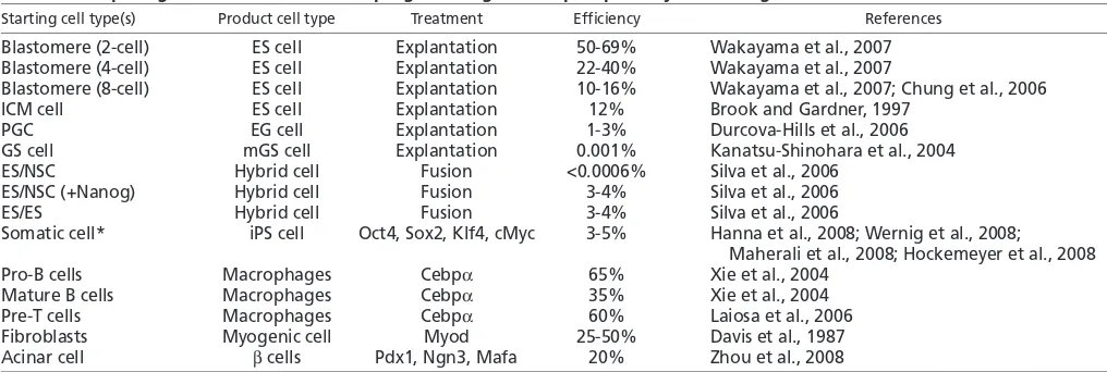
ES cell transcription factor networks
Studies of the transcriptional circuitry that controls the pluripotent state of ES cells might be helpful for understanding the function of the reprogramming factors. Recent genome-wide analyses in ES cells have suggested that the three reprogramming factors Oct4, Sox2 and Klf4, and the transcription factor Nanog specify ES cell identity by transcriptionally activating the self-renewal program and by repressing lineage commitment pathways (Boyer et al.,
Box 5. Retention of an ‘epigenetic memory’ in iPS cells?
Does reprogramming erase all of the epigenetic modifications, the ‘epigenetic memory’, of the somatic donor cell? Interestingly, frog embryos generated by nuclear transfer (NT) and derived from frog somite cells retain Myodexpression in cells that normally do not express Myod, even after 24 cell divisions (Ng and Gurdon, 2008). Similarly, cloned frog embryos derived from endoderm cells express an endodermal marker gene in non-endodermal cells (Ng and Gurdon, 2005). The histone variant H3.3 is deposited through replication-independent mechanisms at actively transcribed loci, thus marking actively transcribed genes (Henikoff and Ahmad, 2005), and is believed to be necessary for the establishment of an epigenetic memory, but detailed mechanisms remain unclear (Ng and Gurdon, 2008). Additional evidence from cloned frogs and mice has indicated that animals derived from different donor cells show different phenotypic and transcriptional abnormalities, consistent with the retention of an epigenetic memory of the donor cell (Boiani et al., 2002; Bortvin et al., 2003; Hochedlinger and Jaenisch, 2002b). However, ES cells derived from blastocysts that have been cloned from different donor cell types are indistinguishable from each other and from fertilization-derived ES cells, based on transcriptional profiling and their ability to give rise to normal mice (Brambrink et al., 2006), indicating that the process of ES cell derivation selects for faithfully reprogrammed cells. So far, no comparative analyses have been reported on iPS cells and mice derived from different donor cells, and genome-wide analyses of fibroblast-derived iPS cells did not reveal any epigenetic aberrations suggestive of an epigenetic memory (Maherali et al., 2007; Mikkelsen et al., 2008; Okita et al., 2007; Wernig et al., 2007). This might indicate that iPS cells, like NT ES cells, have faithfully erased any epigenetic marks present in donor cells. However, one study showed that iPS cell-derived chimeras derived from liver and stomach cells have increased rates of perinatal death compared with iPS chimeras derived from fibroblasts (Aoi et al., 2008). It will be informative to compare the epigenetic and transcriptional patterns of iPS cells derived from a variety of donor cell types to solve the question of whether or not a donor cell’s epigenetic memory is retained.
D
E
V
E
LO
P
M
E
N
2005; Loh et al., 2006) (Fig. 4). Generally, multiple pluripotency transcription factors co-occupy genes in ES cells that are active but are repressed upon differentiation, and encode proteins that are important for ES cell self-renewal and pluripotency (Fig. 4A). By contrast, when bound by only one of these transcription factors, target genes are often transcriptionally repressed in ES cells, and encode regulators of lineage commitment, which become activated upon the induction of differentiation (Fig. 4B).
Two models can explain how the association of a gene with multiple transcription factors could increase its transcriptional output. Protein-protein interactions between these transcription factors could increase the affinity of the proteins for adjacent recognition sites (Chen et al., 1998) or DNA conformations generated by the binding of one transcription factor could favor the binding of another (Panne et al., 2004). In support of the latter, Oct4 binding is required for the recruitment of Smad1 and Stat3 to target sites (Chen et al., 2008). In both modes, steric constraints on the proteins could favor the formation of unique configurations that would create docking surfaces for co-activators required for strong transcriptional activation (Merika et al., 1998). Indeed, the p300 histone acetyltransferase is found at many Oct4 target sites and is recruited to them in an Oct4-dependent manner (Chen et
al., 2008). Alterations in DNA structure induced by the binding of multiple factors could also contribute to the specificity of their binding (Panne et al., 2007), explaining how Oct4 and Sox2 distinguish developmentally appropriate binding sites from those that carry the recognition motif but are irrelevant in the ES cell context.
It has been proposed that the four reprogramming factors do not act on the same set of genes, as cMyc binds to many genes that are not bound by Oct4, Sox2 or Klf4 (Chen et al., 2008; Kim et al., 2008a). Nevertheless, cMyc shares target genes with other transcription factors, including the family member nMyc, which can replace cMyc in reprogramming experiments, and the cell cycle regulator E2F1 and Zfx (Chen et al., 2008) (Fig. 4C), again indicating cooperation among multiple transcription factors. It remains to be shown whether cMyc does indeed have separable functions from the other three reprogramming factors.
How do the transcription factors induce reprogramming? Reprogramming needs to inactivate the somatic cell program and to activate the ES cell-specific transcription programs of self-renewal and pluripotency. One could speculate that the reprogramming factors contribute to both functions, as they can, in
Transcriptional repression
p300 (HAT)
Sox2
Transcriptional activation
Klf4 A Activating function of Oct4, Klf4 and Sox2
Nanog Nr0b1
Nac1
Transcriptional activation
cMyc C cMyc function
nMyc Zfx Smad1 Stat3
B Repressive function of Oct4, Klf4 or Sox2
*Or binding by either Sox2, Klf4, Nanog, Nr0b1 or Nac1.
ES cell targets:
transcriptional regulators of the pluripotent state
Potential function during reprogramming:
activation of pluripotency regulators
ES cell targets:
metabolism and proliferation regulators
Potential function during reprogramming:
activation of the basic energy, proliferation and metabolism program of the embryonic state
ES cell targets:
regulators of differentiation and lineage commitment
Potential function during reprogramming:
repression of somatic cell program
E2F1 HD A C PcG
Oct4
[image:9.612.50.555.59.475.2]Oct4*
Fig. 4. ES cell transcription factor network and implications for reprogramming.(A) The
reprogramming factors Oct4, Sox2 and Klf4 (light blue) often co-bind promoter regions with other transcription factors, including Nanog, Nr0b1 (nuclear receptor subfamily 0, group B, member 1), Esrrb (estrogen-related receptor, beta), Zfp281 (zinc finger protein 281) and Nac1 (nucleus accumbens associated 1; all of which have been purified in large protein complexes with Oct4 or Nanog), as well as with Stat3 and Smad1 (transcription factors downstream of the Bmp4 and Lif signaling pathways that maintain ES cell self-renewal and pluripotency) (Chen et al., 2008; Kim et al., 2008a; Wang et al., 2006). The recruitment of co-activators, such as the histone acetyltransferase (HAT) p300 is often observed (yellow) (Chen et al., 2008). This binding pattern is found in transcriptionally active genes in ES cells. ES cell target groups and implications for reprogramming are also indicated. (B) In ES cells, genes bound by either Oct4, Sox2 or Klf4 are often repressed, potentially through the recruitment of Polycomb group (PcG) proteins or histone deacetylases (HDACs), but become activated upon differentiation (Liang et al., 2008; Lee et al., 2006). (C) cMyc is proposed to bind and activate largely different sets of genes to Oct4, Klf4 and Sox2, but in collaboration with other transcription factors (Kim et al., 2008a; Chen et al., 2008).
D
E
V
E
LO
P
M
E
N
ES cells, be both repressive and activating. Thus, genes that encode somatic cell regulators could be repressed by the binding of the reprogramming factors, while self-renewal and pluripotency genes would be turned on (Fig. 4). Autoregulatory loops, i.e. the binding of factors to their own promoters (Boyer et al., 2005), could provide a platform on which ectopic transcription factors can jump-start the transcription of their endogenous counterparts to a level that is sufficient to maintain their own expression. Furthermore, the ectopic activation of lineage differentiation programs that has been observed in partially reprogrammed cells might reflect a function of Klf4 and Sox2 in normal development (Mikkelsen et al., 2008). These transcription factors are also expressed in neural and epidermal lineages, and could, potentially in combination with other lineage-specific transcription factors, target genes during reprogramming that they would not normally associate with in pluripotent cells.
The reprogramming factors could also have more global functions that do not involve direct transcriptional control, which remain completely unexplored. A few pleiotropic functions have been suggested for cMyc, ranging from the control of initiation of DNA replication (Dominguez-Sola et al., 2007) to global effects on chromatin structure, especially on histone acetylation (Knoepfler, 2008), which could be important for providing the other reprogramming factors access to target sites. In agreement with the latter observation, the inhibition of histone deacetylation can replace cMyc in reprogramming experiments (Huangfu et al., 2008a; Huangfu et al., 2008b). One could also envision that ectopically expressed factors titrate proteins important for somatic cell transcription away from the DNA and sequester them in inactive complexes, thereby acting as differentiation antagonists, as has been described for Myod in muscle specification (Puri et al., 2001). Thus, reprogramming is likely to be more complex than a simple model suggests, and will involve a number of different mechanisms to overcome the epigenetic barriers that are imposed during differentiation.
Overcoming epigenetic barriers
The inefficient activation of Oct4in somatic cells following NT is associated with the embryonic lethality of cloned mouse embryos (Boiani et al., 2002; Bortvin et al., 2004), and Oct4activation is a stringent measure of reprogramming success during iPS cell generation (Meissner et al., 2007; Wernig et al., 2007), which is in agreement with the notion thatOct4is absolutely required for the establishment and maintenance of pluripotency during normal development (Nichols et al., 1998). The Oct4gene undergoes a complex process of inactivation during post-implantation development, which involves multiple layers of repression. Using differentiating ES cells as a model system, it was shown that the binding of transiently acting transcriptional repressors, such as Coup-TF1/2 (chicken ovalbumin upstream promoter-transcription factor) and Gcnf (germ cell nuclear factor) (Ben-Shushan et al., 1995; Fuhrmann et al., 2001), leads to the recruitment of histone deacetylases and the methyltransferase G9a, which in turn triggers de novo methylation of the Oct4promoter by the de novo DNA methyltransferases Dnmt3a and Dnmt3b. Prior to de novo DNA methylation, Oct4can be readily reactivated when differentiating cells are returned to ES cell culture conditions (Feldman et al., 2006), indicating that only DNA methylation stably locks in the repressed state. Differentiation-induced de novo DNA methylation is not limited to Oct4 and appears to be a mechanism for the repression of a larger set of pluripotency genes that includes Nanog,
Zfp42, Gdf3, Tdgf1, Dppa4and Tcl1 (Deb-Rinker et al., 2005;
Farthing et al., 2008; Mohn et al., 2008), suggesting that DNA methylation lowers the chance of these genes being inappropriately activated upon lineage commitment.
The observation that in GS cells many pluripotency genes show demethylation but are not expressed might explain their tendency to spontaneously reprogram into mGS cells upon explantation (see Glossary, Box 1) (Imamura et al., 2006; Kanatsu-Shinohara et al., 2004; Seandel et al., 2007). Interestingly, Oct4continues to be highly methylated in differentiated cells that are deficient in DNA methyltransferase 1 (DNMT1), the main enzyme that enables the inheritance of DNA methylation patterns through mitosis, despite the fact that many other genes become demethylated immediately (Feldman et al., 2006). This is presumably because of the constant recruitment of de novo methyltransferases to the Oct4promoter (Feldman et al., 2006). Consistent with this observation, the reactivation of Oct4, and probably its demethylation, occur at a very late stage of reprogramming (Brambrink et al., 2008; Stadtfeld et al., 2008b). However, not all pluripotency regulators are repressed through the acquisition of DNA methylation in somatic tissues. For example, Sox2 does not acquire this chromatin mark (Mikkelsen et al., 2008; Mohn et al., 2008). Thus, multiple repressive mechanisms function to silence the embryonic program, which need to be overcome during nuclear reprogramming. In agreement with this notion, interfering with the three repressive mechanisms that are implicated in the silencing of Oct4and other pluripotency genes – histone deacetylation, histone methylation and DNA methylation – improves the efficiency of transcription factor-induced reprogramming (Huangfu et al., 2008a; Huangfu et al., 2008b; Mikkelsen et al., 2008; Shi et al., 2008b). Whether these inhibitors function by directly targeting the promoter regions of pluripotency genes or by acting indirectly remains to be determined.
Nucleosome packaging and a repressive chromatin structure might ‘hide’ many of the ES cell-specific targets of the four reprogramming factors and so prevent them from binding early in the reprogramming process. In some instances, the reprogramming factors could possess an intrinsic ability to alter chromatin structure in a manner similar to that of the transcription factors HNF3 (hepatocyte nuclear factor 3) and GATA4 (GATA binding protein 4), which bind to the albumin gene enhancer in silent chromatin and facilitate the opening of an H1-compacted nucleosome array on this enhancer to activate the gene (Cirillo et al., 2002). In other cases, the reprogramming factors might act passively by blocking processes that would normally act to maintain the repressed state of ES cell-specific genes. We will discuss the possibilities for overcoming DNA methylation more specifically below.
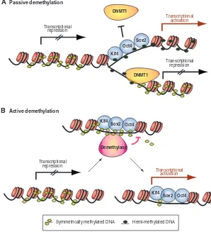
Possible mechanisms leading to DNA demethylation The mechanisms of DNA demethylation in reprogramming and normal development remain largely elusive. Two waves of global demethylation can be distinguished in mammalian development. Upon fertilization, the paternal genome becomes actively demethylated in the zygote, while the maternal genome looses its methylation marks passively during cleavage divisions (Jaenisch and Bird, 2003). During the specification of PGCs, another round of demethylation occurs in order to erase and subsequently re-establish methylation marks associated with imprinted genes. During reprogramming, demethylation may occur passively; that is, the direct binding of reprogramming factors to promoter or enhancer regions might interfere with the methylation of newly synthesized DNA during DNA replication (Fig. 5A). This process
D
E
V
E
LO
P
M
E
N
would be stochastic and would be more likely to occur if multiple transcription factors associate with the target gene (Lin et al., 2000). An example of the localized demethylation that would result from such a physical hindrance of DNA methylation is seen at the glucocorticoid receptor. This receptor can induce localized DNA demethylation, which is required for the subsequent recruitment of other transcription factors to neighboring sites (Thomassin et al., 2001). Similarly, specific factors that contribute to the stable inheritance of methylation patterns could be stochastically impaired, such as Uhrf1 (Ubiquitin-like, containing PHD and RING finger domains, protein), Dnmt3l, Suv39h1/2 (Suppressor of variegation 3-9 homologs), Lsh (Lymphoid-specific helicase), as well as Piwil1/2 (piwi-like homologs 1/2; Fig. 5A) (Bostick et al., 2007; Bourc’his and Bestor, 2006; Kuramochi-Miyagawa et al., 2008; Lehnertz et al., 2003; Sharif et al., 2007; Zhu et al., 2006).
Alternatively, active DNA demethylation mechanisms could be required for the reactivation of pluripotency genes (Fig. 5B). Compelling evidence indicates that DNA demethylation can occur by methylcytosine-specific DNA glycosylases and by the base excision repair machinery in plants (Choi et al., 2002; Gong et al., 2002). A repair-based process might also function in mammals, leading to global DNA demethylation in PGCs (Hajkova et al., 2008). Furthermore, an active enzyme appears to be responsible for the selective DNA demethylation of the paternal mouse genome upon fertilization (Santos et al., 2002), and for Oct4
promoter demethylation after the transplantation of a somatic cell nucleus into the frog oocyte (Simonsson and Gurdon, 2004). However, the nature and existence of demethylating enzymes in mammals remains highly controversial (Barreto et al., 2007;
Bhattacharya et al., 1999; Hendrich et al., 2001; Jin et al., 2008; Kangaspeska et al., 2008; Metivier et al., 2008). Taken together, how pluripotency genes become demethylated at the DNA level – either actively or passively – is still under debate. Partially reprogrammed cells could offer a unique system in which to distinguish between these possibilities and to decipher the role of other chromatin modifications.
In vivo versus in vitro reprogramming
As outlined above, the mechanisms that underlie nuclear reprogramming by NT and transcription factors remain largely elusive. However, both reprogramming events may involve processes that are similar to the ones that operate during germ cell reprogramming. PGCs erase DNA and histone methylation patterns, as well as genomic imprints, and reactivate the X chromosome during their development. Of note, early PGCs express transcription factors and show a chromatin signature that is reminiscent of that in pluripotent cells, correlating with the time window when pluripotent EG cells can be derived (Hajkova et al., 2008). Thus, their methylation and chromatin state must be permissive for their spontaneous conversion into a pluripotent state and may resemble that of nascent iPS cells. Other changes in PGCs include active demethylation of DNA followed by the replacement of histone variants, such as H2A.Z (Hajkova et al., 2008). A comparison of the mechanisms that lead to DNA demethylation and chromatin changes during PGC differentiation and somatic cell reprogramming to pluripotency should be quite informative, and may lead to strategies that improve the efficient and faithful reprogramming of somatic cells by NT or by defined factors.
Klf4 Sox2 Klf4
Transcriptional activation
DNMT1
Transcriptional repression
DNMT1
Transcriptional repression
A Passive demethylation
Oct4 Sox2
Symmetrically methylated DNA Hemi-methylated DNA Transcriptional activation Transcriptional
repression
B Active demethylation
Oct4
Demethylase
Klf4
[image:11.612.51.349.64.391.2]Sox2 Oct4
Fig. 5. Pathways to DNA demethylation of key pluripotency genes.(A) The establishment of symmetric DNA methylation patterns could be prevented passively during replication by the steric hindrance of Dnmt1 due to the stochastic binding of the reprogramming factors to target sites or by inhibiting Dnmt1 function indirectly.
Hemimethylation of the DNA would result in a progressive loss of methylation upon further rounds of cell division. (B) Alternatively, DNA methylation could be actively removed by the recruitment of a demethylating enzyme.