ISSN Online: 2164-5337 ISSN Print: 2164-5329
DOI: 10.4236/wjcd.2019.93018 Mar. 22, 2019 202 World Journal of Cardiovascular Diseases
Assessment of LV Function in Children with
Wilson’s Disease: Speckle Tracking Imaging
Study
Mahmoud Kamel Ahmed, Mohamed Yahia, Rehab Yaseen, Morad Beshay
Department of Cardiology, Menoufia University, Menoufia, EgyptAbstract
Background and Objective: Wilson’s disease is a genetic disorder of copper metabolism that affects liver and other organs including heart. In early stages of myocardial affection, the left ventricle (LV) appears apparently normal when evaluated by traditional two-dimensional (2D) echocardiography. The aim of this study was to detect subclinical LV dysfunction in children with Wilson’s disease using 2D speckle tracking echocardiography. Patients and Methods: Twenty children with Wilson’s disease were compared with age- and sex-matched 20 healthy children. All subjects were evaluated by tradi-tional 2D echocardiography and speckle tracking echocardiography. Results: There were no significant differences between patients and controls regarding conventional echo parameters except for lower E mitral flow and E' annular septal peak velocity in patient group. The regional peak longitudinal strain of apical 4 chamber view was −17.8% ± 4.2% in patients and −20.1% ± 2.3 % in control subjects (P = 0.043), and for apical 2 chambers view, it was −20.1% ± 3.6% in patients and −22.6% ± 3.4% in control subjects (P = 0.034) and it was −18% ± 3.5% in patients and −20.5% ± 3.2% in control subjects (P = 0.025) in apical long axis view. The global peak longitudinal strain was also lower in patients than control group (18.3% ± 3.2%, and 20.85% ± 2.4%) respectively (P = 0.014). There were no significant differences between both groups re-garding circumferential and radial strains (P > 0.05). Conclusions: Despite apparently normal LV systolic function, the children with Wilson’s disease demonstrated significantly lower peak longitudinal strain as an indicator for early affection of LV systolic function.
Keywords
Wilson’s Disease, Speckle Tracking Echocardiography
How to cite this paper: Ahmed, M.K., Yahia, M., Yaseen, R. and Beshay, M. (2019) As-sessment of LV Function in Children with Wilson’s Disease: Speckle Tracking Imag-ing Study. World Journal of Cardiovascular Diseases, 9, 202-211.
https://doi.org/10.4236/wjcd.2019.93018
Received: February 18, 2019 Accepted: March 19, 2019 Published: March 22, 2019
Copyright © 2019 by author(s) and Scientific Research Publishing Inc. This work is licensed under the Creative Commons Attribution International License (CC BY 4.0).
DOI: 10.4236/wjcd.2019.93018 203 World Journal of Cardiovascular Diseases
1. Introduction
Wilson’s disease is an inherited metabolic disease, in which there is inability of the liver to excrete copper in the bile. As a result, there is excessive copper sto-rage in the liver which usually extends to other organs, practically the brain [1] [2]. Copper deposition may extend to the myocardium, resulting in electrocar-diographic changes, arrhythmias, autonomic dysfunction, heart failure and sud-den cardiac death [3][4][5]. The aim of this study was to detect subclinical LV dysfunction in children with Wilson’s disease using two-dimensional speckle tracking strain echocardiography.
2. Methods
2.1. Study Population
This case-control study included 20 children with Wilson’s disease who were re-cruited from national liver institute in the period between February 2018 and October 2018. Wilson’s disease was diagnosed by pediatric hepatologist, de-pending on the presence of jaundice, Kayser Fleischer ring, liver disease (cirr-hotic changes), increased blood levels of copper and decreased blood levels of ceruloplasmin. The diagnosis was confirmed by presence of histological asso-ciated features in liver biopsy. Patients with poor quality echocardiographic im-ages, diabetes mellitus, congenital heart disease, primary cardiac valve disease and atrial fibrillation were excluded.
A twenty age and sex matched healthy children were taken as a control group. The study was approved by the local ethical committee of Menoufia University. Cardiac evaluation was done at cardiology department in Menoufia university hospital.
2.2. Methods
All the study population subjected to full history taking, clinical examination, standard 12 leads ECG.
2.3. Conventional Echocardiography
Echocardiographic examination was done to all participants by using the com-mercially available (Vivid 9, General Electric Healthcare, GE Vingmed, Norway) equipped with a 1.7 - 4 MHz phased-array transducer. echocardiographic imag-ing was obtained in the parasternal long- and short-axis, and apical two, three and four-chamber views using standard transducer positions.
LV end-diastolic and end-systolic diameters and volumes, septal and posterior wall thickness, ejection fraction and left atrial diameter were measured in accordance with the recommendations of the American Society of Echocardiography [6][7].
Pulsed and continuous-wave Doppler was used for valvular assessment. Peak early (E) and late (A) trans-mitral filling velocities were measured from mitral flow.
DOI: 10.4236/wjcd.2019.93018 204 World Journal of Cardiovascular Diseases diastolic mitral flow peak velocity to early diastolic septal annular peak velocity was measured (E/E').
LV Deformation: three cycles from apical views: 4, 2 and 3 chamber views were taken and another three cycles from the short axis view at the level of the LV papillary muscles for measuring circumferential and radial strain.
Frame rate was selected between 40 - 90 or at least 40% of HR. Then after ac-tivation of automated function imaging (AFI), digital data were transferred for off-line analysis, using Vivid Nine System Echo Pac, GE Vingmed, Horton, Norway.
2.4. Statistical Analysis
Data were analyzed using SPSS software version 20.0. Quantitative data ex-pressed as mean and standard deviation Chi-square test student test, Mann whiney U test, Kruskal Walls test were used. The P value < 0.05 was considered significant.
3. Results
Twenty children with Wilson disease (mean age 10.3 ± 2.6 years) and 20 age and sex matched healthy controls (mean age 10.6 ± 2.5 years) were enrolled in this study (Table 1).
3.1. Conventional Echocardiography
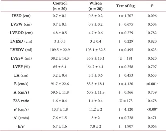
There were no significant differences between the studied groups as regard to LA diameter, LVEDD, LVESD and EF (P values > 0.05).
Trans-mitral E velocities were significantly lower in patients compared to controls (85.5 ± 18.1 cm/s versus 91.7 ± 22.6 cm/s, P < 0.001). Moreover, the septal peak early diastolic velocities (E’) were significantly lower in patients compared to controls (11.2 ± 2 cm/s versus 13.7 ± 1.8 cm/s, P < 0.001). But there was no significant difference between the studied groups as regard E/E’ (P val-ue > 0.05) (Table 2).
3.2. Speckle-Tracking Echocardiography
3.2.1. Segmental, Regional and Global Longitudinal LV Strain
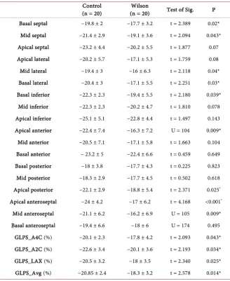
Segmental analysis showed that peak systolic longitudinal strain values of most segments were statistically significantly lower in patients compared to controls (P < 0.05).
DOI: 10.4236/wjcd.2019.93018 205 World Journal of Cardiovascular Diseases
Table 1. Characteristics of the two studied groups. Control
(n = 20) (n = 20) Wilson Test of Sig. P Sex (male), n., % 13 (65%) 12 (60%) χ2 = 0.107 1.000 Age (Y.), mean ± SD 10.6 ± 2.5 10.3 ± 2.6 t = 0.374 0.711 BSA (m2), mean ± SD 1.7 ± 0.2 1.7 ± 0.2 t = 0.129 0.898
BSA: body surface area.
Table 2. Comparison between the two studied groups according to two dimensional, doppler and tissue doppler echocardiographic parameters.
Control
(n = 20) (n = 20) Wilson Test of Sig. P IVSD (cm) 0.7 ± 0.1 0.8 ± 0.2 t = 1.707 0.096 LVPW (cm) 0.7 ± 0.1 0.8 ± 0.2 t = 0.675 0.504 LVEDD (cm) 4.8 ± 0.5 4.7 ± 0.6 t = 0.279 0.782 LVESD (cm) 3 ± 0.5 3 ± 0.4 t = 0.229 0.820 LVEDV (ml) 109.5 ± 22.9 105.1 ± 32.5 t = 0.495 0.623 LVESV (ml) 38.2 ± 14.3 35.9 ± 13.1 U = 181 0.620 LVEF (%) 65 ± 6.4 64.7 ± 4.1 t = 0.258 0.797 LA (cm) 3.2 ± 0.4 3.3 ± 0.6 t = 0.453 0.653 E (cm/s) 91.7 ± 22.6 85.5 ± 18.1 t = 4.120 <0.001* A (cm/s) 59.6 ± 11.8 60.9 ± 11.8 t = 0.366 0.739 E/A ratio 1.6 ± 0.4 1.4 ± 0.4 U = 173 0.478 e’ (cm/s) 13.7 ± 1.8 11.2 ± 2 t = 4.120 <0.00* A’ (cm/s) 7.6 ± 1.5 8 ± 2 t = 0.728 0.471
E/e’ 6.7 ± 1.6 7.8 ± 2 t = 1.907 0.064
IVSD: interventricular septal thickness at end diastole, LVPW: left ventricular posterior wall thickness, LVEDD: left ventricular end diastolic dimension, LVESD: left ventricular end systolic dimension, LVEDV: left ventricular end diastolic volume, LVESV: left ventricular end systolic volume, LVEF: left ventricular ejection fraction. LA: left atrium, E: early peak pulsed doppler velocity on mitral valve, A: late pulsed dopp-ler velocity on mitral valve, e': early tissue velocity of septal annulus. t: Student t-test, U: Mann Whitney test, p: p value for comparing between the studied groups. All variables are expressed by mean ± standard devia-tion. *: Statistically significant at P ≤ 0.05.
3.2.2. Circumferential and Radial Strain of LV
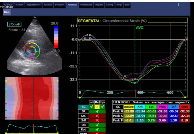
There was no significant difference between the studied groups regarding cir-cumferential LV strain (−16.7% ± 4.5% versus −19.3% ± 5% P > 0.05).
Also, there was no significant difference between the studied groups regarding radial strain of LV (38.5% ± 16% versus 40.3% ± 15.2% P > 0.05) (Table 4,
Fig-ure 2).
4. Discussion
[image:4.595.206.538.223.484.2]DOI: 10.4236/wjcd.2019.93018 206 World Journal of Cardiovascular Diseases
Table 3. Longitudinal strain values of the two studied groups. Control
(n = 20) (n = 20) Wilson Test of Sig. P Basal septal −19.8 ± 2 −17.7 ± 3.2 t = 2.389 0.02*
Mid septal −21.4 ± 2.9 −19.1 ± 3.6 t = 2.094 0.043* Apical septal −23.2 ± 4.4 −20.2 ± 5.5 t = 1.877 0.07 Apical lateral −20.2 ± 5.7 −17.1 ± 5.3 t = 1.759 0.08 Mid lateral −19.4 ± 3 −16 ± 6.3 t = 2.118 0.04* Basal lateral −20.4 ± 3 −17.1 ± 5.5 t = 2.251 0.03* Basal inferior −22.3 ± 2.3 −19.4 ± 5.5 t = 2.180 0.039*
Mid inferior −22.3 ± 2.3 −20.2 ± 4.7 t = 1.810 0.078 Apical inferior −25.1 ± 5.1 −22.8 ± 4.4 t = 1.497 0.143 Apical anterior −22.4 ± 7.4 −16.3 ± 7.2 U = 104 0.009*
Mid anterior −20.5 ± 7.1 −17.1 ± 5.8 t = 1.663 0.104 Basal anterior − 23.2 ± 5 −22.4 ± 6.6 t = 0.459 0.649 Basal posterior −18 ± 3.8 −17.7 ± 4.3 t = 0.225 0.823 Mid posterior −18.3 ± 2.9 −17.7 ± 4.5 t = 0.502 0.618 Apical posterior −22.1 ± 2.9 −18.8 ± 5.4 t = 2.371 0.025* Apical anteroseptal −24 ± 4.2 −17 ± 6.2 t = 4.168 <0.001*
Mid anteroseptal −21.1 ± 6.2 −16.2 ± 6.9 U = 105 0.009* Basal anteroseptal −19.4 ± 6.6 −18 ± 6 U = 174 0.495
GLPS_A4C (%) −20.1 ± 2.3 −17.8 ± 4.2 t = 2.093 0.043* GLPS_A2C (%) −22.6 ± 3.4 −20.1 ± 3.6 t = 2.193 0.034* GLPS_LAX (%) −20.5 ± 3.2 −18 ± 3.5 t = 2.340 0.025* GLPS_Avg (%) −20.85 ± 2.4 −18.3 ± 3.2 t = 2.578 0.014*
GLPS_A4C: apical 4 chamberglobal longitudinal peak systolic strain, GLPS_A2C: apical 2 chamber global longitudinal peak systolic strain, GLPS_LAX: apical long axisglobal longitudinal peak systolic strain,
GLPS_Avg: average global longitudinal peak systolic strain. t: Student t-test, U: Mann Whitney test, p: p
value for comparing between the studied groups. All variables are expressed by mean ± standard deviation. *: Statistically significant at P ≤ 0.05.
various mechanisms; first, it is caused by intra- and extracellular pathological accumulations of copper that lead to infiltrative myocardial diseases [8] [9]. Second, copper ions had toxic effects which may result in damage of myocytes. Copper stimulates the production of interleukin-6 and interleukin-1β with re-sultant oxidative stress [10] [11]. Furthermore, Patients with Wilson’s disease demonstrated a polymorphism in the gene which encoded an interleukon-1 re-ceptor antagonist [12] [13]. Finally, copper activated sphingomyelin, which ac-tivates cardiac apoptosis [14].
DOI: 10.4236/wjcd.2019.93018 207 World Journal of Cardiovascular Diseases
[image:6.595.209.538.318.546.2]Figure 1. Peak longitudinal strain of all apical view in one of the affected children with Wilson disease.
Figure 2. Normal circumferential strain of one of control group.
Table 4. Comparison between the two studied groups according to different parameters. Control
(n = 20) (n= 20) Wilson Test of Sig. P Circumferential Strain −19.3 ± 5 −16.7 ± 4.5 t = 1.743 0.089
Radial Strain 40.3 ± 15.2 38.5 ± 16 t = 0.369 0.714
Variables are expressed by mean ± standard deviation: Student t-test, p: p value for comparing between the studied groups.
[image:6.595.207.539.597.654.2]ap-DOI: 10.4236/wjcd.2019.93018 208 World Journal of Cardiovascular Diseases pear at the earlier stage of the disease [16].
In early stages of the Wilson’s disease, the LV appears apparently normal when evaluated by traditional 2D echocardiographic parameters. Early detection of LV dysfunction by the relatively new echocardiographic technique; speckle tracking echocardiographic, helps in strict control of blood copper levels to pre-vent development of cardiomyopathy. The current study evaluated LV function by speckle tracking echocardiography in 20 children with Wilson’s disease, and compared the results to 20 age and sex matched healthy children. The mean LV ejection fraction did not differ significantly in patients and controls. Trans-mitral E velocities and the septal peak early diastolic velocities (E’) were significantly lower in patients in comparison to control participants, which re-flects impaired LV filling in patients with Wilson’s disease.
Our results agreed with Malgorzata et al. who evaluated LV function in 125 adult patients with Wilson’s disease and reported non-significant differences in LV ejection fractions in comparison to controls, while diastolic dysfunction was only present in patients presented with neurological form rather than hepatic form. They also reported mitral valve disease in some patients with Wilsons dis-ease, in contrast, our study reported no valvular pathology [17].
Similarly, Elkiran et al. studied 22 children with Wilson’s disease, and they reported diastolic dysfunction [18]. In another study, the LV systolic function measured by ejection fraction did not differ significantly between Wilson’s dis-ease and controls, however, diastolic dysfunction was more frequent in patients with Wilson’s disease [19].
Hlubocka Z et al. compared 2D echocardiographic data of 42 patients with Wilson’s disease to 42 healthy subjects. They reported a non-significant trend towards lower LV ejection fraction values in patients with Wilson’s disease (62% versus 64%), the LV diastolic function did not differ in the two groups [20].
In a recent study by Quick et al., 61 patients with Wilson’s disease were eva-luated by cardiac magnetic resonance. They reported no differences between pa-tients and controls with respect to LV ejection fractions, however, there were significant differences between the two groups with respect to late gadolinium enhancement. Regarding such data, they suggested that patients with Wilson’s disease are at risk of developing impaired LV functions [21].
DOI: 10.4236/wjcd.2019.93018 209 World Journal of Cardiovascular Diseases strain rate [23].
5. Conclusion
Despite apparently normal LV systolic function, the patients with Wilson’s dis-ease demonstrated a significant reduction in regional and global LV longitudinal strain as an indicator of early affection of LV systolic function.
6. Limitations
The study was based on a small number of patients as Wilson’s disease is being a rare; therefore, the results need to be verified using a larger sample size. Another limitation is lack of neurological form of the disease, as our patients were re-cruited from a liver tertiary center. In addition, there was a difficulty to assess the drug therapy of Wilson’s disease on myocardial pathology.
Conflicts of Interest
The authors declare no conflicts of interest regarding the publication of this pa-per.
References
[1] Rosencrantz, R. and Schilsky, M. (2011) Wilson’s Disease: Pathogenesis and Clinical Considerations in Diagnosis and Treatment. Seminars in Liver Disease, 31, 245-259. https://doi.org/10.1055/s-0031-1286056
[2] Bennett, J. and Hahn, S.H. (2011) Clinical Molecular Diagnosis of Wilson’s Disease. Seminars in Liver Disease, 13, 233-238.https://doi.org/10.1055/s-0031-1286054
[3] Aggarwal, A. and Bhatt, M. (2013) Update on Wilson Disease. International Review of Neurobiology, 110, 313-348.
https://doi.org/10.1016/B978-0-12-410502-7.00014-4
[4] Kuan, P. (1987) Cardiac Wilson’s Disease. Chest, 91, 579-583. https://doi.org/10.1378/chest.91.4.579
[5] Meenakshi-Sundaram, S., Sinha, S., Rao, M., Prashanth, L.K., Arunodaya, G.R., Rao, S., et al. (2004) Cardiac Involvement in Wilson’s Disease—An Electrocardiographic Observation. The Journal of the Association of Physicians of India, 52, 294-296. [6] Lang, R.M., Badano, L.P., Mor-Avi, V., Afilalo, J., Armstrong, A., et al. (2015)
Recommendations for Cardiac Chamber Quantification by Echocardiography in Adults: An Update from the American Society of Echocardiography and the Euro-pean Association of Cardiovascular Imaging. Journal of the American Society of Echocardiography, 28, 1-39.https://doi.org/10.1016/j.echo.2014.10.003
[7] Nagueh, S.F., Smiseth, O.A., Appleton, C.P., Byrd, B.F., Dokainish, H., et al. (2016) Recommendations for the Evaluation of Left Ventricular Diastolic Function by Echocardiography: An Update from the American Society of Echocardiography and the European Association of Cardiovascular Imaging. European Heart Jour-nal—Cardiovascular Imaging, 17, 1321-1360.https://doi.org/10.1093/ehjci/jew082
[8] Moinuddin, M.J., Figueredo, V. and Amanullah, A.M. (2010) Infiltrative Diseases of the Heart. Reviews in Cardiovascular Medicine, 11, 218-227.
DOI: 10.4236/wjcd.2019.93018 210 World Journal of Cardiovascular Diseases
Classification of the Cardiomyopathies: An American Heart Association Scientific Statement from the Council on Clinical Cardiology, Heart Failure and Transplanta-tion Committee; Quality of Care and Outcomes Research and FuncTransplanta-tional Genomics and Translational Biology Interdisciplinary Working Groups; and Council on Epi-demiology and Prevention. Circulation, 113, 1807-1816.
https://doi.org/10.1161/CIRCULATIONAHA.106.174287
[10] Ansteinsson, V., Refsnes, M., Skomedal, T., Osnes, J.B., Schiander, I. and Lag, M. (2009) Zinc- and Copper-Induced Interleukin-6 Release in Primary Cell Cultures from Rat Heart. Cardiovascular Toxicology, 9, 86-94.
https://doi.org/10.1007/s12012-009-9043-5
[11] Bruha, R., Vitek, L., Marecek, Z., et al. (2012) Decreased Serum Antioxidant Capac-ity in Patients with Wilson Disease Is Associated with Neurological Symptoms. Journal of Inherited Metabolic Disease, 35, 541-548.
https://doi.org/10.1007/s10545-011-9422-5
[12] Gromadzka, G. and Czlonkowska, A. (2011) Influence of IL-1RN Intron 2 Variable Number of Tandem Repeats (VNTR) Polymorphism on the Age at Onset of Neu-ropsychiatric Symptoms in Wilson’s Disease. International Journal of Neuroscience, 121, 8-15.https://doi.org/10.3109/00207454.2010.523131
[13] Gromadzka, G., Kruszynska, M., Wierzbicka, D., et al. (2015) Gene Variants En-coding Proteins Involved in Antioxidant Defense System and the Clinical Expres-sion of Wilson Disease. Liver International, 35, 215-222.
https://doi.org/10.1111/liv.12493
[14] Lang, P.A., Schenck, M., Nicolay, J.P., et al. (2007) Liver Cell Death and Anemia in Wilson Disease Involve Acid Sphingomyelinase and Ceramide. Nature Medicine, 13, 164-170.https://doi.org/10.1038/nm1539
[15] Cheng, N., Wang, H., Dong, J., et al. (2015) Elevated Serum Brain Natriuretic Pep-tide and Matrix Metalloproteinases 2 and 9 in Wilson’s Disease. Metabolic Brain Disease, 30, 1087-1091.https://doi.org/10.1007/s11011-015-9685-x
[16] Ferenci, P., Czlonkowska, A., Merle, U., et al. (2007) Late-Onset Wilson’s Disease. Gastroenterology, 132, 1294-1298.https://doi.org/10.1053/j.gastro.2007.02.057
[17] Buksińska-Lisik, M., Litwin, T., Pasierski, T. and Członkowska, A. (2017) Cardiac Assessment in Wilson’s Disease Patients Based on Electrocardiography and Echo-cardiography Examination. Archives of Medical Science.
https://doi.org/10.5114/aoms.2017.69728
[18] Elkiran, O., Karakurt, C., Selimoglu, A., et al. (2013) Subclinical Diastolic Dysfunc-tion in Children with Wilson’s Disease Assessed by Tissue Doppler Echocardiogra-phy: A Possible Early Predictor of Cardiac Involvement. Acta Cardiologica, 68, 181-187.https://doi.org/10.1080/AC.68.2.2967276
[19] Quick, S., Reuner, U., Weidauer, M., Hempel, C., Heidrich, F.M., Mues, C., Sveric, K.M., et al. (2019) Cardiac and Autonomic Function in Patients with Wilson’s Dis-ease. Orphanet Journal of Rare Diseases, 14, 22.
https://doi.org/10.1186/s13023-019-1007-7
[20] Hlubocká, Z., Marecek, Z., Linhart, A., Kejková, E., Pospísilová, L., Martásek, P. and Aschermann, M. (2002) Cardiac Involvement in Wilson Disease. Journal of Inhe-rited Metabolic Disease, 25, 269-277.https://doi.org/10.1023/A:1016546223327
DOI: 10.4236/wjcd.2019.93018 211 World Journal of Cardiovascular Diseases
[22] Forsey, J., Friedberg, M. and Mertens, L. (2013) Speckle Tracking Echocardiography in Pediatric and Congenital Heart Disease. Echocardiography, 30, 447-459. https://doi.org/10.1111/echo.12131