Alex Berenstein 1
Received July 19. 1979: accepted atler revi-sion October 9, 1979.
'Department of Neuroradiology, Bellevue Hos-pital, and New York University Medical Center. 550 First Ave .. New York, NY 1 001 6.
This article appears in March/ April 1980 AJNR and June 1980 AJR.
AJNR 1:161-166, March/April 1980 0195-6108/80/0102-0161 $00.00 ©American Roentgen Ray Society
Flow-Controlled Silicone
Fluid Embolization
161
Flow-controlled silicone fluid embolization is a technique that can be useful in situations where selective injection cannot be achieved, but where the proximal part of the feeding vessel must be preserved. It consists of a modified double-lumen balloon catheter and silicone fluid made radiopaque with tantalum powder with a predetermined viscosity and vulcanization time. Flow control by manipulation of the balloon in the proximal artery permits the distribution of emboli to desired sites in distal arteries. A case is described and other details offered about this technique that may be valuable for special situations.
Increased availability of delivery systems and embolic agents have enhanced the physician's capabilities in the treatment of complex hemodynamic lesions [1-4 ). Selective catheter placement prior to embolic agent deposition is the ideal situation for the best results and fewest complications. However, situations arise when selective catheterization is impossible due to vessel tortuosity and/or the extent of the lesion. A technique that may be useful in such situations is described in detail.
Technique
Delivery System
The delivery system consists of a modified double-lumen balloon catheter {3) (Meditech Div., Cooper Scientific, Watertown, Mass.) that differs from conv en-tional double-lumen catheters in two ways: (1) the lumen connected to the balloon is oval, following the catheter wall, allowing a larger internal diameter of the injection lumen with no change in overall catheter size, and (2) the distal end of the catheter is not tapered, in order to maintain a constant internal diameter (fig. 1 ). The larger injection lumen facilitates injection of more viscous embolic agents. An introducer sheath (Cook, Bloomington, Ind.) is used to prevent pericatheter leakage. Continuous heparinized saline is perfused at the puncture site between the introducer sheath and the inner catheter. The catheters are available in different sizes. A
7
French catheter has a2.34
mm00
at the shaft and2
.88
mm OD over the balloon tip, with a 1.35 mm10.
A 3 French catheter has a 1 mm OD at the shaft and 1 .35 mm00
over the balloon tip, with an0
.49
mm
10
[3). Embolic Material162
BERENSTEIN AJNR:1, March/April1980A
B
Fig. I .- A, Cross seclion of conventional double-lumen balloon catheter with lwo circular lumens: smaller for balloon, larger or distal lumen for injection. B, Modified double-lumen balloon catheler. Balloon lumen changed Ia oval, following wall of catheler shaft. which increases injeclion lumen by 30%. (Reprinled from [3).)
centistokes viscosity is thoroughly mixed with 1 -2 J.Lm opa-cifying tantalum powder (Kena Metal, Latrobe, Pa.) (1 g/ml silicone mixture). A 1 drop/ml mixture of stannous octate
(Catalyst M, Dow Corning Corp., I.N.D. required) which will
lead to vulcanization in about
20
min is added. If faster vulcanization is desired, the addition of 1 drop/ml of tetra-N-proxysilane[5]
will produce vulcanization in 3-4 min, while 1 drop/3 ml will vulcanize the fluid in about 10 min. An in vitro test of vulcanization time is made before injection and adjustments are made as needed.Catheterization and Embolization
A percutaneous technique is preferred for catheter
inser-tion, but in some instances direct arteriotomy may be used.
After the balloon catheter has been placed at the desired position, the balloon is inflated and flow stasis is angiograph-ically tested. A drop of the silicone mixture is placed beyond the catheter tip with the balloon deflated; the silicone globule will ascend with the blood flow and will stop and vulcanize at the point where the vessel diameter diminishes sufficiently to arrest its flow. If the silicone fluid is not arrested, as may be the case in large arteriovenous communications, the maneuver is repeated with the balloon inflated. By gradual balloon deflation, the radiopaque silicone will ascend. Under fluoroscopic monitoring, the balloon is reinflated when the embolus has reached the desired position, and the material is allowed to vulcanize before the balloon is deflated. The
catheter lumen may be kept patent by pushing the silicone
with saline, from the catheter hub to its tip. More than 1 drop may be injected at a time in a large, multiple vessel lesion. Previous experience in animal models is mandatory for proper control of the technique.
Case Report
A 32-year-old man was first seen at age 1 9 with seizure disorder and a progressive left hemiparesis due to a right frontotemporal
arteriovenous malformation that was partially resected at that time. Postoperatively, the patient did well except for continued paresthe-sia of the lett upper and lower extremities; he was able to continue school and then attended college.
At age 26 he was readmitted for progression in his hemiparesis and a bruit in his head. A silicone sphere embolization was per -formed after surgical exposure of the right external carotid artery in the neck, through which a 4 mm 10 catheter was passed into the right internal carotid artery; 50 emboli, 1.5-3 mm in diameter, were
introduced. The external carotid artery was ligated at the end of the
embolization. Postoperatively, the right eye was blind secondary to an embolus lodging in the ophthalmic artery and ligature of the external carotid artery that decreased collateralization to the right
ophthalmic artery. The flow through the malformation was
signifi-cantly reduced. Neurologically there was no further deterioration; however, there was difficulty in controlling the patient's seizures. The patient suffered a subarachnoid hemorrhage 3 months be-fore the most recent admission and again 1 month later with an increase in seizure activity. A third episode of subarachnoid hem-orrhage occurred 4 days before admission and was documented by a bloody and xanthochromic spinal fluid. The postictal course was characterized by a marked spastic lett hemiparesis, inability to walk, and slow mentation.
Admission physical examination showed a 32-year-old man in slight to moderate distress, alert and oriented, but with mild slow
mentation; both left upper and lower extremities showed marked
muscle wasting in all groups and a left Babinski sign. The patient was blind in the right eye and had 20/20 vision in the left, the remaining cranial nerves were intact. Sensory examination revealed mild hemianesthesia on the lett side of the body; pin sensation was present on the right and absent on the lett. Lett-sided ataxia was noted.
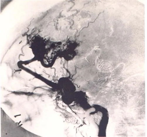
Angiography revealed a large right frontotemporal arteriovenous malformation, supplied by the right anterior cerebral artery (fig. 2A). The right middle cerebral artery filled through leptomeningeal col-laterals from the vertebrobasilar circulation and multiple hypertro -phied muscular branches of the right vertebral artery supplied the right external carotid trunk from which vessels filled the malforma-tion (figs. 28 and 2C). Lett external carotid angiography revealed meningeal and superficial temporal supply (fig. 20) to the
malfor-mation. The left internal carotid circulation did not feed the mal
for-mation directly. The lett vertebral artery also supplied the mal for-mation in its most posterior and medial aspect via the right posterior cerebral artery with drainage to the internal cerebral vein.
First embolization. The left common carotid artery was entered percutaneously and the left middle meningeal artery was selectively catheterized with a conventional double-lumen balloon catheter and a drop of viscous silicone was injected. This drop of silicone stopped in the distal meningeal vessel, and was well anchored to the vessel bifurcation (fig. 3). The postembolization course was uneventful with the exception of mild tenderness in the left temporal region that completely cleared after 5 days.
Second embolization. Three weeks later, the right external
ca-rotid artery (previously ligated 6 years earlier) was surgically
ex-posed in the neck and our modified 7 French double-lumen balloon catheter was selectively placed in the right middle meningeal artery (fig. 4A). Using flow control, emboli were successfully injected to produce distal occlusion of more than 90% of the supply from the right external carotid artery (figs. 48 and 4C). The postembolization course. apart from mild right temporal pain, was uneventful. The pain subsided in about 2 weeks. The neurologic status seemed improved; the left lower extremity spasticity was considerably de-creased. The patient regained control of movement in the left lower extremity and was able to move his toes and ankle. A radionuclide
AJNR:1, March/ April 1980 FLOW CONTROLLED SILICONE EMBOLIZATION 163
164 BERENSTEIN AJNR: 1, March/ April 1980
A
A
8
8
c
Fig. 3.-Fiow-controlled silicone em -boli in left middle meningeal artery. A, Frontal view. Emboli (arrow). B, Lateral view, close-up. Anchored emboli (straight arrow). Prominent vascular groove
(curved arrow).
Fig. 4.- A, Lateral subtraction angiogram of right middle meningeal artery. Modified double-lumen balloon catheter. Balloon (arrow) in proximal part of middle meningeal pedicle. Multiple feeding vessels. (Reprinted from [2].) B. Lateral skull radiograph. Numerous radiopaque silicone emboli (arrows) after InJection w1lh balloon flow control (Reprinted from [2].) C. Lateral subtraction angiogram of right external carotid artery after embolization. Contrast material fills vessels until site of emboli (arrows); no filling of malformation. (Ct. fig. 2C.)
supply (first embolization) and the second embolization of the right
external carotid supply, showed a marked decrease in the flow of
the right side and no gross change on the left (fig. 5).
Th1rd embolization. Percutaneous catheterization of the left ex
-ternal carotid artery via the femoral route was performed. A control
angiogram showed the previously embolized meningeal vessel to
be occluded. On the left side two other feeders were embolized by
the same technique with good results of 80% occlusion of the left
external carotid supply (fig. 6). The procedure was stopped because
some of the fluid silicone passed into the lungs; there were no
clinical manifestation from the pulmonary emboli.
Fourth embolization. The right internal carotid artery was
cathe-terized through the femoral route; repeated attempts to flow-guide
a microballoon catheter lo the right anterior cerebral artery were
unsuccessful due to vessel tortuosity, and silicone spheres were
flow directed to the malformation. A total of 25 1 mm spheres and
65 1.5 mm spheres was injected with a reduction of about 40% in
[image:4.613.39.569.91.577.2]AJNR: 1. March/ April 1980 FLOW CONTROLLED SILICONE EMBOLIZATION 165
A
B
Fig. 5.-A. Preembolization radionuclide flow study. Increased activity in neck area bilaterally (longer solid arrows) represents collateral supply be -tween vertebral artery and external carotid systems. Lett side supply (curved arrows). right side supply (Shorr solid arrows). and activity in brain (open
arrows). Single long arrow demonstrates flow sequence. B. After embolizalion of right side and partial embolization in lett. Decreased activity in neck on lett side (straight arrows) and no activity on right side of neck or in right convexity, but no gross change in left convexity area (curved arrows).
Fig. 6.- A. Frontal skull view after third embolization. Two new feeders from left side occluded (white arrows). Radiopaque silicone also seen in right occipital artery (from second emboliza-tion). (Ct. fig. 4B.) Drop of radiopaque silicone in venous side (open arrow). (Ct. fig. 3A.) B. Lateral subtraction angio-gram after embolization of left external carotid artery. Preservation of proximal vessels (open arrows>. with only minimal filling of malformation (curved arrows). (Cf. fig. 2D.)
(figs. 7 and 8). The patient was discharged 7 days after
emboliza-tion. Plain films of the skull show the radiopaque emboli to be
unchanged. Neurologic status was markedly improved. to the point
of the patient walking out of the hospital assisted only by a short
brace on the left lower extremity and a cane. The patient's clinical
status was stable 1 'h years later. He could walk unassisted and
without lhe brace or cane. He was seizure-free (on the same
medication). and no subarachnoid hemorrhage had recurred.
Discussion
Sana et al. [6] reported the use of viscous silicone (6,500
centistokes viscosity) for the embolization of intracerebral
arteriovenous malformations. We modified his technique by
varying the silicone viscosity from 6,500-25,000 centi -stokes, depending on the vessel to be occluded, by making
the silicone radiopaque with tantalum powder, and using
balloon catheters to control the flow. Due to the multiple
vessel involvement in our case, it became apparent that the
proximal part of the scalp vessels should be preserved to
prevent unwanted skin necrosis. The first embolization dem-onstrated this could be accomplished, and, encouraged by
this result. we undertook some animal experimentation with
this technique before attempting further embolization. At the
same time, we had been working on the development of a modified double-lumen balloon catheter for coaxial
emboli-zation [3] and noted that the injection of more viscous
mixture was facilitated by the increased diameter of the injection lumen.
These modifications permit the operator to vary the vi s-cosity of the silicone mixture for the size of the vessel to be occluded; less viscous material is used for smaller vessels
and higher viscosity for larger vessels. Long vulcanization
time is desired to allow multiple emboli o be introduced before the catheter lumen becomes obstructed. The use of
introducer sheaths at the puncture site permits multiple
catheter change without the need of guide wires, which
cannot be inserted if the catheter is blocked by the vul can-ized silicone.
[image:5.612.40.290.94.456.2]166 BERENSTEIN AJNR:1, March/April1980
Fig. 7.-A, Radionuclide flow scan after fourth embolization. (Ct. fig. 5A.)
the mixture radiopaque, allowing fluoroscopic monitoring of its final position. Graduated control of the flow by the balloon
adds flexibility to the system and gives better control of the
emboli final position, decreasing the amount of material that
will pass to the venous side.
In our case, only branches of the external carotid circu-lation were embolized with this technique. We have used
this technique in the cerebral circulation on one occasion
and in a spinal hemangioma. The indications for this tech-nique are existence of a significant hemodynamic sump
effect and linearity toward the lesion.
At present, curing complex arteriovenous malformations
by intravascular embolization is still not possible in most
patients with inoperable lesions. However, palliative
treat-ment is possible with some improvement or at least an arrest
in the progression of the disease. Repeated embolizations
Fig. a.- Lateral subtraction angiogram alter silicone sphere embolization of right internal carotid supply with reduction of about 40%. Slower flow in lesion (fig. 2 comparable in time sequence).
may be necessary, and the greater versatility in technique
will enhance the possibilities in patients previously unsuited
for this therapeutic method.
ACKNOWLEDGMENTS
We thank Joseph Ransohoff, Norman Chase, and Irvin Kricheff for guidance, encouragement, and support.
REFERENCES
1. Berenstein A, and Kricheff II. Catheter and material selection
for transarterial embolization: technical considerations. I. Cath
-eters. Radiology 1979; 1 32: 61 9-630
2. Berenstein A, Kricheff II. Catheter and material selection for transarterial embolization: technical considerations. II. Materi-als. Radiology 1979;132: 631-639
3. Berenstein A, Kricheff II. A new balloon catheter for coaxial
embolization. Neuroradiology 1979; 18: 239-241
4. Grace OM, Pitt OF, Gold RE. Vascular embolization and occlu-sion by angiographic techiques as an aid or alternative to operation, collective review. Surg G ynecol Obstet 1976; 1 43:
469-482
5. Hila! SK, Michelson JW. Therapeutic percutaneous
emboliza-tion for extra-axial vascular lesions of the head, neck and
spine. J Neurosurg 1975;43: 275-287
6. Sane K, Jimbo M, Saito I. Artificial embolization of inoperable angiomas with polymerizing substance. In: Pia HW, Gleave JRW, Grote E, et al., eds. Cerebral angiomas. Advances in
[image:6.613.310.561.97.332.2] [image:6.613.49.168.98.489.2]