Acta Cryst.(2003). E59, i125±i128 DOI: 10.1107/S1600536803017537 Uwe Kolitsch Fe1.84Mg0.16(PO4)(OH)
i125
inorganic papers
Acta Crystallographica Section E
Structure Reports Online
ISSN 1600-5368
Mg-rich wolfeite, (Fe
II,Mg)
2(PO
4)(OH):
structure refinement and Raman
spectroscopic data
Uwe Kolitsch
UniversitaÈt Wien, Institut fuÈr Mineralogie und Kristallographie, Geozentrum, Althanstrasse 14, A-1090 Wien, Austria
Correspondence e-mail: [email protected]
Key indicators
Single-crystal X-ray study T= 293 K
Mean(P±O) = 0.001 AÊ Disorder in main residue Rfactor = 0.022 wRfactor = 0.065
Data-to-parameter ratio = 17.3
For details of how these key indicators were automatically derived from the article, see http://journals.iucr.org/e.
#2003 International Union of Crystallography Printed in Great Britain ± all rights reserved
Mg-rich wolfeite [diiron(II) hydroxide phosphate],
(FeII,Mg)
2(PO4)(OH), from the Big Fish River area, Yukon Territory, Canada, is isotypic with its MnII-dominant analogue triploidite. The framework structure contains edge- and corner-sharing, distorted MO4(OH) and MO4(OH)2 (M = FeII or FeII,Mg) polyhedra linked by fairly regular PO
4 tetrahedra. All atoms are on general positions. Four of the eight independent Fe sites contain between 9 and 25% Mg substituting for Fe. Two of these four sites show distorted trigonal-bipyramidal coordination, whereas the remaining two sites show distorted octahedral coordination. The average (FeII,Mg)ÐO bond length decreases with increasing Mg content. Average PÐO distances range between 1.538 and 1.543 AÊ. The hydrogen bonds are all strongly bent and weak, with O O distances > 2.73 AÊ, an observation con®rmed by single-crystal Raman spectroscopic data which show ®ve bands due to OÐH stretching vibrations between 3478 and 3557 cmÿ1.
Comment
Wolfeite, (FeII,MnII)
2(PO4)(OH) (Mandarino, 1999), is a rare iron phosphate mineral which occurs as a metasomatic alteration phase in pegmatites, and also rarely in hydrothermal veins, phosphatic nodules in shales, and amphibolite-facies metamorphosed iron formations (Anthonyet al., 2000; Masau
et al., 2000; Stalder & Rozendaal, 2002, and references
therein). Wolfeite is a member of a large family of compounds with the general formula MII
2(XO4)Z, where M = Fe, Mn, Mg, ...;X= P, As, ...; andZ= F, OH, O, Cletc. According to the crystal-chemical classi®cation of Strunz & Nickel (2001), wolfeite belongs to the triploidite group of minerals, whose other members are triploidite [(MnII,FeII)
2(PO4)(OH); Waldrop, 1970], wagnerite [Mg2(PO4)F; Coda et al., 1967], staneÏkite [(Mn,FeII,Mg)FeIII(PO
4)O; Keller et al., 1997] and sarkinite [MnII
2(AsO4)(OH); Dal Negroet al., 1974]. Various synthetic compounds are also isotypic with triploidite [e.g.
-Mg2(PO4)(OH) (Raade & Rùmming, 1986) and
Zn2(PO4)(F,OH) (Taasti et al., 2002)]. All triploidite-type compounds crystallize with space groupP21/aand have similar unit-cell parameters. The common structure type is closely related to that of triplite, (MnII,FeII)
2(PO4)F (space group
I2/a; Waldrop, 1969; alternative setting inC2/c for synthetic triplite by Rea & Kostiner, 1972).
The crystal structure of wolfeite has not been studied thus far, although X-ray powder diffraction data and unit-cell parameters have been provided by Frondel (1949), Antenucci
et al. (1989), and Masau et al. (2000). The present article
reports the results of a single-crystal structure re®nement and of single-crystal Raman spectroscopic studies of a Mg-rich
inorganic papers
i126
Uwe Kolitsch Fe1.84Mg0.16(PO4)(OH) Acta Cryst.(2003). E59, i125±i128wolfeite sample from the Big Fish River area, Yukon Territory, Canada (Robertson, 1982; Robinsonet al., 1992). The article supplements an earlier study of the crystal structure and IR spectra of a Mg-rich satterlyite, (Fe,Mg)12(PO3OH)(PO4)5 -(OH,O)6, from the same locality (Kolitschet al., 2002).
At the Big Fish River area, the mineral is a common constituent of epigenetic phosphatic nodules and forms divergent, columnar aggregates of crude, glassy, light brown to clove-brown crystals up to several centimetres in length. Crystallization occurred at temperatures of about 453 to 473 K, according to ¯uid inclusion studies (Robinson et al., 1992). Previous electron microprobe analyses of wolfeite from this locality yielded an average formula close to (Fe1.65Mg0.20Mn0.15)2(PO4)(OH0.95F0.05) (Robinson et al., 1992). The presently studied sample is from the collection of the author, and its appearance closely ®ts the published descriptions (Robinsonet al., 1992). The chemical composition of the sample has been characterized by semiquantitative SEM±EDS data which revealed major Fe and P, minor Mg and only very small amounts of Mn (ratio Fe:Mn ca. 13:1), and insigni®cant compositional inhomogeneities. The EDS-based chemical composition is in good agreement with the formula subsequently derived from the structure re®nement.
Mg-rich wolfeite is con®rmed to be isotypic with triploidite (MnII,FeII)
2(PO4)(OH); space groupP21/a; Waldrop, 1970). It has a complex framework structure based on edge- and corner-sharing, distorted MO4(OH) and MO4(OH)2 poly-hedra (M = FeII,Mg), corner-linked to PO
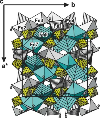
4 tetrahedra (Figs. 1,2). For detailed descriptions of the connectivity, the reader is referred to the previous reports on the isotypic members of the triploidite group (see above). The present paper restricts itself to the Mg distribution and the hydrogen bonding in Mg-rich wolfeite. To facilitate comparisons, the atomic coordinates and the labeling used by Waldrop (1970) for triploidite were adopted for Mg-rich wolfeite, except for the H atoms which had not been located during the earlier study of triploidite. The crystal structure of Mg-rich wolfeite contains eight non-equivalent Fe sites, four P sites, twenty O sites, and four H sites (belonging to OH groups). All atoms are on general positions. The Fe sites Fe1, Fe4, Fe6, and Fe8 are all ®ve-coordinated (with distorted trigonal-bipyramidal geometry), whereas the remaining Fe sites show distorted octahedral coordination (with OH groups in the cis con®guration). Site occupancy re®nements demonstrated that the considerable Mg present in the structure strictly prefers the following four out of the eight Fe sites: Fe5, Fe6, Fe7, and Fe8. The re®ned Fe:Mg ratios at these sites range between approximately 0.76:0.24 (Fe6) and 0.90:0.10 (Fe5) (Table 1). Thus, the Mg substitutes for Fe on two ®ve-coordinated and two six-coordinated sites, and therefore exhibits no preference for a certain coordination environment. A view of the polyhedral arrangement along [001] (Fig. 1) shows that the Mg-bearing polyhedra Fe7O4(OH)2and Fe8O4(OH) (shown in blue) are connected into undulating chains running parallel to [100], by alternately sharing corners and edges. The other two Mg-bearing poly-hedra, Fe5O4(OH)2 and Fe6O4(OH), are connected via a shared corner on a level below the undulating chain (Fig. 2);
Figure 1
View along [001] of the complex framework structure of Mg-rich wolfeite from the Big Fish River area, Yukon Territory, Canada. Edge- and corner-sharing, distortedMO4(OH) (M= FeII,Mg) trigonal bipyramids (striped)
and MO4(OH)2 octahedra (unmarked) are corner-linked to PO4
tetrahedra (yellow, marked with crosses). All Mg-containing polyhedra are shown in blue. The unit cell is outlined.
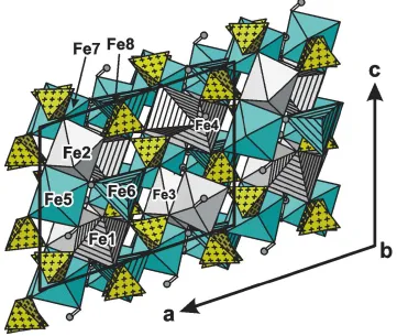
Figure 2
the Fe5±Fe6 vector also runs approximately parallel to [100].
The two polyhedra most rich in Mg, Fe6O4(OH) and
Fe7O4(OH)2, are not connected to each other. Seemingly, the structure thereby avoids mis®t-induced strain in the structure. Average FeÐO distances for the ®ve-coordinated sites are 2.103 (Fe1), 2.109 (Fe4), 2.062 (Fe6), and 2.077 AÊ (Fe8). The six-coordinated sites show average FeÐO distances of 2.188 (Fe2), 2.180 (Fe3), 2.152 (Fe5), and 2.139 AÊ (Fe7). All containing sites have shorter average distances than their Mg-free counterparts. This is consistent with the fact that the commonly observed average [6]FeII±O distance, 2.138 AÊ, is distinctly larger than the corresponding value for Mg, 2.085 AÊ (Baur, 1981). Because FeIIis generally considered an actively distorting cation which prefers distorted octahedra if present, it also comes as no surprise that the six-coordinated Fe sites containing no Mg (Fe2 and Fe3) exhibit a higher bond-length distortion than both their Mg-containing counterparts (Fe5 and Fe7). Hydrothermal syntheses would be necessary to determine how much Mg can substitute for FeIIin synthetic wolfeite under speci®edp±Tconditions. The unit-cell volume of the presently studied Mg-rich wolfeite, 1493.9 (5) AÊ3, is distinctly smaller than previously reported cell volumes for wolfeites very poor in Mg [1521.74 AÊ3(Antenucciet al., 1989) and 1523.0 (5) AÊ3 (Masau et al., 2000)]. The cell edge most strongly affected by the Mg incorporation is theaedge whose length decreases by nearly 3% by comparison to the literature data (Antenucci et al., 1989; Masau et al., 2000). This is convincingly explained by the arrangement along [100] of the Mg-containing polyhedra (cf. Figs. 1 and 2).
The four non-equivalent PO4 tetrahedra all show fairly regular geometries (Table 1). Average PÐO distances are 1.543 (P1), 1.538 (P2), 1.540 (P3), and 1.542 AÊ (P4). The hydrogen bonding scheme in Mg-rich wolfeite is somewhat
unusual. The hydrogen bonds are all weak, with O O
distances between 2.73 and ca. 3.1 AÊ. Each OH group is bonded to three cations (Fe or Mg), i.e. its bond-valence requirements are basically satis®ed, and formally one might
not expect the H atoms to form any hydrogen bond at all. However, the framework topology allows a larger number of weak hydrogen bonds, and, in fact, three of the four OH groups are involved in bifurcated, possibly even trifurcated hydrogen bonds which are strongly bent (Table 2). Only the bond donated by the O20ÐH4 group has a single acceptor atom, O7. None of the four (freely re®ned) H sites shows any unusual displacement parameters, and OÐH distances are within a very narrow range between 0.78 and 0.80 AÊ (Table 2). In isotypic synthetic-Mg2(PO4)(OH) (Raade & Rùmming, 1986), the hydrogen bonds are also weak and strongly bent (OÐHÐO angles range between 116 and 144, similar to the
situation in Mg-rich wolfeite). The hydrogen bonding scheme in triploidite is unknown because positions of H atoms could not be located during the structure determination by Waldrop (1970).
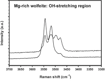
A further, spectroscopic characterization of the hydrogen bonds was obtained by laser Raman spectroscopy. Spectra of a single-crystal fragment were recorded in the range from 4000 to 100 cmÿ1 with a Renishaw M1000 MicroRaman Imaging System using a laser wavelength of 633 nm and excitation through a Leica DMLM optical microscope (spectral resolu-tion 2 cmÿ1, minimum lateral resolution ca 2mm, unpolar-ized laser light, 180 backscatter mode, random sample
orientation). Two representative spectra, given in Fig. 3, were recorded from different, but unspeci®ed cleavage planes. The spectra show four sharp bands (and one shoulder) due to OÐ H stretching vibrations at 3557 (shoulder), 3544, 3518, 3497 (only seen in one spectrum due to orientation effects), and
3478 cmÿ1. Using the correlation of OÐH stretching
frequencies and O O hydrogen bond lengths in minerals by Libowitzky (1999), the observed OÐH stretching frequencies in Mg-rich wolfeite would correspond to approximate O O bond lengths ranging between 2.8 and 3.0 AÊ, in good agree-ment with the results of the structure re®neagree-ment. In the powder infrared spectrum of isotypic synthetic -Mg2 -(PO4)(OH), only two sharp bands at 3595 and 3580 cmÿ1were observed (Raade & Rùmming, 1986), thus indicating weaker hydrogen bonding than in Mg-rich wolfeite.
Experimental
Crystal data
Fe1.84Mg0.16(PO4)(OH)
Mr= 218.63 Monoclinic,P21=a
a= 12.274 (2) AÊ
b= 13.169 (3) AÊ
c= 9.754 (2) AÊ = 108.64 (3)
V= 1493.9 (6) AÊ3
Z= 16
Dx= 3.888 Mg mÿ3 MoKradiation Cell parameters from 5647
re¯ections = 2.0±32.6
= 7.52 mmÿ1
T= 293 (2) K Fragment, yellow 0.180.180.13 mm
Data collection
Nonius KappaCCD diffractometer and!scans
Absorption correction: multi-scan (HKL SCALEPACK; Otwinowski & Minor, 1997)
Tmin= 0.294,Tmax= 0.376
10662 measured re¯ections
5432 independent re¯ections 4267 re¯ections withI> 2(I)
Rint= 0.014 max= 32.6
h=ÿ18!18
k=ÿ19!19
l=ÿ14!14
Acta Cryst.(2003). E59, i125±i128 Uwe Kolitsch Fe1.84Mg0.16(PO4)(OH)
i127
inorganic papers
Figure 3
inorganic papers
i128
Uwe Kolitsch Fe1.84Mg0.16(PO4)(OH) Acta Cryst.(2003). E59, i125±i128Re®nement
Re®nement onF2
R[F2> 2(F2)] = 0.022
wR(F2) = 0.065
S= 1.03 5432 re¯ections 314 parameters
All H-atom parameters re®ned
w= 1/[2(F
o2) + (0.034P)2 + 0.36P]
whereP= (Fo2+ 2Fc2)/3 (/)max= 0.003
max= 0.55 e AÊÿ3
min=ÿ0.67 e AÊÿ3
Extinction correction:SHELXL97 Extinction coef®cient: 0.00357 (15)
Table 1
Selected geometric parameters (AÊ).
Fe1ÐO20 2.0326 (13) Fe1ÐO10 2.0546 (12) Fe1ÐO15i 2.0858 (13)
Fe1ÐO6 2.1591 (12)
Fe1ÐO11 2.1827 (11) Fe2ÐO16i 2.1124 (13)
Fe2ÐO17ii 2.1185 (13)
Fe2ÐO12 2.1372 (11) Fe2ÐO18ii 2.2029 (13)
Fe2ÐO9 2.2526 (12)
Fe2ÐO5ii 2.3059 (12)
Fe3ÐO14iii 2.1054 (13)
Fe3ÐO9 2.1058 (11)
Fe3ÐO19 2.1237 (13)
Fe3ÐO7 2.2139 (12)
Fe3ÐO20 2.2433 (13) Fe3ÐO12 2.2877 (12) Fe4ÐO18 2.0320 (13) Fe4ÐO11 2.0391 (12) Fe4ÐO13iv 2.0660 (13)
Fe4ÐO8 2.1862 (12)
Fe4ÐO10 2.2195 (11) Fe5ÐO17 2.1131 (12)
Fe5ÐO5 2.1223 (13)
Fe5ÐO20iv 2.1269 (14)
Fe5ÐO14 2.1733 (13) Fe5ÐO2v 2.1781 (12)
Fe5ÐO2vi 2.2006 (13)
Fe6ÐO17iii 2.0090 (13)
Fe6ÐO1v 2.0240 (12)
Fe6ÐO6 2.0482 (12)
Fe6ÐO13 2.0665 (13)
Fe6ÐO1 2.1604 (13)
Fe7ÐO4vii 2.0782 (12)
Fe7ÐO19 2.0869 (12) Fe7ÐO16 2.1314 (13)
Fe7ÐO7 2.1464 (13)
Fe7ÐO18i 2.1478 (14)
Fe7ÐO3 2.2437 (13)
Fe8ÐO19viii 2.0167 (13)
Fe8ÐO8 2.0580 (12)
Fe8ÐO3vii 2.0666 (11)
Fe8ÐO15 2.0893 (13)
Fe8ÐO4 2.1543 (12)
P1ÐO7ix 1.5327 (13)
P1ÐO9 1.5397 (12)
P1ÐO2 1.5501 (12)
P1ÐO13 1.5508 (12)
P2ÐO1 1.5343 (12)
P2ÐO14 1.5359 (12)
P2ÐO8x 1.5366 (13)
P2ÐO10 1.5464 (11)
P3ÐO5xi 1.5295 (13)
P3ÐO4 1.5379 (12)
P3ÐO16 1.5405 (12)
P3ÐO11 1.5529 (11)
P4ÐO12 1.5322 (11)
P4ÐO6xii 1.5407 (13)
P4ÐO15ii 1.5476 (12)
P4ÐO3 1.5496 (12)
Symmetry codes: (i) 1
2ÿx;12y;ÿz; (ii) x;1y;z; (iii) 12ÿx;12y;1ÿz; (iv) 1
2ÿx;yÿ12;1ÿz; (v) ÿx;1ÿy;1ÿz; (vi) x;yÿ1;z; (vii) 1ÿx;1ÿy;ÿz; (viii) 1
2ÿx;yÿ12;ÿz; (ix) xÿ12;32ÿy;z; (x) xÿ12;12ÿy;z; (xi) 12x;12ÿy;z; (xii) 1
2x;32ÿy;z.
Table 2
Hydrogen-bonding geometry (AÊ,).
DÐH A DÐH H A D A DÐH A
O17ÐH1 O9i 0.79 (3) 2.23 (3) 2.7343 (17) 122 (2)
O17ÐH1 O1ii 0.79 (3) 2.32 (3) 2.7798 (17) 118 (2)
O17ÐH1 O10ii 0.79 (3) 2.50 (3) 3.1627 (18) 142 (2)
O18ÐH2 O5 0.79 (3) 2.33 (3) 2.8418 (17) 124 (3) O18ÐH2 O4iii 0.79 (3) 2.58 (3) 2.9803 (19) 113 (2)
O19ÐH3 O12 0.78 (3) 2.32 (3) 2.8057 (17) 122 (2) O19ÐH3 O4iv 0.78 (3) 2.39 (2) 2.7978 (17) 114 (2)
O19ÐH3 O11iv 0.78 (3) 2.40 (3) 3.0609 (17) 143 (2)
O20ÐH4 O7 0.80 (2) 2.21 (2) 2.7313 (17) 123 (2)
Symmetry codes: (vi) x;yÿ1;z; (iv) 1
2ÿx;yÿ12;1ÿz; (x) xÿ12;12ÿy;z; (i) 1
2ÿx;12y;ÿz.
H atoms were freely re®ned. The Fe:Mg ratios of the four Mg-containing sites, Fe5, Fe6, Fe7 and Fe8, were freely re®ned, assuming full occupancy of each site. All atomic displacement ellipsoids were regular.
Data collection:COLLECT(Nonius, 2002); cell re®nement:HKL
SCALEPACK(Otwinowski & Minor, 1997); data reduction: HKL
DENZO (Otwinowski & Minor, 1997) and SCALEPACK;
program(s) used to solve structure: SHELXS97 (Sheldrick, 1997); program(s) used to re®ne structure:SHELXL97 (Sheldrick, 1997); molecular graphics:ATOMS(Shape Software, 1999).
Financial support by the German Science Foundation (DFG) and the Austrian Science Foundation (FWF) (grant P15220-N06) is gratefully acknowledged.
References
Antenucci, D., Fontan, F. & Fansolet, A. M. (1989).Powder Diffract.4, 34±35. Anthony, J. W., Bideaux, R. A., Bladh, K. W. & Nichols, M. C. (2000).
Handbook of Mineralogy. Vol. IV: Arsenates, Phosphates, Vanadates. Tucson: Mineral Data Publishing.
Baur, W. H. (1981).Structure and Bonding in Crystals, Vol. II, edited by M. O'Keeffe and A. Navrotsky, pp. 31±52. New York: Academic Press. Coda, A., Giuseppetti, G. & Tadini, C. (1967).Atti Accad. Naz. Lincei, Rend.
Cl. Sci. Fis. Mater. Natur.43, 212±224.
Dal Negro, A., Giuseppetti, G. & Martin Pozas, J. M. (1974).Tschermaks Mineral. Petrogr. Mitt.21, 246±260.
Frondel, C. (1949).Am. Mineral.34, 692±705.
Keller, P., Fontan, F., Velasco-Roldan, F. & Melgarejo i Draper, J. C. (1997).
Eur. J. Mineral.9, 475±482.
Kolitsch, U., Andrut, M. & Giester, G. (2002).Eur. J. Mineral.14, 127±133. Libowitzky, E. (1999).Monatsh. Chem.130, 1047±1059.
Mandarino, J. A. (1999).Fleischer's Glossary of Mineral Species1999. Tucson: The Mineralogical Record Inc.
Masau, M., StaneÏk, J., CÏernyÂ, P. & Chapman, R. (2000).J. Czech. Geol. Soc.45, 159±173.
Nonius (2002).COLLECT. Nonius BV, Delft, The Netherlands.
Otwinowski, Z. & Minor, W. (1997). Methods in Enzymology, Vol. 276,
Macromolecular Crystallography, Part A, edited by C. W. Carter and R. M. Sweet, pp. 307±326. New York: Academic Press.
Raade, G. & Rùmming, C. (1986).Z. Kristallogr.177, 15±26. Rea, J. R. & Kostiner, E. (1972).Acta Cryst.B28, 2525±2529. Robertson, B. T. (1982).Can. Mineral.20, 177±187.
Robinson, G. W., Van Velthuizen, J., Ansell, H. G. & Sturman, B. D. (1992).
Mineral.Rec.23, 4±47.
Sheldrick, G. M. (1997). SHELXS97 and SHELXL97. University of GoÈttingen, Germany.
Shape Software (1999).ATOMS for Windows and Macintosh. Version 5.0.4. Shape Software, Kingsport, TN 37663, USA.
Stalder, M. & Rozendaal, A. (2002).Mineral.Mag.66, 915±927.
Strunz, H. & Nickel, E. H. (2001).Strunz Mineralogical Tables. Stuttgart: E. Schweizerbart'sche Verlagsbuchhandlung.
Taasti, K. I., Christensen, A. N., Norby, P., Hanson, J. C., Lebech, B., Jakobsen, H. J. & Skibsted, J. (2002).J. Solid State Chem.164, 42±50.
supporting information
sup-1
Acta Cryst. (2003). E59, i125–i128supporting information
Acta Cryst. (2003). E59, i125–i128 [doi:10.1107/S1600536803017537]
Mg-rich wolfeite, (Fe
II,Mg)
2(PO
4)(OH): structure refinement and Raman
spectroscopic data
Uwe Kolitsch
S1. Comment
Wolfeite, (FeII,MnII)
2(PO4)(OH) (Mandarino, 1999), is a rare iron phosphate mineral which occurs as a metasomatic
alteration phase in pegmatites, and also rarely in hydrothermal veins, phosphatic nodules in shales, and
amphibolite-facies metamorphosed iron formations (Anthony et al., 2000; Masau et al., 2000; Stalder & Rozendaal, 2002, and
references therein). Wolfeite is a member of a large family of compounds with the general formula MII
2(XO4)Z, where M
= Fe, Mn, Mg, ···; X = P, As, ···; and Z = F, OH, O, Cl etc. According to the crystal-chemical classification of Strunz &
Nickel (2001), wolfeite belongs to the triploidite group of minerals, whose other members are triploidite
[(MnII,FeII)
2(PO4)(OH); Waldrop, 1970], wagnerite [Mg2(PO4)F; Coda et al., 1967], stanekite [(Mn,FeII,Mg)FeIII(PO4)O;
Keller et al., 1997] and sarkinite [MnII
2(AsO4)(OH); Dal Negro et al., 1974]. Various synthetic compounds are also
isotypic with triploidite [e.g.β-Mg2(PO4)(OH) (Raade & Rømming, 1986) and Zn2(PO4)(F,OH) (Taasti et al., 2002)]. All
triploidite-type compounds crystallize with space group P21/a and have similar unit-cell parameters. The common
structure type is closely related to that of triplite, (MnII,FeII)
2(PO4)F (space group I2/a; Waldrop, 1969; alternative setting
in C2/c for synthetic triplite by Rea & Kostiner, 1972).
The crystal structure of wolfeite has not been studied yet, although X-ray powder diffraction data and unit-cell
parameters have been provided by Frondel (1949), Antenucci et al. (1989), and Masau et al. (2000). The present article
reports the results of a single-crystal structure refinement and of single-crystal Raman spectroscopic studies of a Mg-rich
wolfeite sample from the Big Fish River area, Yukon Territory, Canada (Robertson, 1982; Robinson et al., 1992). The
article supplements an earlier study of the crystal structure and IR spectra of a Mg-rich satterlyite, (Fe,Mg)12(PO3OH)
(PO4)5(OH,O)6, from the same locality (Kolitsch et al., 2002).
At the Big Fish River area, the mineral is a common constituent of epigenetic phosphatic nodules and forms divergent,
columnar aggregates of crude, glassy, light brown to clove-brown crystals up to several centimetres in length.
Crystallization occurred at temperatures of about 453 to 473 K according to fluid inclusion studies (Robinson et al.,
1992). Previous electron microprobe analyses of wolfeite from this locality yielded an average formula close to
(Fe1.65Mg0.20Mn0.15)2(PO4) (OH0.95F0.05) (Robinson et al., 1992). The presently studied sample is from the collection of the
author, and its appearance closely fits the published descriptions (Robinson et al., 1992). The chemical composition of
the sample has been characterized by semiquantitative SEM–EDS data which revealed major Fe and P, minor Mg and
only very small amounts of Mn (ratio Fe:Mn ca 13:1), and insignificant compositional inhomogeneities. The EDS-based
chemical composition is in good agreement with the formula subsequently derived from the structure refinement.
Mg-rich wolfeite is confirmed to be isotypic with triploidite (MnII,FeII)
2(PO4)(OH); space group P21/a; Waldrop, (1970).
It has a complex framework structure based on edge- and corner-sharing, distorted MO4(OH) and MO4(OH)2 polyhedra
(M = FeII,Mg), corner-linked to PO
supporting information
sup-2
Acta Cryst. (2003). E59, i125–i128referred to the previous reports on the isotypic members of the triploidite group (see above). The present paper restricts
itself to the Mg distribution and the hydrogen bonding in Mg-rich wolfeite. To facilitate comparisons, the atomic
coordinates and the labeling used by Waldrop (1970) for triploidite were adopted for Mg-rich wolfeite, except for the H
atoms which had not been located during the earlier study of triploidite. The crystal structure of Mg-rich wolfeite
contains eight non-equivalent Fe sites, four P sites, twenty O sites, and four H sites (belonging to OH groups). All atoms
are on general positions. The Fe sites Fe1, Fe4, Fe6, and Fe8 are all five-coordinated (with distorted trigonal-bipyramidal
geometry), whereas the remaining Fe sites show distorted octahedral coordinations (with OH groups in cis configuration).
Site occupany refinements demonstrated that the considerable Mg present in the structure strictly prefers the following
four out of the eight Fe sites: Fe5, Fe6, Fe7, and Fe8. The refined Fe:Mg ratios on these sites range between
approximately 0.76:0.24 (Fe6) and 0.90:0.10 (Fe5) (Table 1). Thus, the Mg substitutes for Fe on two five-coordinated and
two six-coordinated sites, and therefore exhibits no preference for a certain coordination environment. A view of the
polyhedral arrangement along [001] (Fig. 1) shows that the Mg-bearing polyhedra Fe7O4(OH)2 and Fe8O4(OH) (shown in
blue) are connected into indulating chains running parallel to [100] by alternately sharing corners and edges. The other
two Mg-bearing polyhedra, Fe5O4(OH)2 and Fe6O4(OH), are connected via a shared corner on a level below the
undulating chain (Fig. 2); the Fe5—Fe6 vector also runs approximately parallel to [100]. The two polyhedra most rich in
Mg, Fe6O4(OH) and Fe7O4(OH)2, are not connected to each other. Seemingly, the structure thereby avoids misfit-induced
strain in the structure.
Average Fe—O distances for the five-coordinated sites are 2.103 (Fe1), 2.109 (Fe4), 2.062 (Fe6), and 2.077 Å (Fe8).
The six-coordinated sites show average Fe—O distances of 2.188 (Fe2), 2.180 (Fe3), 2.152 (Fe5), and 2.139 Å (Fe7). All
Mg-containing sites have shorter average distances than their Mg-free counterparts. This is consistent with the fact that
the commonly observed average [6]FeII—O distance, 2.138 Å, is distinctly larger than the corresponding value for Mg,
2.085 Å (Baur, 1981). Because Fe is generally considered an actively distorting cation which prefers distorted octahedra
if present, it also comes as no surprise that the six-coordinated Fe sites containing no Mg (Fe2 and Fe3) exhibit a higher
bond-length distortion than both their Mg-containing counterparts (Fe5 and Fe7). Hydrothermal syntheses would be
necessary to determine how much Mg can substitute for FeII in synthetic wolfeite under specified p-T conditions. The
unit-cell volume of the presently studied Mg-rich wolfeite, 1493.9 (5) Å3, is distinctly smaller than previously reported
cell volumes for wolfeites very poor in Mg [1521.74 Å3 (Antenucci et al., 1989) and 1523.0 (5) Å3 (Masau et al., 2000)].
The cell edge most strongly affected by the Mg incorporation is the a edge whose length decreases by nearly 3% by
comparison to the literature data (Antenucci et al., 1989; Masau et al., 2000). This is convincingly explained by the
arrangement along [100] of the Mg-containing polyhedra (cf. Figs. 1 and 2).
The four non-equivalent PO4 tetrahedra all show fairly regular geometries (Table 1). Average P—O distances are 1.543
(P1), 1.538 (P2), 1.540 (P3), and 1.542 Å (P4). The hydrogen bonding scheme in Mg-rich wolfeite is somewhat unusual.
The hydrogen bonds are all weak, with O···O distances between 2.73 and c. 3.1 Å. Each OH group is bonded to three
cations (Fe or Mg), i.e. its bond-valence requirements are basically satisfied, and formally one might not expect the H
atoms to form any hydrogen bond at all. However, the framework topology allows a larger number of weak hydrogen
bonds, and in fact three of the four OH groups are involved in bifurcated, possibly even trifurcated hydrogen bonds which
are strongly bent (Table 2). Only the bond donated by the O20—H4 group has a single acceptor atom, O7. None of the
four (freely refined) H sites shows any unusual displacement parameters, and O—H distances are within a very narrow
range between 0.78 and 0.80 Å (Table 2). In isotypic synthetic β-Mg2(PO4)(OH) (Raade & Rømming, 1986), the
hydrogen bonds are also weak and strongly bent (O—H—O angles range between 116 and 144°, similar to the situation
in Mg-rich wolfeite). The hydrogen bonding scheme in triploidite is unknown because positions of H atoms could not be
supporting information
sup-3
Acta Cryst. (2003). E59, i125–i128A further, spectroscopic characterization of the hydrogen bonds was obtained by laser Raman spectroscopy. Spectra of a
single- crystal fragment were recorded in the range from 4000 to 100 cm−1 with a Renishaw M1000 MicroRaman
Imaging System using a laser wavelength of 633 nm and excitation through a Leica DMLM optical microscope (spectral
resolution ±2 cm−1, minimum lateral resolution ca 2 µm, unpolarized laser light, 180° backscatter mode, random sample
orientation). Two representative spectra, given in Fig. 3, were recorded from different, but unspecified cleavage planes.
The spectra show four sharp bands (and one shoulder) due to O—H stretching vibrations at 3557 (shoulder), 3544, 3518,
3497 (only seen in one spectrum due to orientation effects), and 3478 cm−1. Using the correlation of O—H stretching
frequencies and O···O hydrogen bond lengths in minerals by Libowitzky (1999), the observed O—H stretching
frequencies in Mg-rich wolfeite would correspond to approximate O···O bond lengths ranging between 2.8 and 3.0 Å, in
good agreement with the results of the structure refinement. In the powder infrared spectrum of isotypic synthetic β
-Mg2(PO4)(OH), only two sharp bands at 3595 and 3580 cm−1 were observed (Raade & Rømming, 1986), thus indicating
weaker hydrogen bonding than in Mg-rich wolfeite.
S2. Experimental
Natural sample (see Comment).
S3. Refinement
H atoms were freely refined. The Fe:Mg ratios of the four Mg-containing sites, Fe5, Fe6, Fe7 and Fe8, were freely
supporting information
[image:8.610.130.482.73.491.2]sup-4
Acta Cryst. (2003). E59, i125–i128Figure 1
View along [001] of the complex framework structure of Mg-rich wolfeite from the Big Fish River area, Yukon Territory,
Canada. Edge- and corner-sharing, distorted MO4(OH) (M = FeII,Mg) trigonal bipyramids (striped) and MO4(OH)2
octahedra (unmarked) are corner-linked to PO4 tetrahedra (yellow, marked with crosses). All Mg-containing polyhedra
supporting information
[image:9.610.125.486.74.379.2]sup-5
Acta Cryst. (2003). E59, i125–i128Figure 2
supporting information
[image:10.610.128.483.72.344.2]sup-6
Acta Cryst. (2003). E59, i125–i128Figure 3
Two single-crystal laser-Raman spectra of Mg-rich wolfeite from the Big Fish River area, Yukon Territory, Canada, in the
OH stretching region. See text for details and band positions.
diiron(II) hydroxide phosphate
Crystal data
Fe1.84Mg0.16(PO4)(OH) Mr = 218.63
Monoclinic, P21/a
Hall symbol: -P 2yab a = 12.274 (2) Å b = 13.169 (3) Å c = 9.754 (2) Å β = 108.64 (3)° V = 1493.9 (6) Å3 Z = 16
F(000) = 1692 Dx = 3.888 Mg m−3
Mo Kα radiation, λ = 0.71073 Å Cell parameters from 5647 reflections θ = 2.0–32.6°
µ = 7.52 mm−1 T = 293 K Fragment, yellow 0.18 × 0.18 × 0.13 mm
Data collection
Nonius KappaCCD diffractometer
Radiation source: fine-focus sealed tube Graphite monochromator
ψ and ω scans
Absorption correction: multi-scan
(HKLSCALEPACK; Otwinowski & Minor, 1997)
Tmin = 0.294, Tmax = 0.376
10662 measured reflections 5432 independent reflections 4267 reflections with I > 2σ(I) Rint = 0.014
θmax = 32.6°, θmin = 2.2° h = −18→18
supporting information
sup-7
Acta Cryst. (2003). E59, i125–i128Refinement
Refinement on F2
Least-squares matrix: full R[F2 > 2σ(F2)] = 0.022 wR(F2) = 0.065 S = 1.03 5432 reflections 314 parameters 4 restraints
Primary atom site location: structure-invariant direct methods
Secondary atom site location: difference Fourier map
Hydrogen site location: difference Fourier map All H-atom parameters refined
w = 1/[σ2(F
o2) + (0.034P)2 + 0.36P]
where P = (Fo2 + 2Fc2)/3
(Δ/σ)max = 0.003
Δρmax = 0.55 e Å−3
Δρmin = −0.67 e Å−3
Extinction correction: SHELXL97, Fc*=kFc[1+0.001xFc2λ3/sin(2θ)]-1/4
Extinction coefficient: 0.00357 (15)
Special details
Geometry. All e.s.d.'s (except the e.s.d. in the dihedral angle between two l.s. planes) are estimated using the full covariance matrix. The cell e.s.d.'s are taken into account individually in the estimation of e.s.d.'s in distances, angles and torsion angles; correlations between e.s.d.'s in cell parameters are only used when they are defined by crystal symmetry. An approximate (isotropic) treatment of cell e.s.d.'s is used for estimating e.s.d.'s involving l.s. planes.
Refinement. Refinement of F2 against ALL reflections. The weighted R-factor wR and goodness of fit S are based on F2,
conventional R-factors R are based on F, with F set to zero for negative F2. The threshold expression of F2 > σ(F2) is used
only for calculating R-factors(gt) etc. and is not relevant to the choice of reflections for refinement. R-factors based on F2
are statistically about twice as large as those based on F, and R- factors based on ALL data will be even larger.
Fractional atomic coordinates and isotropic or equivalent isotropic displacement parameters (Å2)
x y z Uiso*/Ueq Occ. (<1)
Fe1 0.18625 (2) 0.479059 (19) 0.19287 (3) 0.00980 (6) Fe2 0.19712 (2) 0.996441 (18) 0.21294 (3) 0.01034 (6) Fe3 0.30503 (2) 0.752291 (19) 0.29253 (3) 0.00998 (6) Fe4 0.31941 (2) 0.269768 (19) 0.30463 (3) 0.00970 (6)
Fe5 0.09673 (2) 0.070820 (19) 0.46928 (3) 0.00829 (8) 0.9027 (17) Mg5 0.09673 (2) 0.070820 (19) 0.46928 (3) 0.00829 (8) 0.0973 (18) Fe6 0.08530 (2) 0.57376 (2) 0.45154 (3) 0.00886 (8) 0.7596 (17) Mg6 0.08530 (2) 0.57376 (2) 0.45154 (3) 0.00886 (8) 0.2404 (17) Fe7 0.39431 (2) 0.67498 (2) 0.03060 (3) 0.00814 (8) 0.8138 (18) Mg7 0.39431 (2) 0.67498 (2) 0.03060 (3) 0.00814 (8) 0.1862 (18) Fe8 0.42084 (2) 0.178485 (19) 0.03902 (3) 0.00827 (8) 0.8806 (18) Mg8 0.42084 (2) 0.178485 (19) 0.03902 (3) 0.00827 (8) 0.1194 (18) P1 0.07895 (3) 0.82096 (3) 0.38003 (4) 0.00565 (8)
supporting information
sup-8
Acta Cryst. (2003). E59, i125–i128O8 0.46690 (10) 0.20262 (9) 0.25864 (12) 0.0113 (2) O9 0.17393 (9) 0.84775 (9) 0.31437 (12) 0.0094 (2) O10 0.17245 (9) 0.36054 (9) 0.32331 (12) 0.0095 (2) O11 0.32337 (9) 0.38398 (8) 0.16515 (12) 0.0092 (2) O12 0.33522 (9) 0.90386 (9) 0.19588 (12) 0.0094 (2) O13 0.11856 (10) 0.72748 (9) 0.48034 (12) 0.0094 (2) O14 0.11776 (10) 0.23476 (9) 0.48261 (12) 0.0093 (2) O15 0.38512 (10) 0.02300 (9) 0.02283 (12) 0.0095 (2) O16 0.37799 (10) 0.51398 (9) 0.01303 (13) 0.0096 (2) O17 0.25351 (10) 0.03131 (9) 0.43671 (12) 0.0102 (2) O18 0.20613 (10) 0.16268 (9) 0.19383 (13) 0.0113 (2) O19 0.24283 (9) 0.71893 (9) 0.06783 (12) 0.0095 (2) O20 0.30215 (10) 0.58247 (9) 0.30905 (12) 0.0112 (2) H1 0.264 (2) −0.026 (2) 0.459 (3) 0.044 (8)* H2 0.155 (2) 0.172 (2) 0.224 (3) 0.061 (10)* H3 0.236 (2) 0.776 (2) 0.044 (3) 0.037 (7)* H4 0.353 (2) 0.5759 (18) 0.276 (3) 0.038 (7)*
Atomic displacement parameters (Å2)
U11 U22 U33 U12 U13 U23
supporting information
sup-9
Acta Cryst. (2003). E59, i125–i128O12 0.0100 (5) 0.0089 (5) 0.0116 (5) 0.0010 (4) 0.0067 (4) 0.0012 (4) O13 0.0107 (5) 0.0076 (5) 0.0091 (5) 0.0002 (4) 0.0021 (4) 0.0022 (4) O14 0.0105 (5) 0.0074 (5) 0.0094 (5) 0.0010 (4) 0.0023 (4) 0.0028 (4) O15 0.0110 (5) 0.0081 (5) 0.0093 (5) 0.0007 (4) 0.0031 (4) 0.0020 (4) O16 0.0101 (5) 0.0076 (5) 0.0107 (5) 0.0009 (4) 0.0028 (4) 0.0033 (4) O17 0.0087 (5) 0.0089 (5) 0.0117 (6) 0.0007 (4) 0.0014 (4) 0.0003 (4) O18 0.0130 (6) 0.0098 (5) 0.0110 (5) −0.0023 (4) 0.0038 (5) −0.0025 (4) O19 0.0076 (5) 0.0089 (5) 0.0109 (5) 0.0000 (4) 0.0013 (4) −0.0002 (4) O20 0.0129 (5) 0.0110 (6) 0.0106 (5) −0.0014 (4) 0.0053 (5) −0.0014 (4)
Geometric parameters (Å, º)
Fe1—O20 2.0326 (13) Fe6—O1 2.1604 (13)
Fe1—O10 2.0546 (12) Fe7—O4vii 2.0782 (12)
Fe1—O15i 2.0858 (13) Fe7—O19 2.0869 (12)
Fe1—O6 2.1591 (12) Fe7—O16 2.1314 (13)
Fe1—O11 2.1827 (11) Fe7—O7 2.1464 (13)
Fe2—O16i 2.1124 (13) Fe7—O18i 2.1478 (14)
Fe2—O17ii 2.1185 (13) Fe7—O3 2.2437 (13)
Fe2—O12 2.1372 (11) Fe8—O19viii 2.0167 (13)
Fe2—O18ii 2.2029 (13) Fe8—O8 2.0580 (12)
Fe2—O9 2.2526 (12) Fe8—O3vii 2.0666 (11)
Fe2—O5ii 2.3059 (12) Fe8—O15 2.0893 (13)
Fe3—O14iii 2.1054 (13) Fe8—O4 2.1543 (12)
Fe3—O9 2.1058 (11) P1—O7ix 1.5327 (13)
Fe3—O19 2.1237 (13) P1—O9 1.5397 (12)
Fe3—O7 2.2139 (12) P1—O2 1.5501 (12)
Fe3—O20 2.2433 (13) P1—O13 1.5508 (12)
Fe3—O12 2.2877 (12) P2—O1 1.5343 (12)
Fe4—O18 2.0320 (13) P2—O14 1.5359 (12)
Fe4—O11 2.0391 (12) P2—O8x 1.5366 (13)
Fe4—O13iv 2.0660 (13) P2—O10 1.5464 (11)
Fe4—O8 2.1862 (12) P3—O5xi 1.5295 (13)
Fe4—O10 2.2195 (11) P3—O4 1.5379 (12)
Fe5—O17 2.1131 (12) P3—O16 1.5405 (12)
Fe5—O5 2.1223 (13) P3—O11 1.5529 (11)
Fe5—O20iv 2.1269 (14) P4—O12 1.5322 (11)
Fe5—O14 2.1733 (13) P4—O6xii 1.5407 (13)
Fe5—O2v 2.1781 (12) P4—O15ii 1.5476 (12)
Fe5—O2vi 2.2006 (13) P4—O3 1.5496 (12)
Fe6—O17iii 2.0090 (13) O17—H1 0.79 (3)
Fe6—O1v 2.0240 (12) O18—H2 0.79 (3)
Fe6—O6 2.0482 (12) O19—H3 0.78 (3)
Fe6—O13 2.0665 (13) O20—H4 0.80 (2)
O20—Fe1—O10 110.19 (5) O17iii—Fe6—O13 94.67 (5)
O20—Fe1—O15i 111.71 (5) O1v—Fe6—O13 91.37 (5)
supporting information
sup-10
Acta Cryst. (2003). E59, i125–i128O20—Fe1—O6 93.30 (5) O17iii—Fe6—O1 83.54 (5)
O10—Fe1—O6 90.32 (5) O1v—Fe6—O1 79.77 (5)
O15i—Fe1—O6 87.07 (5) O6—Fe6—O1 88.33 (5)
O20—Fe1—O11 91.34 (5) O13—Fe6—O1 167.41 (4) O10—Fe1—O11 81.98 (5) O4vii—Fe7—O19 167.68 (5)
O15i—Fe1—O11 97.25 (5) O4vii—Fe7—O16 88.12 (4)
O6—Fe1—O11 172.01 (4) O19—Fe7—O16 102.93 (4) O16i—Fe2—O17ii 160.25 (5) O4vii—Fe7—O7 100.16 (5)
O16i—Fe2—O12 94.22 (5) O19—Fe7—O7 82.54 (5)
O17ii—Fe2—O12 101.40 (5) O16—Fe7—O7 104.39 (5)
O16i—Fe2—O18ii 79.63 (4) O4vii—Fe7—O18i 89.68 (5)
O17ii—Fe2—O18ii 82.13 (5) O19—Fe7—O18i 86.86 (5)
O12—Fe2—O18ii 119.85 (5) O16—Fe7—O18i 80.47 (4)
O16i—Fe2—O9 118.63 (4) O7—Fe7—O18i 169.08 (4)
O17ii—Fe2—O9 77.37 (5) O4vii—Fe7—O3 81.29 (4)
O12—Fe2—O9 75.70 (4) O19—Fe7—O3 87.18 (4) O18ii—Fe2—O9 156.61 (4) O16—Fe7—O3 168.21 (4)
O16i—Fe2—O5ii 89.34 (5) O7—Fe7—O3 82.70 (4)
O17ii—Fe2—O5ii 79.51 (5) O18i—Fe7—O3 94.23 (4)
O12—Fe2—O5ii 162.04 (4) O19viii—Fe8—O8 113.20 (5)
O18ii—Fe2—O5ii 78.10 (4) O19viii—Fe8—O3vii 135.10 (5)
O9—Fe2—O5ii 87.13 (4) O8—Fe8—O3vii 108.71 (5)
O14iii—Fe3—O9 93.68 (5) O19viii—Fe8—O15 93.98 (5)
O14iii—Fe3—O19 161.09 (5) O8—Fe8—O15 102.19 (5)
O9—Fe3—O19 101.17 (5) O3vii—Fe8—O15 92.30 (5)
O14iii—Fe3—O7 89.79 (5) O19viii—Fe8—O4 84.18 (5)
O9—Fe3—O7 160.52 (5) O8—Fe8—O4 85.50 (5) O19—Fe3—O7 80.13 (5) O3vii—Fe8—O4 83.73 (4)
O14iii—Fe3—O20 80.10 (4) O15—Fe8—O4 172.17 (4)
O9—Fe3—O20 123.90 (5) O7ix—P1—O9 109.72 (6)
O19—Fe3—O20 81.90 (4) O7ix—P1—O2 111.08 (7)
O7—Fe3—O20 75.58 (4) O9—P1—O2 111.24 (7) O14iii—Fe3—O12 116.53 (4) O7ix—P1—O13 110.50 (7)
O9—Fe3—O12 75.55 (4) O9—P1—O13 108.53 (6) O19—Fe3—O12 78.89 (4) O2—P1—O13 105.67 (7) O7—Fe3—O12 85.75 (4) O1—P2—O14 109.90 (7) O20—Fe3—O12 155.20 (4) O1—P2—O8x 109.29 (7)
O18—Fe4—O11 108.38 (5) O14—P2—O8x 109.50 (7)
O18—Fe4—O13iv 108.68 (5) O1—P2—O10 108.65 (7)
O11—Fe4—O13iv 142.80 (5) O14—P2—O10 108.72 (6)
O18—Fe4—O8 94.64 (5) O8x—P2—O10 110.77 (7)
O11—Fe4—O8 87.91 (5) O5xi—P3—O4 110.74 (7)
O13iv—Fe4—O8 92.21 (5) O5xi—P3—O16 110.73 (7)
O18—Fe4—O10 88.94 (5) O4—P3—O16 108.70 (7) O11—Fe4—O10 81.42 (5) O5xi—P3—O11 110.20 (6)
O13iv—Fe4—O10 96.16 (5) O4—P3—O11 108.95 (7)
supporting information
sup-11
Acta Cryst. (2003). E59, i125–i128O17—Fe5—O20iv 84.67 (5) O12—P4—O15ii 109.77 (6)
O5—Fe5—O20iv 168.53 (4) O6xii—P4—O15ii 110.48 (7)
O17—Fe5—O14 99.04 (4) O12—P4—O3 111.15 (7) O5—Fe5—O14 102.00 (5) O6xii—P4—O3 109.34 (7)
O20iv—Fe5—O14 81.25 (4) O15ii—P4—O3 105.88 (7)
O17—Fe5—O2v 171.98 (5) Fe6iv—O17—H1 93.8 (18)
O5—Fe5—O2v 99.51 (5) Fe5—O17—H1 106.0 (19)
O20iv—Fe5—O2v 91.58 (5) Fe2vi—O17—H1 92.9 (19)
O14—Fe5—O2v 87.36 (4) Fe4—O18—H2 101 (2)
O17—Fe5—O2vi 88.05 (4) Fe7viii—O18—H2 96 (2)
O5—Fe5—O2vi 83.17 (4) Fe2vi—O18—H2 94 (2)
O20iv—Fe5—O2vi 94.98 (5) Fe8i—O19—H3 96.3 (18)
O14—Fe5—O2vi 171.58 (4) Fe7—O19—H3 103.0 (19)
O2v—Fe5—O2vi 85.21 (4) Fe3—O19—H3 94.5 (19)
O17iii—Fe6—O1v 125.87 (5) Fe1—O20—H4 101.9 (18)
O17iii—Fe6—O6 114.16 (5) Fe5iii—O20—H4 96.9 (18)
O1v—Fe6—O6 116.35 (5) Fe3—O20—H4 92.7 (18)
Symmetry codes: (i) −x+1/2, y+1/2, −z; (ii) x, y+1, z; (iii) −x+1/2, y+1/2, −z+1; (iv) −x+1/2, y−1/2, −z+1; (v) −x, −y+1, −z+1; (vi) x, y−1, z; (vii) −x+1, −y+1, −z; (viii) −x+1/2, y−1/2, −z; (ix) x−1/2, −y+3/2, z; (x) x−1/2, −y+1/2, z; (xi) x+1/2, −y+1/2, z; (xii) x+1/2, −y+3/2, z.
Hydrogen-bond geometry (Å, º)
D—H···A D—H H···A D···A D—H···A
O17—H1···O9vi 0.79 (3) 2.23 (3) 2.7343 (17) 122 (2)
O17—H1···O1iv 0.79 (3) 2.32 (3) 2.7798 (17) 118 (2)
O17—H1···O10iv 0.79 (3) 2.50 (3) 3.1627 (18) 142 (2)
O18—H2···O5 0.79 (3) 2.33 (3) 2.8418 (17) 124 (3) O18—H2···O4x 0.79 (3) 2.58 (3) 2.9803 (19) 113 (2)
O19—H3···O12 0.78 (3) 2.32 (3) 2.8057 (17) 122 (2) O19—H3···O4i 0.78 (3) 2.39 (2) 2.7978 (17) 114 (2)
O19—H3···O11i 0.78 (3) 2.40 (3) 3.0609 (17) 143 (2)
O20—H4···O7 0.80 (2) 2.21 (2) 2.7313 (17) 123 (2)