Original Article
Scanning electron microscopy (SEM) study
on filum terminale with human fetus
Jianfeng Li1*, Jun Zhang1*, Xia Guan2, Chunlei Zhang3, Zheng Shao4, Xinwei Li5, Tieli Dong1, Fuyun Liu5
1Department of Pain Medicine, The Second Affiliated Hospital of Zhengzhou University, Zhengzhou 450014,
China; 2Department of Kidney Disease & Rheumatology, Tongliao City Hospital, Tongliao 028000, China; 3Department of Anesthesia, Henan Provincial People’s Hospital, Zhengzhou 450000, China; 4Department of
Women and Children Health Care, The Third Affiliated Hospital of Zhengzhou University, Zhengzhou 450052, China; 5Department of Pediatric Orthopedic, The Third Affiliated Hospital of Zhengzhou University, Zhenghou
450052, China. *Co-first authors.
Received September 29, 2015; Accepted January 23, 2016; Epub February 15, 2016; Published February 29, 2016
Abstract: The filum terminale plays an important role in the pathophysiology of tethered cord syndrome (TCS). The study on ultrastructure of fetal filum can provide reference standard for diagnosis of TCS. Eleven fresh Chinese human aborted fetuses had their fila measured and removed. Transverse and longitudinal sections of the middle and distal segments of fila were submitted for scanning electron microscopy (SEM) analysis respectively. The bulk of the filum terminale is composed of 1 µm to 5 µm thick spring like longitudinal bundles of collagen separated by 5 µm to 30 µm layer intervals and 1 µm to 5 µm intervals in the layer, although a small quantity of capillaries and other components may be present. A delicate meshwork of collagen transverse fibers connects these bundles and fibers in every bundle. A delicate meshwork of collagen transverse fibers connects these bundles and fibers
in every bundle. A complex tridimensional structure is evidenced on electron microscopy. In conclusion, a complex
tridimensional structure composed by ordered arrangement of spring-like fibers and a small quantity of capillaries should elicit considerable elastic properties to the filum terminale. Its alternation of structure and component may
be involved in TCS closely.
Keywords: Filum terminale, tethered cord syndrome, ultrastructure
Introduction
The filum terminale is a fibrous band, com-posed of two distinct segments (intradural and extradural), that extends from the conus medul-laris to the periosteum of the coccyx [1]. The proximal structure of the filum is similar to spi-nal cord, the middle and distal segments of which are fibrous tissue. In the first three months of the embryo, vertebral column grows the same as spinal cord, after that, the verte-brae exhibits a higher growth rate than does the spinal cord, resulting in an ascending move-ment of the conus medullaris relative to the vertebrae, pia mater on its surface is stretched into linear filum, attatched to the coccyx. The caudal end of full term neonate spinal cord is close to adult, about L1-L2 vetebral level. The filum of adult is about 20 cm long (intradural, 15 cm; extradural, 5 cm), 1-2 mm thick. Atrophy
formation of the filum terminale, the central canal and neural stem cell are noticed in the proximal section of the filum, as described in the filum of various species [2].
The clinical significance of the teminal filum lies in the tethered cord syndrome (TCS), which was first described by Garceau in 1953 [3]. Tethered cord syndrome is a stretch-induced functional disorder of the spinal cord. The mechanical cause of TCS is an inelastic structure, which includes filum terminale disease, (such as fatty filum, tumor, mineral deposition, etc.) myelome-ningocele, lipomyelomemyelome-ningocele, or scar for-mations adherent to the spinal cord or nerve roots, anchoring the caudal end of the spinal cord that prevents cephalad movement of the lumbosacral cord [4-9].
SEM study on filum terminale with human fetus
than the spinal cord or when the spinal cord undergoes forcible flexion and extension. This syndrome is defined as a disorder manifested by motor and sensory deficits in lower limbs, incontinence, and musculoskeletal deformi- ties.
Once diagnosed definitely, undoubtedly, surgi-cal management of TCS is the best choice of all, furthermore, less complication and better effectiveness is attributed to application of new surgical technique [10], whereas the diag-nosis of TCS is still difficult, especially when there is a normal level of the conus medullaris or normal thickness of the filum on radiological examination [11]. The terminal filum has received increasing attention on TCS pathogen-esis with the deepening of study on TCS. The rat experiments conducted by Miklos and his colleagues have revealed that its basic compo-nents (central canal, gray matter, white matter) continued within the filum, uniform small size neurons and glial cells populated the gray mat-ter; small nerve cell surrouding ependymal cell may be nerve stem cell (NSC) [12]. Or rather, NSCs arranged densely in the proximal seg-ment while sparsely in the distal one, as we have previously reported [13, 14]. Tubbs et al. found previously undescribed smooth muscle cells within the filum terminale externum. Furthermore, histological analysis identified adipose, nerve, bone, and cartilage cells. Macroscopically, the filum thicker than 2 mm and its fatty change were distinguished easily on MR investigation or direct visualization dur-ing the surgical intervention, which is more
Specimen origin
Eleven samples (five male, six female) were dis-sected 4 to 8 hours after abortion in second trimester pregnancy. (These had been investi-gated and validated by Ethical Review Committee, and informed consent forms were signed by their parents). The detailed data of the subjects are given in Table 1. There were no abnormalities on the skin of the spine region. None of these subjects involved orthopaedic, neurological, or urinary malformation. Dis- section was initiated with a mediodorsal skin incision, followed by exposure of the spinous processes and laminae of the T11-S5 verte-brae. The initial point of filum was promptly identified as the junction of the terminal filum and the conus medullaris, separated downward until the end of coccyx. The 12th rib and 12th thoracic vertebra were taken as a reference point, length and thickness measurements were obtained with a vernier caliper after the filum was sectioned. No tension was applied to the fila for measurements. Each filum was divided into three sections equally, then, thor-oughly washed in 0.1 mol/L phosphate buff-ered saline (PBS) before fixation.
Scanning electron microscopy
Segments (1 cm) of the middle and distal seg-ments of four fila were perfused with a fixative containing 2.5% glutaral, 4% paraform in 0.1 mol/L PBS for more than 2 h at 4°C. After wash-ing in PBS, they were immersed in 12% dimeth-yl sulfoxide for two 30 min, 25% for two 30 min, and 50% for one 1.5 h, followed by freeze-frac-Table 1. Specimen data
No. (weeks)GA Sex (mm)CRL lengthFT eter (mm) levelFT iniate diam- ameter (mm) ConusFT middle
di-1 12-13 Male 161 28.5 1.10 0.1 L4
2 15-16 Female 182 35.3 1.18 0.15 L4
3 25-26 Female 252 41.4 1.22 0.18 L2-L3
4 29-30 Male 283 45.6 1.24 0.29 L1-L2
5 31-32 Female 302 55.6 1.29 0.28 L1-L2
6 28-29 Female 276 46.8 1.3 0.69 L2
7 30-31 Male 297 54.8 1.26 0.26 L2
8 13-14 Male 168 37.8 1.23 0.21 L4
9 34-35 Male 330 57.3 1.3 0.7 L2
10 33-34 Female 318 56.7 1.3 0.63 L1-L2
11 24-25 Female 249 40.6 1.22 0.36 L2-L3
GA = gestational age; CRL = Crown-rump length; FT = filum terminale.
obvious under light micros-copy [15]. The filum with normal appearance on MRI showed some hyalinization and dilated capillaries [16]. Fontes et al. made a more detailed study of 20 sam-ples of normal adult fila by light and scanning electron microscopies [17], howev-er, it has scarcely been reported about the SEM study on the structure of normal fetal fila has not been reported.
[image:2.612.91.388.84.254.2]turing in liquid nitrogen into cross section, sag-ittal plane, coronal plane; then, they were immersed in 50% dimethyl sulfoxide for 15 min and immersed in 0.1 mol/L PBS for 10 min. Succeedingly, they were dehydrated in an increasing alcohol series (30%, 10 min; 50%, 10 min; 70%, 10 min; 90%, two 10 min; 100%, three 10 min), replaced with amyl acetate for 1 min. Sections were critical-pointdried with liq-uid carbon, gold coated in a Balzers SCD-040 ion sputterer, and examined in a JEOL-JSM840 (Jeol, Tokyo, Japan) scanning electron micro- scope.
GA = gestational age; CRL = Crown-rump leng- th; FT = filum terminale.
Results
Macroscopic description
The conus medullaris was located at the lower end of the spinal cord. Originating at the base of the brain, this thick bundle of nerve tissue passes through the center of the spinal col-umn, penetrating the vertebrae. At its terminal end, at the lumbar spine in the lower back, the spinal cord tapers into a cone shape and then into a narrow bundle of fibrous tissue called the
filum terminale, about 28.5-57.3 mm in length (mean value 45.5 mm). It consists of two parts: The upper part, or filum terminale internum, was about 21.2-43.1 mm long and reached as far as the lower border of S2. It was continuous above with the pia mater and contained within a tubular sheath of the dura mater. In addition, it was surrounded by the nerves forming the cauda equina, from which it can be easily rec-ognized by its bluish-white color. The lower part, or filum terminale externum, closely adhered to the dura mater. It extended downward from the apex of the tubular sheath and was attached to the back of the first segment of the coccyx in a structure.
Electron microscopy ultrastructure
[image:3.612.91.524.72.313.2]SEM study on filum terminale with human fetus
to 5 µm (Figures 2E, 2F, 3C, 3D). The collection of collagen bundles might be about 10 µm wide zone in the same layer (Figures 2E, 2F, 3C). There were some intervals ranging from 1 µm to 5 µm between bundles in the same layer (Figures 2E, 2F, 3C, 3D). Collagen fiber also connected obliquely from one bundle to anoth-er bundle (Figures 1F, 3E). All layanoth-ers and bun-dles from each other were linked with intricate meshwork of finer transverse fibers (Figure 3F). Blood capillaries were occasionally seen within these longitudinal bundles in the cross section (Figure 3C). High power scanning electron microscopy showed clearer structure.
Discussion
Many scholars do have a deep understanding of that elasticity decrease of the filum is an important cause of TCS. Selcuki et al. observed the fila of normal adult corps via light micros-copy; found that normal filum was composed of loose connective tissue, rarely fat cells, and a small quantity of capillaries [16].This research on structure of normal fetal filum is similar to that of mentioned above. However, the differ-ence is that fat cells not being found in our researches.
[image:4.612.91.523.71.312.2]What is the difference of normal and abnormal filum that requires TCS concretely? Selcuki et al. observed normal and abnormal fila with symptomatic TCS, found that the former con-tained more connective tissue than normal, with dense collagen fibers, dilated capillaries with cell infiltration, random fat cells, and some areas of hyalinization; elevated numbers of col-lagen bundles, areas of hyalinization were fre-quently observed throughout the connective tissue in the latter [16]. The high amounts of dense collagen fibers cause the filum to lose its elastic properties, ultimately leading to conduc-tion of a tethering effect to the conus medul-laris [17]. Liu et al. found that the presence of abundant collagen bundles and adipocytes, sparse or invisible elastic and reticular fibers inside the FT [18]. Ozkan et al. compared path-ological fila with normal fetal ones by light microscopy found adipose tissue, fibrosis, hya-linization, meningothelial proliferation and elas-tic fibers in FT samples of TCS, none of these findings were observed in fetal samples [19]. Fontes et al. selected fila of 20 so-called nor-mal adult corps, submitted to light and scan-ning electron microscopy, concluded that the bulk of the filum is composed of 5 to 20 µm thick longitudinal bundles of Type 1 collagen Figure 2. Middle stage, SEM picture of FT belonging to specimen no. 6 with female, 28~29 gestational weeks. The cross-sections of filum are presented in different multiples (A-F); That shows collagen fiber bundles were stratified
separated 3 to 10 µm intervals [17]. A delicate meshwork of predominantly Type 3 collagen transversal fibers connects these bundles, in addition to abundant longitudinally oriented elastic and elaunin fibers are found inside col-lagen bundles, and thought this elastic proper-ty played an important role in pathogeny, diag-nosis and treatment of TCS, especially pathog-eny explanation of TCS with normal conus posi-tion. Royo-Salvador et al.thought special neu-rology syndrome such as syringomyelia, scolio-sis and Chiari malformation may occur second-ary to elasticity decrease of the filum, but there was a controversy [20].
The main finding of the present study demon-strates the filum is not a simple longitudinal connective cord, but a complex tridimensional structure. Curving and longitudinal fiber bun-dles are in layers, some intervals between
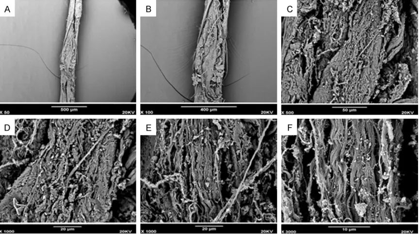
[image:5.612.89.524.70.377.2]lay-ers or bundles, but the latter is smaller than the former, in which sparse, transverse and deli-cate reticular fibers are found to connect longi-tudinal fibers or bundles, as well as some col-lagen connected between bundles and layers. High electron microscopy shows collagen fibers are linked with more transverse fibers. It has not been reported which involves collagen bun-dles are in layers, or intersect with each other between bundles and layers, and collagen fibers linked by more transverse fibers. This fur-ther indicates that the filum is a delicate, com-plicate and arranged orderly elastic structure, the decrease of which may affect its elasticity, resulting in TCS. That finer transverse fibers in this research maybe no other than type 3 col-lagen as Fontes et al. described, the diameter of type 1 or 3 collagen may be identical or close to that of elastic fibers. Under the scanning microscopy, we found spring-like structure of Figure 3. Late stage, SEM picture of FT belonging to specimen no. 10 with female, 33~34 gestational weeks. Crapy pia mantle surrounding longitudinal spring-like collagen bundles and pia mantle surrounding collagen arranged like tree rings (A, B). Some intervals range from about 5 µm to 30 μm between layers, another intervals range from
about 1 µm to 5 μm between collagen bundles of the same lay, the diameter of collagen bundle is about 1 μm-5 μm, the collection of collagen bundles become about 10 μm wide zone (C), capillaries and red blood cell in the upper left corner of C. Longitudinal collagen bundles arrange like spring, about 1 μm-5 μm interval between bundles (Figures 1D, 1E and 3D). Collagen bundle are composed of many longitudinal fibers. Collagen fibers also connect obliquely
SEM study on filum terminale with human fetus
longitudinal fiber bundles of human fetuses more distinct, more delicate, more explicit, by comparison with the fila of normal adult corps, which perhaps relates to freshness of speci-mens or promptness of their collection and fixa-tion, or maybe because of obvious difference of the both, or abnormalities of their specimens proper (coni of 4 samples be located below L2, the fila thickness of 2 samples more than 2 mm), this reflects a better elastic property. This research found there was high concentra-tion of elastic fibers, accompany with longitudi-nal collagen bundles. Few tissues (except the dermis, vascular wall, and lung, etc.) in the human body exhibit this amount of elastic structures; even fewer possess a single longitu-dinal alignment within parallel bundles. The flexibilities of elastic fibers could make the organization have certain deformation, the toughness of collagen fibers could make the organization keep certain shapes, and the con-comitance of the both makes up of connective tissue, make the organization to keep certain shapes and certain adjustable elasticity. This aligned longitudinally and curving fibrous struc-ture confirms important elastic property of the filum, and one of its important physical func-tions: as an elastic buffer which stops spinal cord stretch injury for the developing spinal col-umn or an excessive flexion; conversely, elastic-ity decrease of the filum could injury spinal cord. In addition, transverse reticular fiber con-sists of type 3 collagen, and keeps longitudinal fibers bundles stable like mesh scaffolds. When the filum suffers to inflammation, or reshapes itself in the development, transverse reticular fibers will lose its function, make the arranged structure destroyed, that might also affect elasticity of the filum.
In summary, normal fetal filum is not a simple fibrous band, but a delicate, complex, tridimen-sional structure, containing longitudinally ori-ented elastic, collagen fibers and transverse reticular fibers, and a small quantity of blood vessels, short of adipose tissue, which reflects the filum of considerable elastic properties. Clinically, its alternation of structure and com-ponent may be involved in TCS closely, which has great significance especially for treatment of TCS with normal conus position.
Acknowledgements
We are grateful to all the participants in this study.
Disclosure of conflict of interest None.
Address correspondence to: Dr. Fuyun Liu, Depart-
ment of Pediatric Orthopedic, The Third Affiliated Hospital of Zhengzhou University, Kangfu Front
Street, Zhenghou 450052, Henan Province, China. E-mail: [email protected]; Tieli Dong, Depart-
ment of Pain Medicine, The Second Affiliated Hos-pital of Zhengzhou University, Zhengzhou 450014,
Henan Province, China. E-mail: [email protected]
References
[1] Hansasuta A, Tubbs RS, Oakes WJ. Filum ter -minale fusion and dural sac termination: study in 27 cadavers. Pediatr Neurosurg 1999; 30: 176-179.
[2] Lemire RJ, Loeser JD, Alvord EC Jr. Normal and abnormal development of the human nervous system. Hagerstown: Harper & Row; 1975. pp. 71-83.
[3] Garceau GJ. The filum terminale syndrome (the
cord-traction syndrome). J Bone Joint Surg Am 1953; 35A: 711-716.
[4] Parag SM, Nazeer A, Anuradha P M, Nawal M.
AI Moosawi. Retrospective magnetic reso-nance imaging evaluation of fatty filum
termi-nale in Kuwaiti population. J Nat Sci Biol Med
2015; 6: 85-88.
[5] Wu Y, Huang B, Liang C. Solitary fibrous tumor
of filum terminale. Acta Radiol Short Rep 2012; 1.
[6] Alessandro L, Roberto T, Nicola M, Pierluigi R,
Manila A, Maurizio S, Roberto D. Paraganglioma
of the filum terminale: case report. World J Surg Oncol 2009; 7: 95.
[7] De Jong L, Calenbergh FV, Menten J, van Loon J, De Vleeschouwer S, Plets C, Didgar M, Sciot
R, Goffin J. Ependymomas of the filum termi -nale: the role of surgery and radiotherapy. Surg Neurol Int 2012; 3: 76.
[8] Mantia R, Di Gesù M, Vetro A, Mantia F, Palma
S, Iovane A. Shortness of filum terminale rep
-resents an anatomical specific feature in fibro -myalgia: a nuclear magnetic resonance and clinical study. Muscles Ligaments Tendons J 2015; 5: 33-37.
[9] Cacciotti G, Novegno F, Fiume D. Calcium pyro-phosphate dihydrate deposition disease of the
filum terminale. Eur Spine J 2013; 22:
501-505.
[10] Magrassi L, Chiaranda I, Minelli M, Grimod G, Locatelli D, Arienta C. Total endoscopic ap-proach to the caudal in a patient with a tight
filum. Minim Invasive Neurosurg 2008; 51:
350-353.
[11] Bao N, Chen ZH, Gu S, Chen QM, Jin HM, Shi
analysis based on positioning of the conus and absence or presence of lumbosacral lipoma. Childs Nerv Syst 2007; 23: 1129-1134. [12] Miklos R, Erika L, Csaba B. The caudal end of
the rat spinal cord: transformation to and
ultra-structure of the filum terminale. Brain Res
2004; 1028: 133-139.
[13] Sun KM, Liu FY, Xia B. Study on the proximal structure of fetus filum terminale. Chin J Prakt
Med 2008; 35: 62-63.
[14] Si PC, Liu FY, Xia B. Distribution of neural stem
cells in the terminal fila. J Clin Pediatr Surg
2005; 4: 425-429.
[15] Tubbs RS, Murphy RL, Kelly DR, Lott R, Salter EG, Oakes WJ. The filum terminale externum. J
Neurosurg Spine 2005; 3: 149-152.
[16] Selcuki M, Vatansever S, Inan S, Erdemli E, Bağdatoğlu C, Polat A. Is a filum terminale with a normal appearance really normal? Childs
Nerv Syst 2003; 19: 3-10.
[17] Fontes RB, Saad F, Soares MS, de Oliveira F,
Pinto FC, Liberti EA. Ultrastructural study of the filum terminale and its elastic fibers.
Neurosurgery 2006; 58: 978-984.
[18] Liu FY, Li JF, Guan X, Luo XF, Wang ZL, Dang
QH. SEM study on filum terminale with teth -ered cord syndrome. Childs Nerv Syst 2011; 27: 2141-2144.
[19] Tehli O, Hodaj I, Kural C, Solmaz I, Onguru O, Izci Y. A comparative study of histopathological analysis of filum terminale in patients with
tethered cord syndrome and in normal human fetuses. Pediatr Neurosurg 2011; 47: 412-416. [20] Royo-Salvador MB, Sole-Llenas J, Domenech