Original Article
The condylar morphology in adult females of skeletal
class II division 1 malocclusion with various vertical
skeletal features: a study by cone beam
computed tomography
Yu Song1*, Xiaoyan Zhang2*, Yang Gao1, Fengchun Hou1, Yanling Yu1
1Department of Orthodontics, Qingdao Stomatological Hospital, Qingdao 266001, China; 2Department of
Orthodontics, School of Stomatology, Capital Medical University, Beijing 100050, China. *Equal contributors and
co-first authors.
Received October 22, 2015; Accepted March 31, 2016; Epub May 15, 2016; Published May 30, 2016
Abstract: The purpose of this study was to evaluate the condylar morphology parameters using cone beam com-puted tomography (CBCT). 180 adult female patients with skeletal Class II division 1 were recruited in this study. Then the participants were divided into three groups according to the values of GoGn-SN and S-Go/N-Me. Mimics 10.01 software was used to reconstruct the CBCT images and measure the parameters including condylar volume, surface area, morphological index, inner-outer and front-back diameters on the sagittal and coronal sections. Our results showed that there were no significant difference by condylar morphological parameters in patients with the same vertical skeletal pattern (P>0.05). All the measured parameters in the low angle group were significantly different with those in the average angle group (P<0.05). Similarly, all the measured parameters except the mor-phological index in the low angle group were significantly different with those in the high angle group (P<0.05). Significant difference was found between the high angle group and the low angle group regarding the parameters including condylar MI, inner-outer and front-back diameters (P<0.05). In conclusion, our study demonstrated that
there was significant difference regarding the condylar morphology among females of skeletal Class II division 1 malocclusion with various vertical skeletal features. The data was important and valuable for radiological diagnosis, orthodontic treatments and orthognathic surgery.
Keywords: Skeletal class II division 1 malocclusion, cone beam computed tomography, condyle morphology, verti-cal skeletal pattern
Introduction
Temporomandibular joint (TMJ) plays an impor-tant role in maintaining good masticating func-tion as well as the balance of oral and maxillo-facial system. Condyle is an indispensable part of TMJ and can response to functional simula-tions constantly. Therefore, its morphology and volume can reflect the adaptations to the influ-ential factors. The functional adaptation prop-erty of condyle is essential for maintaining the long term stability of TMJ after orthodontic or orthognathic surgery [1, 2].
Due to the limitations of the two-dimensional images, it was difficult to investigate the struc-ture and morphology of condyle in vivo. How- ever, with the development of technology in the
The incidence of skeletal Class II malocclusions is especially high. In addition, the variations among different vertical skeletal features are great. Currently whether different vertical skel-etal features have any influence on the mor-phology and structure of the condyle in the patients with skeletal Class II division 1 maloc-clusion is controversial.
In the present study, we aimed to determine whether there was any difference regarding the morphological parameters of condyle between adult females of skeletal Class II divi-sion 1 maloccludivi-sion with various vertical skel-etal features.
Materials and methods Study population
The study was approved by the Ethnic Com- mittee of Qingdao Stomatological Hospital and the written consent was obtained from all par-ticipants. 180 females who received orthodon-tic treatment in the Department of Orthodonorthodon-tic, Qingdao Stomatological Hospital from Jan 2011 to Dec 2014 were recruited in the study. All the subjects were diagnosed as skeletal Class II division 1 malocclusion. The average age of the patients was 26.1 y (age range, 18-35 y) The inclusion criteria were as follows: 1. The patients had taken CBCT images before treatments; 2. Females diagnosed as skeletal Class II division 1 malocclusion and with ANB angle greater than 5°; 3. No TMJ disorder indi-cated; 4. The bilateral symmetry of facial was required, without mandibular deviations and occlusion asymmetry; 5. No prior orthodontic treatment, trauma, infection and surgical treat-ment history indicated. Then the 180 subjects were selected from the patient pool randomly and divided into three groups (low angle, aver-age angle, and high angle) according to the GoGn-SN and S-Go/N-Me values [4]. The clas-sification standard was as follows: high angle group (GoGn-SN>37.7°, S-Go/N-Me<62%); aver- age angle group (27.3°<GoGn-SN<37.7°, 62%<S-Go/N-Me<68%) and low angle group (GoGn-SN<27.3°, S-Go/N-Me>68%). There were 60 cases in each group respectively and patients in each group were matched by age. CBCT imaging
The NNT Viewer CBCT (QR SRL Company, Verona, Italy) with imaging parameters of 120 kV, 8mA was used to collect all the participants’ images. The CBCT images were taken under natural head position. The subjects were re-
quired to relax their facial muscles, close the lips naturally and breathe peacefully. In addi-tion, the posterior teeth were intercuspally placed. The shooting time lasted for 20 sec-onds and CBCT images were saved in DICOM format. The CBCT was operated by the same radiologist in this study.
Measurement of condylar volume and surface area
Different layers of CBCT images in DICOM for-mat were imported into Mimics 10.01 software (Materialise NV Technologielaan, Leuven, Belgium) by selecting the order “import image”. 3-dimensional reconstruction of condyle was performed according to previous methods [5, 6]. The gray values of the CBCT images in the software were defined as bone density values (gray value: -1024~1650; threshold value: 542~3071).
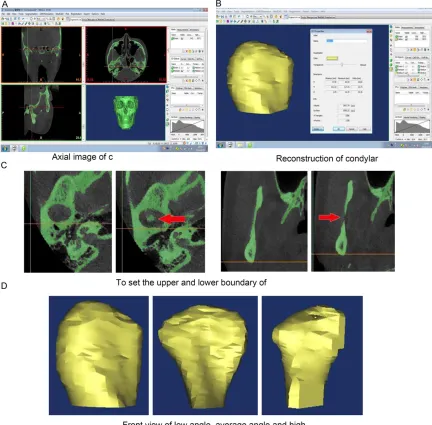
After setting the parameters, the images that above the condyle were deleted by pressing the “Edit Masks” button at the horizontal sections. Similarly, the posterior sections of condylar images were removed in sagittal planes (Figure 1A). Then the images in the coronal sections were amplified by rolling the computer mouse. The first white image that presented in the joint zone was set for the upper boundary of condyle (Figure 1C). The area that the most concave point of coracoid disappeared was defined as condylar lower boundary (Figure 1C). Both hard and soft tissues around condyle within this scope were removed by selecting “Edit Masks” order. Finally, 3-dimensional images of condyle were reconstructed by choosing the “Calculate 3D from Mask” order (Figure 1B, 1D). The con-dylar volume and surface area was calculated automatically by double clicking post recon-structive condylar image. All images were reconstructed twice by the same person for the measurement. The duration between two reconstructions was 1 week. The error of mea-surement was calculated by the following for-mula: Ve X1 X2 /2n2
=/
^
-h
.Measurement of condylar back-front and inner-outer diameters
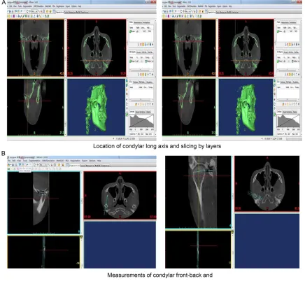
con-dyle. In addition, the slicing line was perpen-dicular to condylar long axis with left-right sym-metry. There were totally 18 slicing layers and the slicing thickness was 0.3 mm. A group of tilted images that perpendicular to the long axis of condyle was reconstructed (Figure 2A). The center tilted image was selected and then amplified. The most front and back points of the image were located, and the connections between them were measured. The maximum measured value was set for back-front diame-ter of condyle. Similarly, a group of tilted imag-es parallel to condylar long axis was recon-structed with the same method, and the inner-outer diameter of condyle was calculated (Figure 2B).
Statistical analysis
[image:3.612.90.522.74.499.2]All the data in the study were expressed as means ± standard deviation (SD). Paired t test was performed to compare the left side and ride side condylar morphological parameters in patients with the same vertical skeletal pat-tern. One-way ANOVA was conducted to com-pare the condylar morphological parameters in patients with different vertical skeletal pat-terns. If any difference was found, further com-parison between the two respective groups was performed. The software of SPSS version 21.0 for Windows (SPSS Inc, IL, USA) was used for statistical analysis. Differences were con-sidered statistically significant when P was less than 0.05.
Results
The values of GoGn-SN and S-Go/N-Me values in different groups
The values of GoGn-SN and S-Go/N-Me were used to divide the participants into three groups. No significant difference regarding age was found among different groups (P>0.05). The average values of GoGn-SN and
[image:4.612.90.526.73.475.2]S-Go/N-We first compared the difference between the left and the right side condylar morphology of the patients with the same vertical skeletal pat-tern. The parameters including condylar vol-ume, condylar surface area, condylar morphol-ogy index (volume/surface area, MI), condylar inner-outer diameter and front-back diameter were measured. The results showed that no significant difference was found regarding the Figure 2. Reconstruction process of condylar front-back and inner-outer diameters.
Table 1. The GoGn-SN and S-Go/N-Me values of different groups
Groups N Age (year) GoGn-SN Angle (°) S-Go/N-Me (%) High angle 60 26.31±3.90 42.82±2.57 58.82±1.83 Average angle 60 24.86±4.01 32.91±2.81 66.28±1.23 Low angle 60 25.89±3.89 24.37±2.56 72.73±3.36
Me of three groups were summa-rized in Table 1.
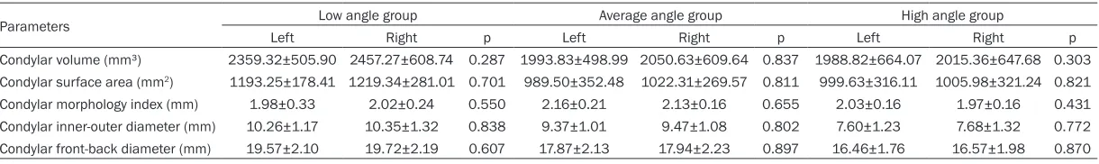
[image:4.612.91.362.538.593.2]8308 Int J Clin Exp Med 2016;9(5):8304-8311 Table 2. Comparison of the left and ride side condylar morphological parameters in patients with the same vertical skeletal pattern
Parameters Low angle group Average angle group High angle group
Left Right p Left Right p Left Right p
Condylar volume (mm³) 2359.32±505.90 2457.27±608.74 0.287 1993.83±498.99 2050.63±609.64 0.837 1988.82±664.07 2015.36±647.68 0.303
Condylar surface area (mm2) 1193.25±178.41 1219.34±281.01 0.701 989.50±352.48 1022.31±269.57 0.811 999.63±316.11 1005.98±321.24 0.821
Condylar morphology index (mm) 1.98±0.33 2.02±0.24 0.550 2.16±0.21 2.13±0.16 0.655 2.03±0.16 1.97±0.16 0.431
Condylar inner-outer diameter (mm) 10.26±1.17 10.35±1.32 0.838 9.37±1.01 9.47±1.08 0.802 7.60±1.23 7.68±1.32 0.772
left-right morphology of condyle in all the par-ticipants (Table 2) (P>0.05), indicating the con-dylar symmetry in the investigated subjects. Comparison of condylar morphological param-eters in patients with different vertical skeletal patterns
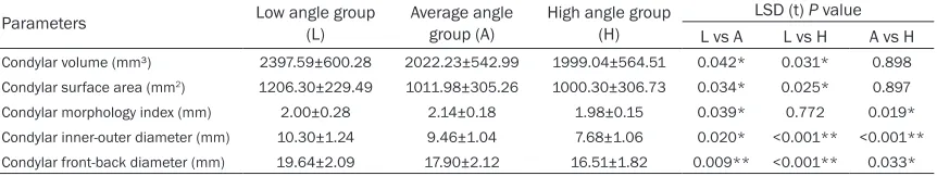
We then compared the condylar morphology of subjects with different vertical skeletal fea-tures. As it was shown in Table 3, all the mea-sured parameters in the low angle group were significantly different from those in the average angle group (*P<0.05; **P<0.01). There was significant difference between high angle group and low angle group regarding the following parameters including condylar MI, inner-outer and front-back diameters (*P<0.05; **P<0.01). However, no significant difference was found between the above two groups regarding con-dylar volume and concon-dylar surface area (P>0.05). What was more, our results showed that the condylar back-front diameters and inner-outer diameters were significantly differ-ent among each group (*P<0.05; **P<0.01). The patients in the low angle group had both longest condylar back-front and inner-outer diameters while the high angle group had the shortest.
Discussion
TMJ, one of the most complex joints in the body, is a bi-arthroidal hinge joint that allows the complex movements necessary for many important functions such mastication, swallow and speech. In addition, TMJ is closely corre-lated with development of craniofacial struc-tures. As the condyle is the growth and devel-opment center of the mandible, its morphology can indirectly reflects the reconstructive pro-cess. The condyle also plays a major role in deciding the size of mandible. Previous study has shown that the condylar volume was not
only closely associated with the final size of mandible, but also with the maxilla-mandible relationship [7]. Patients with skeletal Class II division 1 malocclusion often have the features of developmental disorders in the sagittal direction and vertically dysplasia simultane-ously. In addition, sagittal deformity was often exacerbated by vertically dysplasia. The aim of our study was to explore the potential relation-ship between condylar morphology and various
vertical skeletal features of females who were diagnosed as skeletal Class II division 1 maloc-clusion. Due to the limitations of techniques, it was impossible to conduct the 3-dimension measurement of the condyle in vivo in the past. However, the application of CBCT in the field of clinical dentistry in the recent years has made the measurement become reality. The Mimics software we adopted to reconstruct the mor-phology of condyle is highly reliable and repro-ducible. It enables us to measure the relative parameters of condyle on the basis of the same anatomical position and section, thus it can detect the bone structure characters and dif-ferences of TMJ.
[image:6.612.92.523.99.180.2]In the present study, we divided the female patients who were age matched into three groups based on the skeletal vertical features. This experimental design eliminated the differ-ence in potential influential factors including age and gender. Our results showed that no sig-nificant difference was found between the left and ride side condylar morphology in patients with the same vertical skeletal pattern, indicat-ing that the condyles of the participants were symmetry. One of our strict inclusion criteria that the bilateral symmetry of facial was required might be also a reason accounting for the condylar symmetry in patients with the same vertical skeletal pattern. However, the parameters that can reflect condylar morphol-ogy were significant different among the three Table 3. Comparison of condylar morphological parameters in patients with different vertical skeletal patterns
Parameters Low angle group (L) Average angle group (A) High angle group (H) LSD (t) P value
L vs A L vs H A vs H
Condylar volume (mm³) 2397.59±600.28 2022.23±542.99 1999.04±564.51 0.042* 0.031* 0.898 Condylar surface area (mm2) 1206.30±229.49 1011.98±305.26 1000.30±306.73 0.034* 0.025* 0.897
groups who had different vertical skeletal pat-terns, suggesting that condylar morphology was closely correlated with the vertical skeletal pattern.
Large amounts of studies have been performed to investigate the TMJ joints of patients with skeletal Class II division 1 malocclusion. However, the results were various due to the difference in the research methods as wells as sample size [8]. Currently it was generally accepted that the morphological variations in condyle were mainly resulting from different types of malocclusion and the various burdens that exerted on TMJ. Due to the special maxilla-mandible relationship and occlusion features of patients with skeletal Class II malocclusion, the tissues of TMJ underwent continued recon-struction to adapt to the functional changes. However, Katsavrias et al reported that no sig-nificant morphological difference of TMJ which received burdens was detected in different types of malocclusion [9]. Consistent with our study, Saccucci et al also revealed that the con-dylar volume and surface area of the patients in low angle group were greater than that of high and average angle group [10]. Our results also revealed that no significant difference regarding the condylar volume and surface area was detected between patients in high angle group and average angle group. Two pos-sible reasons might be responpos-sible for this find-ing. Firstly, it is possible that the condyles of patients in the high angle group tend to grow in the upper-front direction and thus have rela-tively slim and long figures. Secondly, the func-tional reconstruction of condyle in patients with skeletal Class II malocclusion might be another reason. As the anterior guidance function is lacking in high angle group patients, the occlu-sal burden is mainly concentrated in the poste-rior teeth area. Moreover, the morphology of condyle in the high angle group patients was long and slim, which made the threshold values of posterior teeth that could withstand the ver-tical pressure decreased. Therefore the force intermaxillary traction must be paid special attention to avoid causing unexpected TMJ inju-ry when treating this type of patients. We induced that the significant difference of con-dylar morphology in females of skeletal Class II division 1 malocclusion with various vertical skeletal features might be correlated with vari-ous factors including bone density, masticating
muscle strength, genetic factors and adaptive reconstructions.
The condylar morphology of TMJ was influenced by many factors such as changes in masticat-ing strength, genetics and facial bio-type [11]. Enomoto et al reported that the condylar width and volume of mice that fed with hard diet were greater than the mice that fed soft diets or hard and soft diets alternately, indicating changes in mastication force could markedly affect the growth of mandibular condylar cartilage as well as the morphology of mandible. Similarly, Kurusu et al showed that occlusal force played an important role in determining the maxillofa-cial morphology and mandibular condyle mor-phology [12].
The mastication muscle alignment of patients with high angle is much more tilting than that of patients with low angle. In addition, the mus-cle’s horizontal section area of high angle patients is smaller. Therefore, this type of patients has weaker oral functions. As the front-rear distance between the central occlu-sion and centric relation position is rather long, the movement pattern of mandible is mainly horizontal. In contrast, the patients with low angle have stronger oral functions, greater masticating muscle strength and occlusal force. The mandibular movement pattern of low angle patients is majorly vertical. Therefore they have powerful masseter and the TMJ can withstand stronger loading force, which increas-es the adaptations of condylar morphology. To the best of our knowledge, this was the first study that systematically compared the condy-lar morphology of females of skeletal Class II division 1 malocclusion with various vertical skeletal features using the advanced CBCT technique.
In conclusion, our CBCT results showed that
there was significant difference regarding the
condylar morphology among females of skele-tal Class II division 1 malocclusion with various
vertical skeletal features, which will provide important and valuable data for radiological diagnosis, orthodontic treatments and orthog-nathic surgery.
Disclosure of conflict of interest
Address correspondence to: Yanling Yu, Department of Orthodontics, Qingdao Stomatological Hospital, Qingdao City Districts, No. 17, Dexian Road, Qingdao 266001, China. Fax: 0532-82796465; E-mail: [email protected]
References
[1] Alexiou K, Stamatakis H, Tsiklakis K. Evaluation of the severity of temporomandibular joint os-teoarthritic changes related to age using cone beam computed tomography. Dentomaxillofac Radiol 2009; 38: 141-147.
[2] Fuentes MA, Opperman LA, Buschang P, Bellinger LL, Carlson DS, Hinton RJ. Lateral functional shift of the mandible: Part II. Effects on gene expression in condylar cartilage. Am J Orthod Dentofacial Orthop 2003; 123: 160-166.
[3] Cattaneo PM, Bloch CB, Calmar D, Hjortshøj M, Melsen B. Comparison between conventional and cone-beam computed tomography-gener-ated cephalograms. Am J Orthod Dentofacial Orthop 2008; 134: 798-802.
[4] Sadek MM, Sabet NE, Hassan IT. Alveolar bone mapping in subjects with different vertical fa-cial dimensions. Eur J Orthod 2015; 37: 194-201.
[5] Schlueter B, Kim KB, Oliver D, Sortiropoulos G. Cone beam computed tomography 3D recon-struction of the mandibular condyle. Angle Orthod 2008; 78: 880-888.
[6] Tecco S, Saccucci M, Nucera R, Polimeni A, Pagnoni M, Cordasco G, Festa F, Iannetti G. Condylar volume and surface in Caucasian young adult subjects. BMC Med Imaging 2010; 10: 1-10.
[7] Krisjane Z, Urtane I, Krumina G, Bieza A, Zepa K, Rogovska I. Condylar and mandibular mor-phological criteria in the 2D and 3D MSCT im-aging for patients with Class II division 1 subdi-vision malocclusion. Stomatology 2007; 9: 67-71.
[8] Pullinger AG, Seligman DA, Gornbein JA. A mul-tiple logistic regression analysis of the risk and relative adds of temporomandibular disorders as a function of common occlusion features. J Dent Res 1993; 72: 968-979.
[9] Katsavrias EG, Halazonetis DJ. Condyle and fossa shape in Class II and Class III skeletal patterns: a morphometric tomographic study. Am J Orthod Dentofacial Orthop 2005; 128: 337-346.
[10] Saccucci M, Polimeni A, Festa F, Tecco S. Do skeletal cephalometric characteristics corre-late with condylar volume, surface and shape? A 3D analysis. Head Face Med 2012; 8: 15. [11] Enomoto A, Watahiki J, Yamaguchi T, Irie T,
Tachikawa T, Maki K. Effects of mastication on mandibular growth evaluated by microcomput-ed tomography. Eur J Orthod 2010; 32: 66-70. [12] Kurusu A, Horiuchi M, Soma K. Relationship