publisher policies. Please scroll down to view the document
itself. Please refer to the repository record for this item and our
policy information available from the repository home page for
further information.
To see the final version of this paper please visit the publisher’s website.
Access to the published version may require a subscription.
Author(s): Catherine Paradis-Bleau, Adrian Lloyd, François
Sanschagrin, Tom Clarke, Ann Blewett, Timothy DH Bugg and Roger C
Levesque
Article Title: Phage display-derived inhibitor of the essential cell wall
biosynthesis enzyme MurF
Year of publication: 2008
Open Access
Research article
Phage display-derived inhibitor of the essential cell wall biosynthesis
enzyme MurF
Catherine Paradis-Bleau*
1, Adrian Lloyd
2, François Sanschagrin
1,
Tom Clarke
2, Ann Blewett
2, Timothy DH Bugg
2and Roger C Levesque
1Address: 1Département de Biologie Médicale, Université Laval, Sainte-Foy, Québec G1K 7P4 , Canada and 2Department of chemistry, University
of Warwick, Coventry, CV4 7AL, United Kingdom
Email: Catherine Paradis-Bleau* - [email protected]; Adrian Lloyd - [email protected];
François Sanschagrin - [email protected]; Tom Clarke - [email protected]; Ann Blewett - [email protected]; Timothy DH Bugg - [email protected]; Roger C Levesque - [email protected]
* Corresponding author
Abstract
Background: To develop antibacterial agents having novel modes of action against bacterial cell wall biosynthesis, we targeted the essential MurF enzyme of the antibiotic resistant pathogen Pseudomonas aeruginosa. MurF catalyzes the formation of a peptide bond between D-Alanyl-D-Alanine (D-Ala-D-Ala) and the cell wall precursor uridine 5'-diphosphoryl N-acetylmuramoyl-L-alanyl-D-glutamyl-meso-diaminopimelic acid (UDP-MurNAc-Ala-Glu-meso-A2pm) with the concomitant hydrolysis of ATP to ADP and inorganic phosphate, yielding UDP-N -acetylmuramyl-pentapeptide. As MurF acts on a dipeptide, we exploited a phage display approach to identify peptide ligands having high binding affinities for the enzyme.
Results: Screening of a phage display 12-mer library using purified P. aeruginosa MurF yielded to the identification of the MurFp1 peptide. The MurF substrate UDP-MurNAc-Ala-Glumeso-A2pm was synthesized and used to develop a sensitive spectrophotometric assay to quantify MurF kinetics and inhibition. MurFp1 acted as a weak, time-dependent inhibitor of MurF activity but was a potent inhibitor when MurF was pre-incubated with UDP-MurNAc-Ala-Glu-meso-A2pm or ATP. In contrast, adding the substrate D-Ala-D-Ala during the pre-incubation nullified the inhibition. The IC50 value of MurFp1 was evaluated at 250 μM, and the Ki was established at 420 μM with respect to the mixed type of inhibition against D-Ala-D-Ala.
Conclusion: MurFp1 exerts its inhibitory action by interfering with the utilization of D-Ala-D-Ala by the MurF amide ligase enzyme. We propose that MurFp1 exploits UDP-MurNAc-Ala-Glu-meso-A2pm-induced structural changes for better interaction with the enzyme. We present the first peptide inhibitor of MurF, an enzyme that should be exploited as a target for antimicrobial drug development.
Background
The bacterial cell wall biosynthesis pathway represents the most validated source of antibacterial targets. The
path-way encodes essential and highly conserved enzymes with no eukaryotic counterparts, the inhibition of which lead-ing to bacterial cell death [1]. The first step of the pathway
Published: 19 December 2008
BMC Biochemistry 2008, 9:33 doi:10.1186/1471-2091-9-33
Received: 30 September 2008 Accepted: 19 December 2008 This article is available from: http://www.biomedcentral.com/1471-2091/9/33
© 2008 Paradis-Bleau et al; licensee BioMed Central Ltd.
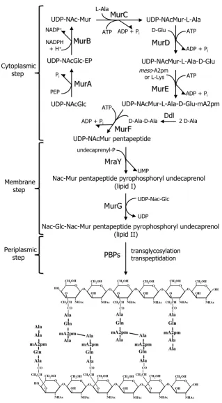
is catalyzed by the cytoplasmic enzymes MurA through MurF, which synthesize UDP-N -acetylmuramyl-pen-tapeptide (Figure 1). Membrane translocases MraY and MurG then add the undecaprenyl-phosphate lipid carrier and N-acetylglucosamine to form lipid II. This precursor is translocated to the periplasm and linked to the growing cell wall polymer by the transglycosylation and transpeptidation actions of penicillin-binding proteins (PBPs). The cell wall layer, composed of alternating units of UDP-N-acetylglucosamine and UDP-N-acetylmuramic acid cross-linked via short peptide chains (Figure 1), maintains cell shape and integrity [2].
While several clinically useful antibiotics interfere with this pathway [3,4], no antibacterial agents target the ATP-dependent Mur ligase enzymes (MurC, MurD, MurE and MurF) that perform the non-ribosomal stepwise addition of the five amino acids forming the cell wall peptide moi-ety (Figure 1). This under-exploitation may be partially explained by the lack of commercially available nucle-otide substrates for studying these enzymes [3,5].
We selected MurF as a specific target. MurF catalyzes the formation of a peptide bond between D-Ala-D-Ala and the nucleotide precursor UDP-N -acetylmuramoyl-L-ala-nyl-D-glutamyl-meso-diaminopimelic acid (UDP-Mur-NAc-Ala-Glu-meso-A2pm) with the concomitant hydrolysis of ATP to ADP and inorganic phosphate, yield-ing UDP-N -acetylmuramoyl-L-alanyl-D-glutamyl-meso-diaminopimelyl-D-alanyl-Dalanine [6]. While the roles of MurC, MurD and MurE may be substituted in a single step by the muropeptide ligase Mpl involved in cell wall recy-cling [7], MurF remains the sole D-Ala-D-Ala adding enzyme [8,9]. The MurF active site is highly conserved among all medically relevant bacteria [10]. Strict limita-tion to D-amino acid substrates [5] also makes MurF an especially attractive target for the development of antibac-terial agents. These amino acids are metabolized only in prokaryotes [11], and D-Ala-D-Ala plays a critical role in cell wall cross-linking [12]. Furthermore, normally func-tioning MurF is essential for proper cell division, optimal expression of methicillin resistance in Staphylococcus aureus [9,13] and glycopeptide resistance mechanism [14,15].
Since MurF acts on a dipeptide to form bonds of a highly distinctive type, we investigated the possibility of inhibit-ing this enzyme with peptide ligands from a phage display library. Phage display screening allows the selection of peptides having specific binding affinities for a targeted protein and has proven useful for identification of various enzyme inhibitors including MurC and MurD [16-18]. The results we present herein constitute the first report of a peptide inhibitor of MurF. We focused this effort on the Gram-negative bacterium Pseudomonas aeruginosa, an
ubiquitous opportunistic pathogen responsible for a vari-ety of chronic nosocomial infections such as lung infec-tion in cystic fibrosis patients [19]. P. aeruginosa is one of the most difficult microorganisms to combat due to high level of resistance to most antibiotics [20].
Results
Purification of biologically active MurF enzyme
The purified MurF protein was visualized as a single 52 kDa band on SDS-PAGE (data not shown). N-terminal sequencing of the first 15 amino acid residues confirmed its identity as P. aeruginosa MurF ligase [Swiss-Prot: Q9EY48, PIR: SF001562] (100% identical to the pub-lished sequence, including the initial Met). MurF biologi-cal activity was confirmed by mass spectrometric identification of the cytoplasmic cell wall precursor UDP-N-acetylmuramyl pentapeptide synthesized in vitro in the presence of the purified enzymes MurA, MurB, MurC, MurD and MurE [21].
Affinity selection of MurF binding peptides
As shown in Table 1, each of the three rounds of bio-pan-ning selected only a fraction of the phage input, which decreased from approximately 1011 down to 107 plaque forming units (PFUs). The third round gave a lower phage recovery compared to the two previous rounds, indicating a strong selection of specific MurF-interacting peptide sequences at this step. Each elution strategy employed at the end of the third round gave similar phage recovery yields. Indeed, the competitive elution conditions using specific MurF substrates were as effective as the well-known glycine-HCl elution strategy.
Schematic representation of the bacterial cell wall biosynthesis pathway
Figure 1
Schematic representation of the bacterial cell wall biosynthesis pathway. The cytoplasmic, membrane and periplas-mic steps of the pathway are shown, along with the structure of the cell wall layer product, composed of alternating units of UDP-N-acetylglucosamine and UDP-N-acetylmuramic acid cross-linked via short peptide chains.
UDP-NAcMur-L-Ala
ATP ADP + Pi L-Ala
MurC
MurC
UDP-NAcMur-L-Ala-D-Glu
ATP
ADP + Pi D-Glu
MurD
MurD
UDP-NAcGlc UDP-NAcGlc-EP PEP PiMur
Mur
A
A
UDP-NAc-Mur
Mur
Mur
B
B
NADPH + H+ NADP+
ATP
ADP + Pi
MurE
MurE
meso-A2pm or L-LysUDP-NAcMur-L-Ala-D-Glu-mA2pm
D-Ala-D-Ala Ddl 2 D-Ala ADP + Pi
MurF
MurF
UDP-NAcMur pentapeptide undecaprenyl-P UMPMraY
Nac-Mur pentapeptide pyrophosphoryl undecaprenol (lipid I)
UDP-Nac-Glc
UDP
MurG
Nac-Glc-Nac-Mur pentapeptide pyrophosphoryl undecaprenol (lipid II) transglycosylation transpeptidation ATP
PBPs
O OCH2OH
NHAc O
CH2OH
HO
NHAc OH
O O
CH3CH
O O
CH2OH
NHAc O
CH2OH
NHAc OH
O O
CH3CH
O OH
CH2OH
NHAc O
CH2OH
NHAc OH
O O
CH3CH
CO CO CO
O O
CH2OH
NHAc O
CH2OH
HO
NHAc OH
O O
CH3CH
O O
CH2OH
NHAc O
CH2OH
NHAc OH
O O
O OH
CH2OH
NHAc O
CH2OH
NHAc OH O O Ala Gln Ala CO
CH3CH
CO
CH3CH
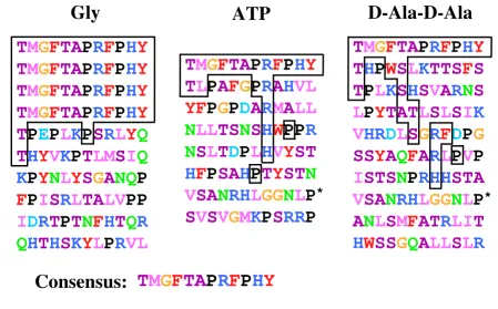
groups. Among the ATP elution group, two sequences pre-sented a "YST" motif also absent in the other elution groups. The vast majority of the sequences eluted with D-Ala-D-Ala presented hydrophobic residues near small res-idues. The D-Ala-D-Ala elution group recovered more small amino acids than the other groups. Overall, the
competitive elutions yielded to more sequence diversity than the glycine-HCl elution.
Bioinformatic analysis
The theoretical pI of MurFp1 was 8.44, indicating that the peptide would be positively charged at physiological pH. MurFp1 contains a single amino acid (R) that would bear this positive charge and no amino acid bearing a counter-balancing negative charge. The ProtParam tool estimated the half-life of MurFp1 at more than 10 hours in
Escherichia coli in vivo and at 7.2 hours in mammalian reticulocytes in vitro. The secondary structure prediction tools indicated with relative certainty a random coil struc-ture. The fasts34 program did not identify any relevant protein in the UniProt database having similarities to MurFp1.
Synthesis and purification of UDP-MurNAc-Ala-Glu-meso-A2pm
[image:5.612.54.295.109.243.2]Analysis of UDP-MurNAc-Ala-Glu-meso-A2pm synthe-sized by the P. aeruginosa enzymes MurA, MurB, MurC,
Table 1: Phage titers obtained after each round of bio-panning using MurF and the 12-mer library
Phage input Eluted phages Elution (%)
Round 1
Gly-HCl 4 × 10¹⁰ 8.2 × 10⁶ 2 × 10⁻²
Round 2
Gly-HCl 1.5 × 10¹¹ 4.1 × 10⁷ 2.7 × 10⁻²
Round 3
Gly-HCl 2 × 10¹³ 1.1 × 10⁷ 5.5 × 10⁻⁵
ATP 1.3 × 10⁷ 6.5 × 10⁻⁵
D-Ala-D-Ala 5.6 × 10⁶ 2.8 × 10⁻⁵
Peptide sequences selected after the third round of bio-panning against MurF
Figure 2
Peptide sequences selected after the third round of bio-panning against MurF. Acidic amino acids (D, E) are in tur-quoise blue, polar amino acids (Q, N) are in light green, basic amino acids (K, R, H) are in blue, hydrophobic amino acids (I, L, M, V) are in pink, hydrophobic aromatic amino acids (F, Y, W) are in red, small amino acids (A, S, C, T) are in magenta, G is in orange, and P is in black (classified according to the Venn diagram for defining the relationships between amino acids). The MurFp1 consensus sequence and related conserved motifs are boxed in black, and the star indicates the other peptide sequence recovered more than once.
T
M
G
F
TA
P
R
F
P
H
Y
T
M
G
F
TA
P
R
F
P
H
Y
T
M
G
F
TA
P
R
F
P
H
Y
T
M
G
F
TA
P
R
F
P
H
Y
T
P
E
P
LK
P
S
RL
Y
Q
T
H
Y
VK
P
T
LM
S
IQ
K
P
Y
NL
Y
S
G
A
NQ
P
F
P
I
S
RL
TA
LV
PP
I
D
R
T
P
T
N
F
H
T
QR
QH
T
H
S
K
Y
L
P
RVL
T
M
G
F
TA
P
R
F
P
H
Y
T
L
P
A
F
G
P
R
A
HVL
YF
P
G
P
D
A
RM
A
LL
NLL
TS
N
S
H
W
PP
R
N
S
L
T
D
P
LHV
Y
ST
H
F
P
SA
H
P
T
Y
ST
N
V
SA
NRHL
GG
NL
P
*S
V
S
V
G
MK
P
S
RR
P
T
M
G
F
TA
P
R
F
P
H
Y
T
H
P
W
S
LK
TTS
F
S
T
P
LK
S
H
S
V
A
RN
S
L
P
Y
TAT
L
S
L
S
IK
VHR
D
L
S
G
R
FD
P
G
SS
Y
A
Q
F
A
RL
P
V
P
I
STS
N
P
RHH
STA
V
SA
NRHL
GG
NL
P
*A
NL
S
M
F
AT
RLI
T
H
W
SS
G
Q
A
LL
S
LR
Gly
ATP
D-Ala-D-Ala
[image:5.612.77.527.356.635.2]MurD and MurE by FPLC anionic exchange indicated a purity of 95%. The structure of the purified UDP-MurNAc-Ala-Glu-meso-A2pm product was confirmed by mass spectrometry (data not shown).
Characterization of MurF activity
The controls performed without MurF or without any one of the substrates did not yield any phosphate from ATP (data not shown), indicating that the reaction mixture was devoid of contaminating ATPase activities that might interfere with the assay. The optimal ATP concentration was 1 mM and MurF reached noticeable activity with 100
μM of UDP-MurNAc-Ala-Glu-meso-A2pm (data not
shown). Steady state kinetic analysis indicated that MurF obeyed Michaelis-Menten kinetics, reaching a maximal specific activity of 4.4 ± 0.4 μmole/min/mg at 5 mM D-Ala-D-Ala with a Km of 115 ± 10 μM (Table 2). Addition of NaCl to the reaction buffer improved MurF activity by a factor of 1.75 (data not shown). The enzyme was very sensitive to the buffer pH, with maximal activity at pH 8.6. The kcat value indicated that each MurF active site per-formed about 100 ligations of D-Ala-D-Ala to UDP-Mur-NAc-Ala-Glu-meso-A2pm and hydrolyzed about 100 ATP molecules per min, releasing the same amount of ADP and inorganic phosphate in accordance with the known stoichiometric relationship (Table 2) [22].
Inhibitory kinetics of MurFp1
MurFp1 was first shown to be a weak time-dependent inhibitor of MurF. The inhibition increased as a function of pre-incubation time, following an overall linear rela-tionship. At a concentration of 2 mM, MurFp1 inhibited 50% of MurF activity subsequent to a 30-minute pre-incu-bation time (Figure 3).
Adding UDP-MurNAc-Ala-Glu-meso-A2pm or ATP dur-ing the pre-incubation step unexpectedly increased the inhibitory action of MurFp1, an effect less pronounced for ATP (data not shown). MurF reaction velocity in the absence of inhibitor was similar with or without UDP-MurNAc-Ala-Glu-meso-A2pm or ATP in the pre-incuba-tion step. MurFp1 inhibited MurF eight times more effi-ciently when UDP-MurNAc-Ala-Glu-A2pm was added to the 30-minute pre-incubation step, giving an IC50 value of 250 ± 10 μM. The inhibition curve displayed a linear
dose-response trend having variable slopes as shown in Figure 4.
The presence of D-Ala-D-Ala during the pre-incubation step nullified the inhibition of MurF by MurFp1 (data not shown). MurF reaction velocity in the absence of inhibitor was nearly identical with or without D-Ala-D-Ala in the pre-incubation step. The Ki of MurFp1 was determined with respect to the D-Ala-D-Ala substrate, with a 30 min pre-incubation of MurF with
UDP-MurNAc-Ala-Glu-Table 2: Kinetic characterization of the MurF enzyme velocities in respect to the D-Ala-D-Ala substrate
Kinetic parameters MurF
Specific activity (μmol/min/mg) 4.4 ± 0.1
kcat (min-1) 100 ± 15
Km (μM) 115 ± 10
kcat/Km (min-1 μM-1) 0.85 ± 0.15
Inhibition of MurF activity by MurFp1 as a function of pre-incubation time
Figure 3
Inhibition of MurF activity by MurFp1 as a function of pre-incubation time. Experiments were done following different pre-incubation times of MurF with 2 mM of MurFp1.
IC50 determination for the inhibition of MurF activity by MurFp1
Figure 4
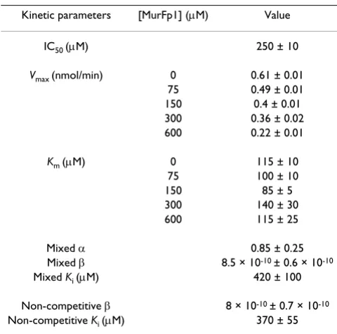
A2pm and MurFp1. The Km values of MurF for D-Ala-D-Ala remained almost the same for each MurFp1 concen-tration used to determine the Ki while the Vmax of MurF decreased significantly as a function of MurFp1 concentra-tion (Figure 5A and Table 3). At 600 μM MurFp1, the Vmax
of MurF was three times lower than for the uninhibited reaction (Table 3). The slopes and the y-intercept of the fitted lines of the Lineweaver-Burk plot varied with MurFp1 concentration (Figure 5B), indicating a catalytic and specific component in the inhibition. The common intersection point of the fitted lines of the Lineweaver-Burk plot was positioned at the left of the ordinate and below the abscissa (Figure 5B), indicating a reversible mixed type of inhibition against D-Ala-D-Ala. This inhibi-tion type was further identified as the most suitable model of inhibition, based on statistical convergence to the experimental data according to the Runs Test, giving the highest R2 value (0.97), the lowest Akaike value (-275) and the lowest standard deviation of the residuals (0.15). The non-competitive type of inhibition was the second best model, giving statistical values close to the mixed model of inhibition (data not shown). The Ki value of MurFp1 was 420 ± 100 μM for the mixed type of inhibi-tion against D-Ala-D-Ala, and 370 ± 55 μM for the non-competitive model. The a parameter indicated that MurFp1 binding to the MurF-D-Ala-D-Ala complex (ES) did not significantly change MurF affinity for D-Ala-D-Ala. The β parameter indicated that the rate constant of ES breaking down to enzyme and product dropped when MurFp1 was bound to ES (Table 3).
A non-specific 12-mer peptide pre-incubated for 30 min with MurF with or without UDP-MurNAc-Ala-Glu-A2pm did not inhibit MurF activity (data not shown). The reac-tions were performed in the presence of excess bovine serum albumin, suggesting that MurFp1 did not interact non-specifically with proteins.
Discussion
Phage display screening of MurF yielded to the identifica-tion of the peptide inhibitor MurFp1. The reducidentifica-tion of the contact time between the phages and MurF during the third round of bio-panning allowed the selection of spe-cific MurF-interacting peptides. This selection pattern was also observed for phage display screening of the amide ligase MurD [18]. In contrast, selection of specific MurC-interacting peptides occurred earlier in the phage display screening [17]. Phage display screenings of P. aeruginosa
proteins FtsA, FtsZ, MurC, MurD and MurF done under similar conditions did not identify any redundant sequences [17,18,23,24].
The ATPase activity of MurF has been shown to depend on the presence of both D-Ala-D-Ala and UDP-MurNAc-Ala-Glu-A2pm [22]. We developed a simple, rapid, sensitive, reliable and inexpensive assay to quantify MurF ATPase activity using the Lanzetta reagent. In opposite to other methods used to measure MurF enzymatic activity [6,8,14,22,25-36], our assay does not involve any radioac-tive isotope or additional enzyme, making high-through-put screening for MurF inhibitors more suitable. The maximal specific activity measured with this assay (4.4
μmole/min/mg) is comparable to the published value for the P. aeruginosa MurF enzyme (3.41 μmole/min/mg) [34]. The Km value determined for D-Ala-D-Ala with P. aeruginosa MurF (115 μM) is in the same range as values published previously for this enzyme and for its E. coli,
Bacillus and Streptococcus faecalis equivalents [6,14,25-27,32,37]. This Km value is roughly equal to the concentra-tion of the D-Ala-D-Ala intracellular pool in E. coli, estab-lished at 200 μM [6,29].
It has been shown that the binding order for substrates to Mur ligases begins with ATP, followed by the nucleotide substrate and ending with the amino acid [6,38]. It has been further demonstrated that the nucleotide substrate binds efficiently to MurD without ATP, inducing confor-mational changes in the C-terminal and N-terminal domains of the protein [39]. To explain how MurFp1 could be a stronger inhibitor following pre-incubation with UDP-MurNAc-Ala-Glu-meso-A2pm, we hypothesize that binding of the nucleotide substrate to MurF induces a structural change that optimizes binding and/or activity of MurFp1. E. coli MurF crystal structure indicates that UDP-MurNAc-Ala-Glu-meso-A2pm interacts mainly with the N-terminal domain, ATP binds principally to the
cen-Table 3: Kinetic characterization of MurF inhibition by MurFp1 with respect to D-Ala-D-Ala.
Kinetic parameters [MurFp1] (μM) Value
IC50 (μM) 250 ± 10
Vmax (nmol/min) 0 0.61 ± 0.01
75 0.49 ± 0.01 150 0.4 ± 0.01 300 0.36 ± 0.02 600 0.22 ± 0.01
Km (μM) 0 115 ± 10 75 100 ± 10 150 85 ± 5 300 140 ± 30 600 115 ± 25
Mixed α 0.85 ± 0.25 Mixed β 8.5 × 10⁻¹⁰ ± 0.6 × 10⁻¹⁰
Mixed Ki (μM) 420 ± 100 Non-competitive β 8 × 10⁻¹⁰ ± 0.7 × 10⁻¹⁰
[image:7.612.54.294.472.710.2]tral domain and D-Ala-D-Ala interacts at the C-terminus [40]. The N-terminal domain of MurF binds the uracyl ring of UDP but does not present the typical nucleotide-binding fold. Indeed, part of the central domain of MurF extends out towards the N-terminal domain to bind the pyrophosphate portion of the nucleotide substrate [40]. This suggests a conformational change upon UDP-Mur-NAc-Ala-Glu-meso-A2pm binding and could explain why ATP binding to the central domain also increased inhibi-tion by MurFp1. To our knowledge, we have presented the first compound having Mur ligase inhibitory action that is enhanced by pre-incubation with the nucleotide sub-strate. However, appropriately substituted phosphinate inhibitors of ATP-dependent amide-forming enzymes depend on their phosphorylation via the ATP substrate. These ATP-dependent phosphinate analogues exhibit a transition-state time-dependent inhibition closely mim-icking the tetrahedral intermediate involved in Mur ligase reactions [3].
Based on classical interpretation of our kinetic and statis-tical data, MurFp1 is a reversible mixed inhibitor. MurFp1 acted as a competitive inhibitor since the inhibition was reversed by addition of D-Ala-D-Ala during pre-incuba-tion, and the slopes of fitted lines from Lineweaver-Burk plot increased as a function of MurFp1 concentration, revealing an increase in inhibitor binding strength [41,42]. Like a non-competitive inhibitor that interferes with substrate utilization without directly competing for the substrate binding site, MurFp1 inhibits MurF at high or low substrate concentrations, the Vmax of MurF
decreas-ing as a function of MurFp1 concentration and the y-inter-cept of Lineweaver-Burk plot varying as a function of MurFp1. MurFp1 appears not to alter MurF affinity for D-Ala-D-Ala but rather inhibit breakdown of the enzyme-substrate complex to enzyme and product. Indeed, MurFp1 acted more like a non-competitive inhibitor than a competitive inhibitor. MurFp1 must bind elsewhere than at the D-Ala-D-Ala binding site. However, phage dis-play and kinetic data indicated that D-Ala-D-Ala released MurFp1 from the MurF surface. We speculate that D-Ala-D-Ala interaction with MurF can enhance conformational changes that displace MurFp1. It should be noted that the glycine elution yielded to a better selection of MurFp1 than the D-Ala-D-Ala competitive elution. Furthermore, reversion of the inhibition is only noticeable when D-Ala-D-Ala is added to the pre-incubation step without the two remaining substrates. In a physiologic environment, such nullification of the inhibition would not be expected as MurFp1 inhibits MurF at high D-Ala-D-Ala concentra-tions. Since D-Ala:D-Ala ligases (DDl, see Figure 1) are highly sensitive to feed-back inhibition by their D-Ala-D-Ala product [37], the inhibitory potential of MurFp1 may be amplified in physiologic conditions.
MurFp1 could exert inhibition by sequestering D-Ala-D-Ala or interfering with MurF structural changes necessary for substrate proximity and amide bond formation [40]. It will be of primary interest to investigate the exact binding site of MurFp1 and whether it affects MurF conformation by co-crystallization of the MurF-MurFp1 complex with each individual substrate. This will yield valuable
infor-Kinetics of MurF inhibition by MurFp1 with respect to D-Ala-D-Ala
Figure 5
Kinetics of MurF inhibition by MurFp1 with respect to D-Ala-D-Ala. A) Michaelis-Menten and B) Lineweaver-Burk plots for MurF activity with respect to the D-Ala-D-Ala substrate showing inhibition by MurFp1 at 0 μM (●), 75 μM (■), 150
mation for further directed optimization of the inhibitor by medicinal chemistry and peptidomimetism [43].
Conclusion
As a first step into the development of novel inhibitors tar-geting unexploited bacterial targets, we have identified the first peptide inhibitor of the essential MurF amide ligase enzyme using a phage display approach. The nucleotide substrate UDP-MurNAc-Ala-Glu-meso-A2pm was synthe-sized and used to develop a sensitive MurF enzymatic assay. The 12-mer peptide MurFp1 acted as a time-dependent inhibitor of MurF activity and its inhibitory potential increased significantly when MurF was pre-incu-bated with UDP-MurNAc-Ala-Glu-meso-A2pm. MurFp1 presumably exploits UDP-MurNAc-Ala-Glu-meso-A2pm-induced structural changes for better interaction with the enzyme. The peptide inhibits the utilization of D-Ala-D-Ala by MurF, preventing the breakdown of the enzyme-substrate complex to enzyme and product with an IC50 value of 250 μM and a Ki value of 420 μM. The discovery of this unique inhibitor opens the path to the rational design of a new generation of antibacterial agents inhibit-ing the bacterial cell wall biosynthesis.
Methods
MurF enzyme preparation
The MurF protein was over-expressed, purified and sequenced as previously described [21]. All reagents were from Sigma Aldrich (Oakville, Ontario, Canada) unless otherwise indicated. Phosphate in the MurF preservation buffer was removed by dialyzing 5 ml against 3 L of buffer A (25 mM Bis-Tris Propane pH 8.0, 1 mM β -mercaptoeth-anol, 2.5 mM MgCl2) three times at 4°C. Purified MurF was visualized on SDS-PAGE using a SYPRO ® Orange staining (Bio-Rad Inc., Mississauga, Ontario, Canada) and the protein concentration was determined by the Bradford method (BioRad Inc., Mississauga, Ontario, Canada) using bovine serum albumin as standard. The phosphate-free MurF protein was aliquoted and preserved at -20°C with glycerol a final concentration of 10%.
Affinity selection of phage displayed peptides against MurF
Phage display screening was performed with the PH.D.-12 phage library (New England Biolabs, Mississauga, Ontario, Canada). Peptide permutations are fused to the minor coat protein pIII of phage M13 via the flexible linker Gly-Gly-Gly-Ser. Three rounds of bio-panning were performed with increasing specificity obtained by increas-ing the Tween concentration durincreas-ing washes and decreas-ing the time of contact between the phages and the targeted protein as previously described [17,23]. Phages encoding selected peptides were eluted at the end of the third round using 100 μl of 0.2 M glycine-HCl (pH 2.2) [17], and 100 μl of 1 mM ATP or 1 mM D-Ala-D-Ala for
30 min. The DNA of ten phages was prepared and sequenced with a -96 gIII primer (New England Biolabs) for each elution condition [23].
Bioinformatic analysis of the MurFp1 sequence
The consensus peptide sequence (TMGFTAPRFPHY) iden-tified by phage display bio-panning was named MurFp1. The MurFp1 peptide sequence was characterized using dif-ferent bioinformatic analysis algorithms of the ExPasy server including the Compute pI/Mw and ProtParam tools [44] as well as the PSIpred and SSpro secondary structure prediction tools [45]. The MurFp1 sequence was also ana-lyzed using the fasts34 program from the FASTA software package, previously identified as the most efficient tool for identifying proteins with similarities to small peptide sequences [18,46].
Synthesis and purification of UDP-MurNAc-Ala-Glu-meso-A2pm
Synthesis of the UDP-MurNAc-Ala-Glu-meso-A2pm sub-strate was performed as previously described [47] except that the reactions starting from UDP-N-acetylglucosamine were carried out in a single mixture using the combined activities of P. aeruginosa enzymes MurA, MurB, MurC, MurD and MurE. The product was purified from the reac-tion mixture by first removing the Mur enzymes by ultra-filtration. The ultrafiltrate was then purified by anion exchange chromatography at room temperature on DEAE-Sephacel pre-equilibrated in 10 mM ammonium acetate at pH 7.5, using a 10–800 mM ammonium acetate (pH 7.5) gradient for elution. UDP-MurNAc-Ala-Glu-meso-A2pm was identified by a pyruvate kinase/lactate dehy-drogenase coupled assay for D-Ala-D-Ala-dependent ADP generation by MurF. Fractions thus found to contain UDP-MurNAc-Ala-Glu-meso-A2pm were freeze-dried from water three times to remove the ammonium acetate. Product purity was assessed by FPLC analytical anion exchange on MonoQ™ using a 0–0.5 M ammonium ace-tate gradient. Structure of the purified UDP-MurNAc-Ala-Glu-meso-A2pm was characterized using negative ion electrospray mass spectrometry, and the synthesized product was stored frozen in water.
Characterization of MurF activity by spectrophotometric assay
rea-gent. After an additional 5 min for colour development, 100 μl of 34% (w/v) sodium citrate was added to stop the reaction [48]. The optical density at 660 nm was immedi-ately measured with a Cary spectrophotometer (Varian, Mississauga, Ontario, Canada). The amount of inorganic phosphate released by MurF was determined by compari-son to the linear portion of a phosphate standard curve with a minimum R2 value of 0.99. The phosphate content of each component in the reaction was subtracted from the total phosphate measured, negative controls were done without enzyme or substrate, and assays were done in triplicate. Different concentrations of each substrate and NaCl were tested to determine optimal conditions for the study of the activity of MurFp1.
The maximal specific activity, the catalytic constant (kcat), the Michaelis-Menten constant (Km) and the enzyme effi-ciency (kcat/Km) were determined for MurF with respect to D-Ala-D-Ala. Kinetic parameters were determined by non-linear regression analysis with a square matrix of enzyme velocities following the Michaelis-Menten equations using the Enzyme Kinetics Module version 1 of SigmaPlot version 8.
Kinetics of MurF inhibition by MurFp1
MurFp1 was synthesized and purified as previously described [17]. Peptide purity (> 95%) was analyzed by HPLC and molecular mass (1424.68 daltons) was con-firmed by MALDI-TOF mass spectroscopy. The peptide was dissolved in buffer B (200 mM Tris-HCl pH 8.0) at a final concentration of 100 mM and the pH was adjusted to 7.0.
Inhibition of MurF enzymatic activity by MurFp1 was measured by the spectrophotometric assay described above. The substrates were added after 30 min of pre-incu-bation of MurF with MurFp1 and the specific activity of MurF was compared with and without MurFp1. Tests were also done in which UDP-MurNAc-Ala-Glu-A2pm was added for pre-incubation with MurF and MurFp1. The concentration of MurFp1 required to reach 50% inhibi-tion (IC50) was determined and pre-incubations for 0, 5, 10, 30 and 60 min with 2 mM MurFp1 were tested. The effect of non-specific peptides was analysed on MurF enzymatic activity, with and without UDP-MurNAc-Ala-Glu-A2pm in the pre-incubation step.
To identify potential interactions between MurFp1 and the MurF substrates, each substrate was added for pre-incubation with MurF, with or without 2 mM MurFp1. The MurFp1 inhibitory constant (Ki) was determined for D-Ala-D-Ala using reaction velocity data obtained with a 30 min pre-incubation of MurF with UDP-MurNAc-Ala-Glu-A2pm and MurFp1. Inhibitor concentrations tested were 0, 75, 150, 300 and 600 μM with D-Ala-D-Ala
con-centrations of 50, 100, 250, 500 and 1000 μM, using fixed optimal concentrations of the remaining substrates.
Kinetic data were fitted to the appropriate model equa-tions using the Sigmaplot Enzyme Kinetics Module. Steady-state inhibition kinetic parameters were deter-mined by nonlinear regression analysis using the least squares method as a reference to ensure accurate dataset fitting and to generate error estimates for each individual observation. The Michaelis-Menten plot of MurF initial velocity as a function of substrate concentration was used to determine the apparent kinetic parameters Vmax and Km with respect to D-Ala-D-Ala using equation 1 [41,42]:
v = Vmax * [S]/(Km + [S]) (1)
The type of inhibition was identified using the Lineweaver-Burk plot of reciprocal MurF initial velocity to reciprocal D-Ala-D-Ala concentration for the various MurFp1 concentrations. Competitive Ki value was deter-mined using the Dixon graphic re-plotting of reciprocal initial velocity versus MurFp1 concentration for each sub-strate concentration, which provides the inhibition com-ponents regardless of the inhibition type according to equation 2:
v = Vmax*((1+β*I/(α*Ki))/(1+I/(α*Ki)))/(1+Km/S)*(1+I/
Ki)/(1+I/(α*Ki))) (2)
where Vmax is the maximum initial velocity for the unin-hibited MurF reaction, I is MurFp1, Km is the Michaelis constant for the uninhibited reaction, α is the Km factor change when MurFp1 is bound to the MurF-D-Ala-D-Ala complex (ES) and β is the Kp (the rate constant when ES breaks down to enzyme and product) factor change when MurFp1 is bound to ES [41,42].
The non-competitive Ki was calculated using equation 3:
v = Vmax/((1+Km/S)*(1+I/Ki)/(1+I*β/Ki)) (3)
Authors' contributions
CPB performed all the described scientific manipulations with the exception of the synthesis and purification of UDP-MurNAc-Ala-Glu-meso-A2pm, achieved by AD, TC, AB and DHB. The kinetic analysis of MurF inhibition by MurFp1 was done by CPB with the help of FS. RCL con-ceived the study. CPB, FS and RCL oriented the study. CPB interpreted the data, prepared the figures and tables, and wrote the manuscript under the supervision of RCL. All authors read and approved the final manuscript.
Acknowledgements
This work was funded by The Canadian Bacterial Diseases Network via the Canadian Centers of Excellence, a FCAR infrastructure team grant to R. C. Levesque and a FRSQ studentship to Catherine Paradis-Bleau.
References
1. Projan SJ: New (and not so new) antibacterial targets – from where and when will the novel drugs come? Curr Opin Pharma-col 2002, 2:513-522.
2. Young KD: Bacterial shape. Mol Microbiol 2003, 49:571-580. 3. El Zoeiby A, Sanschagrin F, Levesque RC: Structure and function
of the Mur enzymes: development of novel inhibitors. Mol Microbiol 2003, 47:1-12.
4. Silver LL: Novel inhibitors of bacterial cell wall synthesis. Curr Opin Microbiol 2003, 6:431-438.
5. Bugg TD, Walsh CT: Intracellular steps of bacterial cell wall peptidoglycan biosynthesis: enzymology, antibiotics, and antibiotic resistance. Nat Prod Rep 1992, 9:199-215.
6. Anderson MS, Eveland SS, Onishi HR, Pompliano DL: Kinetic mech-anism of the Escherichia coli UDPMurNAc-tripeptide D-ala-nyl-D-alanine-adding enzyme: use of a glutathione S-transferase fusion. Biochemistry 1996, 35:16264-16269.
7. Mengin-Lecreulx D, van Heijenoort J, Park JT: Identification of the mpl gene encoding UDP-N-acetylmuramate: L-alanyl-gamma-D-glutamyl-meso-diaminopimelate ligase in Escherichia coli and its role in recycling of cell wall peptidog-lycan. J Bacteriol 1996, 178:5347-5352.
8. Lugtenberg EJ, v Schijndel-van Dam A: Temperature-sensitive mutants of Escherichia coli K-12 with low activities of the L-alanine adding enzyme and the D-alanyl-D-L-alanine adding enzyme. J Bacteriol 1972, 110:35-40.
9. Sobral RG, Ludovice AM, de Lencastre H, Tomasz A: Role of murF in cell wall biosynthesis: isolation and characterization of a murF conditional mutant of Staphylococcus aureus. J Bacteriol
2006, 188:2543-2553.
10. Baum EZ, Crespo-Carbone SM, Abbanat D, Foleno B, Maden A, Gold-schmidt R, Bush K: Utility of muropeptide ligase for identifica-tion of inhibitors of the cell wall biosynthesis enzyme MurF.
Antimicrob Agents Chemother 2006, 50:230-236.
11. Walsh CT: Enzymes in the D-alanine branch of bacterial cell wall peptidoglycan assembly. J Biol Chem 1989, 264:2393-2396. 12. Holtje JV: Growth of the stress-bearing and
shape-maintain-ing murein sacculus of Escherichia coli. Microbiol Mol Biol Rev
1998, 62:181-203.
13. Sobral RG, Ludovice AM, Gardete S, Tabei K, De Lencastre H, Tomasz A: Normally functioning murF is essential for the optimal expression of methicillin resistance in Staphylococ-cus aureus. Microb Drug Resist 2003, 9:231-241.
14. Bugg TD, Wright GD, Dutka-Malen S, Arthur M, Courvalin P, Walsh CT: Molecular basis for vancomycin resistance in Enterococ-cus faecium BM4147: biosynthesis of a depsipeptide pepti-doglycan precursor by vancomycin resistance proteins VanH and VanA. Biochemistry 1991, 30:10408-10415.
15. Reynolds PE: Structure, biochemistry and mechanism of action of glycopeptide antibiotics. Eur J Clin Microbiol Infect Dis
1989, 8:943-950.
16. Christensen DJ, Gottlin EB, Benson RE, Hamilton PT: Phage display for target-based antibacterial drug discovery. Drug Discov Today 2001, 6:721-727.
17. El Zoeiby A, Sanschagrin F, Darveau A, Brisson JR, Levesque RC: Identification of novel inhibitors of Pseudomonas aeruginosa MurC enzyme derived from phage-displayed peptide librar-ies. J Antimicrob Chemother 2003, 51:531-543.
18. Paradis-Bleau C, Beaumont M, Boudreault L, Lloyd A, Sanschagrin F, Bugg TD, Levesque RC: Selection of peptide inhibitors against the Pseudomonas aeruginosa MurD cell wall enzyme. Pep-tides 2006.
19. Davies JC: Pseudomonas aeruginosa in cystic fibrosis: patho-genesis and persistence. Paediatr Respir Rev 2002, 3:128-134. 20. Pierce GE: Pseudomonas aeruginosa, Candida albicans, and
device-related nosocomial infections: implications, trends, and potential approaches for control. J Ind Microbiol Biotechnol
2005, 32:309-318.
21. El Zoeiby A, Sanschagrin F, Havugimana PC, Garnier A, Levesque RC: In vitro reconstruction of the biosynthetic pathway of
pepti-doglycan cytoplasmic precursor in Pseudomonas aerugi-nosa. FEMS Microbiol Lett 2001, 201:229-235.
22. Comb DG: The enzymatic addition of D-alanyl-D-alanine to a uridine nucleotide-peptide. J Biol Chem 1962, 237:1601-1604. 23. Paradis-Bleau C, Sanschagrin F, Levesque RC: Peptide inhibitors of
the essential cell division protein FtsA. Protein Eng Des Sel 2005, 18:85-91.
24. Paradis-Bleau C, Sanschagrin F, Levesque RC: Identification of Pseudomonas aeruginosa FtsZ peptide inhibitors as a tool for development of novel antimicrobials. J Antimicrob Chemother
2004, 54:278-280.
25. Egan A, Lawrence P, Strominger JL: Enzymatic synthesis of the peptide in bacterial ridine nucleotides. V. Co ++ -dependent reversal of peptide bond formation. J Biol Chem 1973, 248:3122-3130.
26. Neuhaus FC, Struve WG: Enzymatic Synthesis of Analogs of the Cell-Wall Precursor. I. Kinetics and Specificity of Uridine Diphospho-N-Acetylmuramyl-L-Alanyl-D-Glutamyl-L-Lysine:D-Alanyl-D-Alanine Ligase (Adenosine Diphosphate) from Streptococcus Faecalis R. Biochemistry 1965, 4:120-131. 27. Michaud C, Blanot D, Flouret B, Van Heijenoort J: Partial
purifica-tion and specificity studies of the glutamate-adding and D-alanyl-D-alanine-adding enzymes from Escherichia coli K12.
Eur J Biochem 1987, 166:631-637.
28. Dementin S, Bouhss A, Auger G, Parquet C, Mengin-Lecreulx D, Dideberg O, van Heijenoort J, Blanot D: Evidence of a functional requirement for a carbamoylated lysine residue in MurD, MurE and MurF synthetases as established by chemical res-cue experiments. Eur J Biochem 2001, 268:5800-5807.
29. Mengin-Lecreulx D, Flouret B, van Heijenoort J: Cytoplasmic steps of peptidoglycan synthesis in Escherichia coli. J Bacteriol 1982, 151:1109-1117.
30. Mengin-Lecreulx D, Blanot D, van Heijenoort J: Replacement of diaminopimelic acid by cystathionine or lanthionine in the peptidoglycan of Escherichia coli. J Bacteriol 1994, 176:4321-4327.
31. Lugtenberg EJ: Studies on Escherichia coli enzymes involved in the synthesis of uridine diphosphate-N-acetyl-muramyl-pen-tapeptide. J Bacteriol 1972, 110:26-34.
32. Duncan K, van Heijenoort J, Walsh CT: Purification and charac-terization of the D-alanyl-D-alanine-adding enzyme from Escherichia coli. Biochemistry 1990, 29:2379-2386.
33. Eveland SS, Pompliano DL, Anderson MS: Conditionally lethal Escherichia coli murein mutants contain point defects that map to regions conserved among murein and folyl poly-gamma-glutamate ligases: identification of a ligase super-family. Biochemistry 1997, 36:6223-6229.
34. Azzolina BA, Yuan X, Anderson MS, El-Sherbeini M: The cell wall and cell division gene cluster in the Mra operon of Pseu-domonas aeruginosa: cloning, production, and purification of active enzymes. Protein Expr Purif 2001, 21:393-400.
35. Gu YG, Florjancic AS, Clark RF, Zhang T, Cooper CS, Anderson DD, Lerner CG, McCall JO, Cai Y, Black-Schaefer CL, Stamper GF, Hajduk PJ, Beutel BA: Structure-activity relationships of novel potent MurF inhibitors. Bioorg Med Chem Lett 2004, 14:267-270. 36. Comess KM, Schurdak ME, Voorbach MJ, Coen M, Trumbull JD, Yang
H, Gao L, Tang H, Cheng X, Lerner CG, McCall JO, Burns DJ, Beutel BA: An ultraefficient affinity-based high-throughout screen-ing process: application to bacterial cell wall biosynthesis enzyme MurF. J Biomol Screen 2006, 11:743-754.
37. Miller DJ, Hammond SM, Anderluzzi D, Bugg TDH: Aminoalkyl-phosphinate inhibitors of D-Ala-D-Ala adding enzyme. J Chem Soc Perkin Trans 1998, 1:131-142.
38. Bouhss A, Mengin-Lecreulx D, Blanot D, van Heijenoort J, Parquet C: Invariant amino acids in the Mur peptide synthetases of bac-terial peptidoglycan synthesis and their modification by site-directed mutagenesis in the UDP-MurNAc:L-alanine ligase from Escherichia coli. Biochemistry 1997, 36:11556-11563. 39. Bertrand JA, Fanchon E, Martin L, Chantalat L, Auger G, Blanot D, van
Heijenoort J, Dideberg O: "Open" structures of MurD: domain movements and structural similarities with folylpolygluta-mate synthetase. J Mol Biol 2000, 301:1257-1266.
Publish with BioMed Central and every scientist can read your work free of charge "BioMed Central will be the most significant development for disseminating the results of biomedical researc h in our lifetime."
Sir Paul Nurse, Cancer Research UK
Your research papers will be:
available free of charge to the entire biomedical community peer reviewed and published immediately upon acceptance cited in PubMed and archived on PubMed Central yours — you keep the copyright
Submit your manuscript here:
http://www.biomedcentral.com/info/publishing_adv.asp
BioMedcentral
41. Cornish-Bowden A: Fundamentals of enzyme kinetics London; Boston: Butterworths; 1979.
42. Segal IH: Enzyme kinetics – behavior and analysis of rapid equilibrium and steady-state enzyme systems John Wiley and Sons, Inc; 1993. 43. Nefzi A, Dooley C, Ostresh JM, Houghten RA: Combinatorial
chemistry: from peptides and peptidomimetics to small organic and heterocyclic compounds. Bioorg Med Chem Lett
1998, 8:2273-2278.
44. Walker JM: The proteomics protocols handbook Totowa, N.J.: Humana Press; 2005.
45. Jones DT: Protein secondary structure prediction based on position-specific scoring matrices. J Mol Biol 1999, 292:195-202. 46. Mackey AJ, Haystead TA, Pearson WR: Getting more from less: algorithms for rapid protein identification with multiple short peptide sequences. Mol Cell Proteomics 2002, 1:139-147. 47. Reddy SG, Waddell ST, Kuo DW, Wong KK, Pompliano DL:
Prepar-ative Enzymatic Synthesis and Characterization of the Cyto-plasmic Intermediates of Murein Biosynthesis. Journal of the American Chemical Society 1999, 121:1175-1178.