ORIGINAL RESEARCH
ADULT BRAIN
Principal Component Analysis of Diffusion Tensor Images to
Determine White Matter Injury Patterns Underlying
Postconcussive Headache
X A. Ghodadra,XL. Alhilali, andXS. Fakhran
ABSTRACT
BACKGROUND AND PURPOSE: Principal component analysis, a data-reduction algorithm, generates a set of principal components that are independent, linear combinations of the original dataset. Our study sought to use principal component analysis of fractional anisot-ropy maps to identify white matter injury patterns that correlate with posttraumatic headache after mild traumatic brain injury.
MATERIALS AND METHODS: Diffusion tensor imaging and neurocognitive testing with the Immediate Post-Concussion Assessment and Cognitive Test were performed in 40 patients with mild traumatic brain injury and 24 without posttraumatic headache. Principal compo-nent analysis of coregistered fractional anisotropy maps was performed. Regression analysis of the major principal compocompo-nents was used to identify those correlated with posttraumatic headache. Finally, each principal component that correlated with posttraumatic headache was screened against other postconcussive symptoms and demographic factors.
RESULTS:Principal component 4 (mean, 7.1⫾10.3) correlated with the presence of posttraumatic headache in mild traumatic brain injury (odds ratio per SD, 2.32; 95% CI, 1.29 – 4.67;P⫽.01). Decreasing principal component 4 corresponded with decreased fractional anisotropy in the midsplenium and increased fractional anisotropy in the genu of the corpus callosum. Principal component 4 identified patients with posttraumatic headache with an area under the receiver operating characteristic curve of 0.73 and uniquely correlated with posttraumatic headache and no other postconcussive symptom or demographic factors.
CONCLUSIONS: Principal component analysis can be an effective data-mining method to identify white matter injury patterns on DTI that correlate with clinically relevant symptoms in mild traumatic brain injury. A pattern of reduced fractional anisotropy in the splenium and increased fractional anisotropy in the genu of the corpus callosum identified by principal component analysis can help identify patients at risk for posttraumatic headache after mild traumatic brain injury.
ABBREVIATIONS:FA⫽fractional anisotropy; ImPACT⫽Immediate Post-Concussion Assessment and Cognitive Test; mTBI⫽mild traumatic brain injury; PC⫽ principal component; PCA⫽principal component analysis; PCS⫽postconcussive symptoms; PTH⫽posttraumatic headache
M
ild traumatic brain injury (mTBI), commonly referred to as “concussion,” is a far-reaching disease, affecting up to 1.7 million individuals in the United States annually.1Many of thesepatients have chronic neurologic symptoms that profoundly im-pact daily life,2with approximately 15% of patients having
persis-tent neurologic symptoms beyond 3 months.3This “miserable
minority” with persistent symptoms such as headache, fatigue,
photophobia, nausea, and visual deficits4include many
individ-uals in the prime of life with notable repercussions on the quality of life and productivity.5
Among postconcussion symptoms (PCS), posttraumatic headache (PTH) is one of the most frequent, enduring, and debil-itating symptoms, with estimates of up to 90% prevalence follow-ing mTBI.6The morbidity associated with PTH is high, with
poorer neurocognitive test performance, exacerbation of other PCS, and longer recovery times.7Unfortunately, imaging of mTBI
and PTH has proved to be difficult because routine CT and MR imaging findings are often negative in these patients. However, recently, diffusion tensor imaging has emerged as a powerful MR imaging technique to identify disruption of major white matter fiber tracts in the brain after trauma8,9and has detected white
matter injuries underlying several postconcussion symptoms.1,10
While DTI continues to show promise as an imaging tool in de-tecting injuries underlying mTBI and PCS, analysis of the large
Received June 26, 2015; accepted July 2.
From the Department of Radiology, University of Pittsburgh Medical Center, Pitts-burgh, Pennsylvania.
Abstract previously presented at: American Society of Neuroradiology Annual Meeting and the Foundation of the ASNR Symposium, April 25–30, 2015; Chicago, Illinois.
Please address correspondence to Anish Ghodadra, MD, University of Pittsburgh Medical Center, Department of Radiology, 200 Lothrop St, Suite 201 East Wing, Pittsburgh, PA 15213; e-mail: [email protected]; @AGhodadraMD
DTI datasets traditionally involves complex voxelwise techniques such as tract-based spatial statistics.11Analytic methods of this
type have many advantages; however, at best, these methods sim-ply identify regions of the brain in which DTI metrics, such as fractional anisotropy (FA), are different among groups of sub-jects. As a result, assessment of the correlation among brain re-gions is more difficult.
In an effort to overcome the limitations of traditional voxel-wise methods, we sought to use principal component analysis (PCA) to analyze FA changes in patients with mTBI. PCA is a data-reduction algorithm that generates a set of new variables or principal components (PCs) that are orthogonal linear combina-tions of the original dataset to maximally explain the variance of the dataset.12This process allows a large number of redundant
variables to be condensed into a relatively few new variables, or PCs, that give the most information about the data. These PCs can then be analyzed to determine which ones correlate with out-comes of interest. The purpose of our study was to use PCA of FA maps to identify white matter injury patterns that correlate with PTH after mTBI.
MATERIALS AND METHODS
Study PopulationOur institutional review board approved this study, with a waiver of informed consent. All studies included were performed as stan-dard of care, and results were retrospectively reviewed.
We searched our electronic medical record to retrospectively identify MR imaging studies with DTI performed for mTBI. Ra-diology reports from January 1, 2006, to March 1, 2013, were searched by using keywords “concussion,” “mild traumatic brain injury,” and “diffusion tensor imaging.” Inclusion criteria were 10 –50 years of age, witnessed closed head trauma, no focal neu-rologic deficit, loss of consciousness of⬍1 minute, posttraumatic amnesia of⬍30 minutes, and English language proficiency. Ex-clusion criteria were a prior neuropsychiatric illness (2 patients), abnormal CT or conventional brain MR imaging findings (3 pa-tients), history of substance abuse (3 papa-tients), lack of DTI (4 patients), lack of neurocognitive assessment (6 patients), total symptom score of zero (3 patients), or inability to affine align FA images (2 patients). Demographic data collected from the elec-tronic medical record included age and sex, type of trauma (sports injury versus non-sports injury), and any history of a prior con-cussion as diagnosed by an athletic trainer, neuropsychologist, or other medical personnel at any facility.
Neuropsychological and neurocognitive testing was per-formed by a neuropsychologist with⬎14 years of experience in treating patients with mTBI. Computerized neurocognitive test-ing was performed with the Immediate Post-Concussion Assess-ment and Cognitive Testing (ImPACT), in which a total symptom score was calculated for each patient using a 7-point Likert survey encompassing 22 PCS. Patients were classified as having head-aches on the basis of the International Headache Society guide-lines13following the postconcussion clinical examination. Time
to recovery was defined as when the ImPACT total symptom score was zero or the patient stated that he or she was asymptomatic.
Imaging
Conventional MR imaging and DTI were performed with a 1.5T unit (Signa; GE Healthcare, Milwaukee, Wisconsin) and a stan-dard head coil. Despite the relatively long time span of this study, all patients and controls included in this study underwent the same imaging sequences on the same system, as follows: sagittal and axial T1-weighted (TR, 600 ms; TE, minimum; section thick-ness, 5 mm; NEX, 1), axial proton attenuation–weighted (TR, 2000 –2500 ms; TE, minimum; section thickness, 5 mm; NEX 1), T2-weighted (TR/TE, 2000 –2500/84 –102 ms; section thickness, 5 mm; NEX, 1), fluid-attenuated inversion recovery (TR/TE, 9000 – 10,000/149 ms; TI, 2200 ms), and diffusion-weighted (single-shot echo-planar sequence; TR, 10,000 ms; TE, minimum; section thickness, 5 mm; matrix, 128⫻128). T2*-weighted gradient re-called-echo (TR/TE, 4400/21 ms; NEX, 1; 90° flip angle; section thickness, 3 mm) or susceptibility-weighted (TR/TE, 37/23 ms; NEX, 1; 15° flip angle; section thickness, 2.4 mm) sequences were performed. The FOV ranged from 200 to 240 mm.
DTI was performed with a single-shot echo-planar sequence (TR/TE, 4000/80 ms; NEX, 2; section thickness, 5 mm; 128⫻128 matrix; FOV, 260 mm). Diffusion gradients were set in 25 non-collinear directions by using 2 b-values (b⫽0 and 1000 s/mm2).
Image Analysis
Fractional anisotropy maps were generated as a measure of white matter integrity by using the fMRI of the Brain Diffusion Toolbox (http://fsl.fmrib.ox.ac.uk/fsl/fslwiki/FDT) as part of the fMRI of the Brain Software Library (FSL; http://www.fmrib.ox.ac.uk/fsl). The FA maps included both gray and white matter and were reg-istered to the Montreal Neurological Institute atlas by using a 12-parameter affine transformation.
A mean FA map was created from the 64 coregistered FA im-ages. For each subject, a new 3D matrix was calculated by sub-tracting the subject’s FA map from the mean FA image. The re-sulting volumes were subjected to PCA, in which each voxel represented a variable. To facilitate interpretation of the PCs, a mean FA map was generated. Additional maps showing the effect of increasing and decreasing the value of each principal compo-nent by 2 SDs were then generated. Analysis was conducted by using a custom script in Matlab (MathWorks, Natick, Massachusetts).
Statistical Analysis
To prevent overfitting, we performed a multistep regression ysis. Initially, the first 20 PCs were screened with univariate anal-yses for correlation with the presence of PTH. Subsequently, for-ward stepwise nominal regression with the PCs whose univariate
discrimina-tion (area under the receiver operating characteristic curve⫽ 0.5), acceptable discrimination (0.7ⱕarea under the receiver oper-ating characteristic curveⱕ0.8), excellent discrimination (0.8ⱕarea under the receiver operating characteristic curveⱕ0.9), and out-standing discrimination (area under the receiver operating charac-teristicⱖ0.9).14Statistical analysis was conducted with the software
package JMP 11 (SAS Institute, Cary, North Carolina).
RESULTS
Sixty-four patients with mTBI were included in our study, of which 40 (63%) had PTH. The mean age was 17.4⫾5.0 years, with 69% male. There was no statistically significant difference in mean age (17.6 versus 17.1 years,P⫽.70) or percentage male (54% versus 78%,P⫽.09) between patients with and without PTH. These data are summarized inTable 1.The median time to presentation was not significantly different in patients with and without PTH (22 versus 24 days,P⫽.79). All patients had normal brain MR imaging findings.
Univariate analysis of the first 20 principal components dem-onstrated only PC 4 (mean, 7.1⫾10.3) correlated with the pres-ence of PTH in patients with mTBI. The odds ratio per SD of PC 4 was 2.32, (95% CI, 1.29 – 4.67;P⫽.01).Table 2summarizes the results of univariate analysis of the correlation between PTH and each principal component.
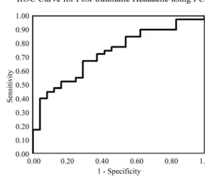
Receiver operating characteristic analysis for PC 4 demon-strated acceptable performance with an area under the curve of
0.73 (Fig. 1).Figure 2shows the effect of changing principal com-ponent 4 on the average FA map by 2 SDs. Lower values of PC 4 primarily indicated relatively decreased FA in the splenium and increased FA in the genu of the corpus callosum and corre-sponded to increased risk of postconcussive headache. Addition-ally, there were more subtle decreases within the corticospinal tract with decreasing PC 4. This principal component uniquely correlated with the presence of PTH and did not correlate with any other postconcussive symptom or demographic factor.
DISCUSSION
Our results show that PCA of DTI in patients with mTBI can successfully identify patterns of FA that correlate with PTH. PCA of FA maps revealed a unique principal component that corre-lated with increased risk of PTH in patients with mTBI. Decreased values of this principal component, PC 4, most prominently cor-responded to decreased FA in the midsplenium of the corpus callosum and increased FA in the genu.
Previous studies of mTBI by using DTI have used 3 major analysis techniques: selection of a priori ROIs, voxelwise analysis (whole-brain or tract-based spatial statistics), and histogram analysis.15While these techniques have shown promise in the
analysis of DTI data, histogram analysis cannot identify focal re-gions of injury and voxelwise analysis and ROI analysis essentially treat each voxel/region as a unique variable that is tested against a clinical outcome of interest. These methods fail to capture the relationship among multiple regions of the brain. Furthermore, despite attempts at compensation for multiple comparisons, there is a higher risk of type 1 statistical errors.
Our technique builds on previous work investigating changes in white matter integrity in patients with mTBI with postconcus-sion syndrome that used voxel-based approaches.1,10,15,16These
reports have demonstrated white matter abnormalities in patients with mTBI relative to controls17-19; however, these differences
have not correlated with symptoms.20Similarly, attempts to
cor-relate structural injuries with postconcussive cognitive perfor-Table 1: Demographics by study groupa
No PTH PTH PValue
Age (yr) 17.08 (2.99) 17.58 (5.87) .7
ImPACT total symptom score
37.46 (24.87) 29.20 (24.65) .2
% Male 254% 278% .09
% Prior concussion 29% 250% .12
Time to recovery (days) 56 (70) 55 (57) .98 a
[image:3.594.313.518.54.230.2]Data are mean (SD) unless otherwise noted.
Table 2: Descriptive statistics and results of univariate regression analysis of PCs and PTH
PC Mean (SD)
Regression
Coefficient PValue
1 1.35 (13.62) ⫺0.002 .897
2 37.81 (11.67) ⫺0.006 .785
3 7.59 (11.58) ⫺0.036 .141
4a 7.09 (10.32) 0.081 .010
5 ⫺14.15 (9.68) 0.010 .712
6 30.59 (9.61) ⫺0.014 .621
7 ⫺17.36 (9.11) ⫺0.011 .699
8 11.23 (9.02) ⫺0.051 .105
9 13.66 (8.83) ⫺0.033 .294
10 10.29 (8.84) ⫺0.003 .917
11 ⫺4.83 (8.82) 0.012 .685
12 3.99 (8.55) 0.053 .112
13 11.26 (8.30) ⫺0.044 .196
14 ⫺16.63 (8.15) ⫺0.026 .419
15 ⫺1.70 (8.03) 0.015 .645
16 ⫺5.50 (8.02) ⫺0.037 .274
17 ⫺3.74 (7.92) 0.016 .627
18 ⫺0.33 (7.65) ⫺0.051 .152
19 ⫺5.82 (7.61) 0.048 .180
20 ⫺9.34 (7.74) ⫺0.013 .691
a
Principal component 4 was the only statistically significant predictor for posttrau-matic headache.
[image:3.594.53.285.170.393.2]mance have shown mixed results without a strong anatomic/ pathologic correlation, with deficits detected in regions as diverse as the occipital cortex and corticospinal tracts.21-25This finding
suggests that not all regions with differences in FA between pa-tients and controls are necessarily symptomatic.1Reduced FA in 1
region alone may not, in and of itself, be clinically relevant. Given the complex interconnectivity within the brain, it may not be sufficient to simply identify individual, isolated regions of FA variance. Rather, it would be more meaningful to view changes in FA in 1 region in the context of potentially related changes else-where in the brain. In short, identifying unique patterns of FA changes in multiple brain regions is needed, rather than merely identifying isolated regions of FA variance.
Our application of PCA to FA maps provides a novel analysis method of complex DTI data as a means of identifying patterns of change in FA. These data-reduction techniques allow distilling complex relationships between variables, in this case FA through-out the brain, to a relatively small set of principal components. Each principal component describes a unique way in which the variables “move” together. Furthermore, to aid in recognition of disease in a clinical setting, these PCs can be visualized in an image (eg,Fig 2).
The finding of a unique principal component that corre-sponds to FA abnormalities in the genu and splenium of the cor-pus callosum offers a means of stratifying patients with postcon-cussive syndrome into groups with high and low risk of the development of posttraumatic headache. Identification of pa-tients at risk for the development of PTH would potentially allow
early intervention in an attempt to im-prove symptoms and outcome, because early comprehensive treatment of PTH has been shown to significantly reduce both the frequency of PTH and head-ache-related disability.7
Additionally, the areas of injury un-derlying PTH identified by PCA may help elucidate the pathophysiology un-derlying headache after mTBI. Identify-ing injury in the splenium is not surpris-ing after mTBI because it is at high risk of direct impact by the falx cerebri and tentorium during trauma.26 However,
most interesting, abnormalities of this region are also seen in other forms of chronic headache, such as migraine.27,28
In fact, in nontraumatic migraine, ab-normalities of the splenium are associ-ated with a more chronic disease course, greater headache frequency, and comor-bid neuropsychiatric conditions.15
Lower values of PC 4 also corre-sponded to increased FA in the genu of the corpus callosum. This could repre-sent compensatory increases in FA in the genu related to the decreased FA/injury identified in the splenium. Compensa-tory increases in FA in areas of the cor-pus callosum in response to other cal-losal injuries have been seen in many pathologies, including schizophrenia, white matter injury from prematurity, and age-related white matter loss.29-31The fact that PC 4 represents not
only decreased FA in the splenium but also increased FA in the genu may indicate that injury to the splenium only results in PTH when it is substantial enough to trigger compensatory increases in FA in the genu. This pattern of FA identified by PC4 highlights the importance of detecting not simply differences in FA but relation-ships of changes in FA in different brain regions. PC 4 is not only a marker of FA in the splenium and genu, but rather it represents a unique pattern of FA in the entire brain and describes a complex relationship among these values. Further analysis of FA in the genu and splenium may yield more refined markers for identifi-cation of patients at risk for developing PTH.
While our study shows the potential of PCA in FA analysis, there are a few limitations of our findings. First, our study population consisted of a single-center retrospective cohort; thus, larger, multicenter prospective studies are needed to con-firm these findings. Second, while our cohort is relatively large, given the relatively large number of voxels and the complexity of the FA structure, larger sample sizes would allow generation of more refined principal components. Furthermore, the reg-istration process, while robust, introduces its own noise into the FA maps that can bias the principal components, particu-larly along edges. Finally, our analysis focused on a single time point after initial injury. The relationship we found between FA in the corpus callosum and PTH could be confounded by a more longitudinal process of injury. Applying this PCA tech-FIG 2.Effect of changing each principal component by⫾2 SDs on fractional anisotropy in a
[image:4.594.56.374.46.288.2]nique to a longitudinal dataset would provide a more robust analysis and could shed light on the pathophysiology of post-concussive headache.
CONCLUSIONS
PCA can be used as a data-mining method to identify white mat-ter injury patmat-terns on DTI that correlate with clinically relevant symptoms in mTBI. PCA of FA maps in patients with mTBI iden-tified a pattern of reduced FA in the splenium and increased FA in the genu of the corpus callosum that correlates with postconcus-sive headache in patients with mTBI. Our results suggest that analysis of the FA patterns in the corpus callosum may offer a means of identifying patients at risk for the development of PTH and thus allow early treatment.
REFERENCES
1. Fakhran S, Yaeger K, Alhilali L.Symptomatic white matter changes in mild traumatic brain injury resemble pathologic features of early Alzheimer dementia.Radiology2013;269:249 –57CrossRef Medline 2. Bohnen N, Jolles J, Twijnstra A.Neuropsychological deficits in pa-tients with persistent symptoms six months after mild head injury.
Neurosurgery1992;30:692–95; discussion 695–96CrossRef Medline 3. Shenton ME, Hamoda HM, Schneiderman JS, et al.A review of
mag-netic resonance imaging and diffusion tensor imaging findings in mild traumatic brain injury.Brain Imaging Behav2012;6:137–92 CrossRef Medline
4. Ganti L, Khalid H, Patel PS, et al.Who gets post-concussion syn-drome? An emergency department-based prospective analysis.Int J Emerg Med2014;7:31CrossRef Medline
5. Emanuelson I, Andersson Holmkvist E, Bjo¨rklund R, et al.Quality of life and post-concussion symptoms in adults after mild traumatic brain injury: a population-based study in western Sweden.Acta Neurol Scand2003;108:332–38CrossRef Medline
6. Kontos AP, Elbin RJ, Lau B, et al.Posttraumatic migraine as a pre-dictor of recovery and cognitive impairment after sport-related concussion.Am J Sports Med2013;41:1497–504CrossRef Medline 7. Erickson JC.Treatment outcomes of chronic post-traumatic
head-aches after mild head trauma in US soldiers: an observational study.Headache2011;51:932– 44CrossRef Medline
8. Bazarian JJ, Zhong J, Blyth B, et al.Diffusion tensor imaging detects clinically important axonal damage after mild traumatic brain injury: a pilot study. J Neurotrauma 2007;24:1447–59CrossRef Medline
9. Wilde EA, McCauley SR, Hunter JV, et al.Diffusion tensor imaging of acute mild traumatic brain injury in adolescents.Neurology2008; 70:948 –55CrossRef Medline
10. Alhilali LM, Yaeger K, Collins M, et al.Detection of central white matter injury underlying vestibulopathy after mild traumatic brain injury.Radiology2014;272:224 –32CrossRef Medline
11. Smith SM, Jenkinson M, Johansen-Berg H, et al.Tract-based spatial statistics: voxelwise analysis of multi-subject diffusion data. Neuro-image2006;31:1487–505CrossRef Medline
12. Jolliffe I.Principal component analysis.In:Wiley StatsRef: Statistics Reference Online.New York: John Wiley & Sons; 2014CrossRef 13. Headache Classification Subcommittee of the International
Head-ache Society. The International Classification of Headache Disorders: 2nd edition. Cephalalgia 2004:24(suppl 1):9 –160 CrossRef Medline
14. Hosmer DW Jr, Lemeshow S, Sturdivant RX.Applied Logistic Regres-sion, 3rd Edition. New York: John Wiley & Sons; 2013
15. Hulkower MB, Poliak DB, Rosenbaum SB, et al.A decade of DTI in traumatic brain injury: 10 years and 100 articles later.AJNR Am J Neuroradiol2013;34:2064 –74CrossRef Medline
16. Zhang K, Johnson B, Pennell D, et al.Are functional deficits in concussed individuals consistent with white matter structural alterations: combined FMRI & DTI study.Exp Brain Res2010; 204:57–70CrossRef Medline
17. Kasahara K, Hashimoto K, Abo M, et al.Voxel- and atlas-based anal-ysis of diffusion tensor imaging may reveal focal axonal injuries in mild traumatic brain injury: comparison with diffuse axonal in-jury.Magn Reson Imaging2012;30:496 –505CrossRef Medline 18. Messe´ A, Caplain S, Paradot G, et al.Diffusion tensor imaging and
white matter lesions at the subacute stage in mild traumatic brain injury with persistent neurobehavioral impairment.Hum Brain Mapp2011;32:999 –1011CrossRef Medline
19. Yallampalli R, Wilde EA, Bigler ED, et al.Acute white matter differ-ences in the fornix following mild traumatic brain injury using dif-fusion tensor imaging.J Neuroimaging2013;23:224 –27CrossRef Medline
20. Lange RT, Iverson GL, Brubacher JR, et al.Diffusion tensor imaging findings are not strongly associated with postconcussional disor-der 2 months following mild traumatic brain injury.J Head Trauma Rehabil2012;27:188 –98CrossRef Medline
21. Niogi SN, Mukherjee P, Ghajar J, et al.Structural dissociation of attentional control and memory in adults with and without mild traumatic brain injury.Brain2008;131:3209 –21CrossRef Medline 22. Lipton ML, Gulko E, Zimmerman ME, et al.Diffusion-tensor
imag-ing implicates prefrontal axonal injury in executive function im-pairment following very mild traumatic brain injury.Radiology
2009;252:816 –24CrossRef Medline
23. Levin HS, Wilde E, Troyanskaya M, et al.Diffusion tensor imaging of mild to moderate blast-related traumatic brain injury and its se-quelae.J Neurotrauma2010;27:683–94CrossRef Medline
24. Little DM, Kraus MF, Joseph J, et al.Thalamic integrity underlies executive dysfunction in traumatic brain injury.Neurology2010;74: 558 – 64CrossRef Medline
25. Wada T, Asano Y, Shinoda J.Decreased fractional anisotropy eval-uated using tract-based spatial statistics and correlated with cogni-tive dysfunction in patients with mild traumatic brain injury in the chronic stage.AJNR Am J Neuroradiol2012:33:2117–22CrossRef Medline
26. Yaeger K, Alhilali L, Fakhran S.Evaluation of tentorial length and angle in sleep-wake disturbances after mild traumatic brain injury.
AJR Am J Roentgenol2014;202:614 –18CrossRef Medline
27. Li XL, Fang YN, Gao QC, et al.A diffusion tensor magnetic reso-nance imaging study of corpus callosum from adult patients with migraine complicated with depressive/anxious disorder.Headache
2011;51:237– 45CrossRef Medline
28. Yu D, Yuan K, Qin W, et al.Axonal loss of white matter in migraine without aura: a tract-based spatial statistics study.Cephalalgia2013; 33:34 – 42CrossRef Medline
29. Xydis V, Astrakas L, Drougia A, et al.Myelination process in preterm subjects with periventricular leucomalacia assessed by magnetiza-tion transfer ratio.Pediatr Radiol2006;36:934 –39CrossRef Medline 30. Kim SN, Park JS, Jang JH, et al.Increased white matter integrity in the corpus callosum in subjects with high genetic loading for schizophrenia.Prog Neuropsychopharmacol Biol Psychiatry2012;37: 50 –55CrossRef Medline