warwick.ac.uk/lib-publications
A Thesis Submitted for the Degree of PhD at the University of Warwick
Permanent WRAP URL:
http://wrap.warwick.ac.uk/86933
Copyright and reuse:
This thesis is made available online and is protected by original copyright.
Please scroll down to view the document itself.
Please refer to the repository record for this item for information to help you to cite it.
Our policy information is available from the repository home page.
by
Shona Moore
Submitted to the University of Warwick
for the degree of
Doctor of Philosophy
June 2016
Liverpool School of Tropical Medicine
Contents
List of Figures . . . vii
List of Tables . . . ix
Abbreviations . . . x
Acknowledgements . . . xiii
Declaration of authorship . . . xiv
List of publications . . . xv
Abstract . . . xvi
1 Introduction 1 1.1 The Immune System . . . 2
1.1.1 Innate immunity . . . 2
1.1.2 Adaptive immunity . . . 3
1.1.3 The structure and function of antibodies . . . 5
1.2 Fc Fusion Technology . . . 15
1.3 Parasite Fc-Binding Proteins . . . 18
1.4 Immunity and Malaria . . . 20
1.4.1 Lifecycle ofPlasmodium falciparum . . . 20
1.4.2 Innate immunity and malaria . . . 22
1.4.3 Adaptive immunity and malaria . . . 25
1.5 Plasmodium falciparum Immune Evasion . . . 27
1.5.1 Sequestration of parasites . . . 28
1.5.2 Rosetting . . . 28
1.5.3 Natural IgM Fc-binding . . . 29
1.6 Plasmodium falciparum Vaccine Development . . . 30
1.6.1 Current progress in vaccine development . . . 30
1.6.2 Potential vaccine candidates . . . 32
1.7 MSPDBL1 and MSPDBL2 . . . 35
1.7.1 Duffy-Binding-Like protein domains . . . 35
1.8 Summary . . . 38
1.9 Aims . . . 40
2 Materials and Methods 41 2.1 Materials . . . 42
2.1.1 Oligonucleotides . . . 42
2.1.2 Plasmids . . . 43
2.1.3 Competent Cells . . . 43
2.1.4 Antibodies . . . 43
2.1.5 Kits . . . 44
2.1.7 Restriction Enzymes and Buffers . . . 46
2.1.8 General Buffers . . . 46
2.1.9 Gel Electrophoresis Buffers . . . 46
2.1.10 ELISA Buffers . . . 46
2.1.11 SDS-Page Electrophoresis Buffers . . . 47
2.1.12 Western Blot Buffers . . . 47
2.1.13 Fast protein liquid chromatography (FPLC) Buffers . . . 47
2.1.14 Antibiotics . . . 47
2.1.15 Cell culture media . . . 47
2.1.16 Microbiological media . . . 48
2.1.17 Software . . . 48
2.2 Molecular Biology Methods . . . 49
2.2.1 Primers . . . 49
2.2.2 PCR Protocol . . . 49
2.2.3 Purification of PCR product from solution . . . 49
2.2.4 Restriction Digest . . . 50
2.2.5 DNA gel electrophoresis . . . 50
2.2.6 Extraction of DNA from agarose gel . . . 50
2.2.7 Vector de-phosphorylation . . . 51
2.2.8 Ligation Protocol . . . 51
2.2.9 Making competentE.coli cells . . . 51
2.2.10 Transformation of competent E.coli cells . . . 51
2.2.11 Mini-prep . . . 52
2.2.12 Screening Positive Colonies . . . 52
2.2.13 Sequencing . . . 53
2.2.14 Midi-prep and Maxi-prep . . . 54
2.2.15 Nanodrop . . . 54
2.2.16 Glycerol stocks . . . 55
2.3 Tissue Culture Methods . . . 55
2.3.1 Preparation and maintenance of cells . . . 55
2.3.2 Splitting Cells . . . 55
2.3.3 Transfection of cells . . . 55
2.3.4 Production of monoclonal population . . . 56
2.3.5 Freezing cells for long-term storage . . . 56
2.4 Methods for Protein Purification and Analysis . . . 56
2.4.1 Fast protein liquid chromatography FPLC . . . 56
2.4.2 Immunoblot Analysis . . . 57
2.4.3 Ultrafiltration . . . 57
2.4.4 Size Exclusion Chromatography . . . 57
2.4.6 SDS-Polyacrylamide gel electrophoresis (SDS-PAGE) . . . 58
2.4.7 Coomassie Blue staining . . . 59
2.4.8 Western blot . . . 60
2.4.9 Enzyme-linked immunoabsorbent assay (ELISA) . . . 60
3 Production of Recombinant MSPDBL1 and MSPDBL2 DBL Domains 61 3.1 Background . . . 62
3.2 Objectives . . . 68
3.3 Methods . . . 69
3.3.1 DNA preparation . . . 69
3.3.2 Amplification of DNA . . . 73
3.3.3 TOPO Ligation to the 3.4 DBL and 3.8 DBL domains . . . 74
3.3.4 Transformation of competent cells with pCR2.1-TOPO-3.4 and pCR2.1-TOPO-3.8 constructs . . . 75
3.3.5 DNA preparation for sub-cloning into pFuse construct . . . 76
3.3.6 pFUSE ligation to the 3.4 DBL and 3.8 DBL domains . . . 78
3.3.7 Transformation of competent cells with pFUSE-3.4 and pFUSE-3.8 . 78 3.3.8 Expression of 3.4 and 3.8 DBL recombinant protein . . . 80
3.3.9 ELISA for Fc detection . . . 81
3.3.10 IgM-binding ELISA . . . 82
3.3.11 Western blot . . . 83
3.3.12 Gel filtration of recombinant DBL domains . . . 83
3.4 Results . . . 84
3.4.1 Production of pCR2.1-TOPO-DBL constructs . . . 84
3.4.1.1 Restriction Digest . . . 84
3.4.1.2 Sequencing Results . . . 85
3.4.2 Production of pFUSE constructs . . . 89
3.4.2.1 Screening of positive colonies . . . 89
3.4.2.2 Sequencing Results . . . 90
3.4.2.3 Restriction Digest . . . 94
3.4.3 Expression of recombinant MSPDBL1 and MSPDBL2 DBL domains 95 3.4.3.1 Immunoblotting . . . 95
3.4.3.2 FPLC protein purification . . . 96
3.4.4 Characterisation of recombinant protein . . . 98
3.4.5 Functional analysis of DBL domains . . . 101
3.4.5.1 The recombinant DBL-Fc domains bind human IgM . . . . 101
3.4.5.2 Malarial MSPDBL1 and MSPDBL2 bind the Cμ4 domain of human IgM . . . 103
4 Analysis of Known IgM-Binding DBL domains 107
4.1 Background . . . 108
4.1.1 IgM-binding DBL domains . . . 110
4.2 Objectives . . . 113
4.3 Methods . . . 114
4.3.1 PCR for random mutagenesis . . . 114
4.3.2 DNA preparation for sub-cloning of mutated pFuse constructs . . . 115
4.3.3 Restriction digest of pFUSE plasmid . . . 116
4.3.4 pFUSE ligation to the mutated 3.4 DBL and 3.8 DBL domains . . . 116
4.3.5 Transformation of competent cells with mutated pFUSE-hIgG1-Fc2-3.4-DBL and pFUSE-hIgG1-Fc2-3.8-DBL . . . . 116
4.3.6 Expression and purification of mutant 3.4 and 3.8 DBL-Fc fusion recombinant protein library . . . 117
4.3.7 IgM-binding ELISA . . . 117
4.3.8 Western blot analysis . . . 117
4.3.9 Gel filtration of recombinant mutant DBL domains . . . 118
4.3.10 Sequence analysis . . . 118
4.3.11 Structural analysis . . . 119
4.3.12 Protein-protein interaction site prediction . . . 120
4.4 Results . . . 121
4.4.1 Generation of Library of MSPDBL mutants . . . 121
4.4.1.1 Random mutagenesis . . . 121
4.4.2 Selection of mutants for expression . . . 124
4.4.2.1 Expression of recombinant DBL mutants . . . 130
4.4.2.2 Characterisation of recombinant DBL mutants . . . 132
4.4.2.3 IgM-binding analysis of MSPDBL Mutants . . . 136
4.4.3 Protein-Protein Interaction Prediction (PPIP) analysis . . . 139
4.4.4 Sequence analysis of IgM-binding domains . . . 141
4.4.5 Structural analysis of IgM-binding DBL domains . . . 151
4.4.5.1 Structural analysis of IgM binding and non-binding MSPDBL domain isolates . . . 151
4.4.5.2 Structural analysis of all known IgM binding and non-binding DBL domains . . . 151
4.5 Summary . . . 153
5 Improving the DBL-Fc fusion constructs 155 5.1 Background . . . 156
5.1.1 Extended hinge Fc-fusion construct . . . 158
5.1.2 Location of the DBL domain . . . 160
5.3 Methods . . . 162
5.3.1 pFMCS-hIgG1-Fc2 (modified pFUSE-hIgG1-Fc2 plasmid) . . . 162
5.3.2 pFMCS-4HF-hIgG1-Fc2 (extended flexible hinge construct) . . . 164
5.3.3 Plasmid Synthesis . . . 165
5.3.4 Sub-cloning the four DBL domains (short and full-length, 3.4 and 3.8) into the pFMCS-4HF-hIgG1-Fc2 construct . . . 165
5.3.5 Expression of recombinant protein . . . 167
5.3.6 IgM-binding ELISA . . . 167
5.3.7 Western blot analysis . . . 167
5.3.8 Gel filtration of recombinant proteins . . . 167
5.4 Results . . . 168
5.4.1 Sub-cloning the four pFMCS-4HF-hIgG1-Fc2-DBL constructs . . . . 168
5.4.2 Producing full-length DBL domains in pFMCS-IgG1-Fc2 . . . 168
5.4.3 Expression of the DBL-Fc fusion recombinant proteins . . . 170
5.4.3.1 Summary of DBL-Fc fusion proteins produced . . . 170
5.4.3.2 FPLC Protein purification . . . 172
5.4.4 Characterisation of recombinant protein . . . 174
5.4.5 Recombinant DBL-Fc fusion proteins containing the extended hinge show improved hIgM binding . . . 175
5.5 Summary . . . 177
6 Discussion 179 6.1 Overview . . . 180
6.2 Expression of merozoite DBL domains as Fc fusion proteins . . . 186
6.3 Accuracy of the molecular simulation of DBL-IgM binding . . . 190
6.4 The role of helix 2a in IgM binding . . . 191
6.5 Minimal IgM-binding region . . . 193
6.6 Merozoite DBL-Fc fusion proteins as reagents for IgM purification . . . 194
6.7 Merozoite DBL-Fc fusion proteins as malaria vaccines . . . 194
6.8 Merozoite DBL-Fc fusion proteins as therapeutics for the treatment of IgM-related diseases . . . 194
6.9 Concluding remarks . . . 196
7 References 197 Appendices 225 A Plasmid Maps . . . 225
A.1 pCR2.1-TOPO . . . 225
A.2 pCR2.1-TOPO-DBL3.4-short . . . 227
A.3 pCR2.1-TOPO-DBL3.8-short . . . 229
A.5 pFUSE-hIgG1-Fc2-DBL3.4-short . . . 234
A.6 pFUSE-hIgG1-Fc2-DBL3.8-short . . . 236
A.7 pFMCS . . . 238
A.8 pFMCS-3.4DBL-FL . . . 240
A.9 pFMCS-3.8DBL-FL . . . 242
A.10 pFMCS-3.4DBL-short . . . 244
A.11 pFMCS-3.8DBL-short . . . 246
A.12 pFMCS-hIgG1-Fc2 . . . 248
A.13 pFMCS-hIgG1-Fc2-3.4DBL-FL . . . 250
A.14 pFMCS-hIgG1-Fc2-3.8DBL-FL . . . 252
A.15 pFMCS-4HF-hIgG1-Fc2 . . . 254
A.16 pFMCS-4HF-hIgG1-Fc2-3.4DBL-short . . . 256
A.17 pFMCS-4HF-hIgG1-Fc2-3.8DBL-short . . . 258
A.18 pFMCS-4HF-hIgG1-Fc2-3.4DBL-FL . . . 260
A.19 pFMCS-4HF-hIgG1-Fc2-3.8DBL-FL . . . 262
B IgM-binding of the IgG-Fc was not detected by sandwich ELISA . . . 264
C Summary of all MSPDBL1 and MSPDBL2 mutant DBL domains . . . 265
D Structural analysis of MSPDBL1 and MSPDBL2 mutant DBL domains . . 269
E Sequence analysis of MSPDBL1 and MSPDBL2 mutant DBL domains . . . 274
F Stability analysis of MSPDBL1 and MSPDBL2 mutant DBL domains . . . 276
G Protein-protein interaction prediction (PPIP) . . . 279
H Homology block analysis . . . 281
List of Figures
1.1 Schematic representation of an antibody . . . 5
1.2 Schematic representation of pentameric Immunoglobulin M . . . 8
1.3 Schematic representation of Fc fusion protein . . . 16
1.4 Life cycle of Plasmodium falciparum within the mosquito and human hosts. 21 1.5 Ribbon diagram showing the structure of MSPDBL2 . . . 36
2.1 DNA Molecular Markers . . . 50
2.2 BioRad gel filtration standards . . . 58
2.3 Protein Markers . . . 59
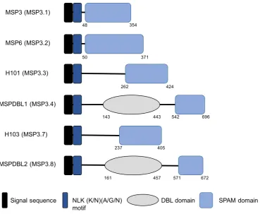
3.1 Structure of the MSP3 family of merozoite surface proteins . . . 63
3.2 Structure of the DBL domains from MSPDBL1/2 . . . 64
3.3 Alignment of the wild type amino acid sequences of DBL 3.4 and DBL 3.8 with N-linked glycan sites removed. . . 69
3.4 Location of the 3.4 DBL domain within the full length MSPDBL1 aa sequence 71 3.5 Location of the 3.8 DBL domain within the full length MSPDBL2 aa sequence 72 3.6 pCR2.1-TOPO-DBL restriction digest . . . 85
3.7 Alignment of pCR2.1-TOPO-DBL3.4 nucleotides . . . 86
3.8 Alignment of pCR2.1-TOPO-DBL3.8 nucleotides . . . 87
3.9 Alignment of pCR2.1-TOPO-DBL constructs with DBL amino acid sequences 88 3.10 Restriction digest screening of pFUSE transformation. . . 89
3.11 pFUSE-hIgG1-Fc2-DBL3.4 sequencing results . . . 91
3.12 pFUSE-hIgG1-Fc2-DBL3.8 sequencing results . . . 92
3.13 Translated amino acid sequence of the pFUSE colonies confirmed. . . 93
3.14 Restriction digests confirm pFUSE-hIgG1-Fc2-DBL3.4 and pFUSE-hIgG1-Fc2-DBL3.8 constructs . . . 94
3.15 Immunoblot for selection of pFUSE cell lines. . . 95
3.16 FPLC elution profile of MSPDBL3.4 and MSPDBL3.8 . . . 96
3.17 pFUSE FPLC fraction immunoblot . . . 97
3.18 Characterisation of DBL-Fc fusion proteins . . . 99
3.19 Size exclusion profiles of recombinant 3.4 DBL and 3.8 DBL proteins . . . . 100
3.20 ELISA analysis of wild-type DBL IgM-binding . . . 102
3.21 Schematic of IgG/IgM domain swap mutants. . . 103
3.22 MSPDBL1 and MSPDBL2 bind the Cμ4 domain of human IgM . . . 104
4.1 Model of IgM highlighting Cμ4 domain . . . 108
4.2 Binding of IgM to Plasmodium falciparum erythrocyte membrane protein 1 (PfEMP1). . . 111
4.3 Mutazyme PCR mutated products of the MSP3.4 and MSP3.8 DBL domains.121 4.4 Restriction digest of amplified 3.4 and 3.8 DBL domains. . . 122
4.6 Model of DBL-IgM binding . . . 124 4.7 Mutated cysteine residues in 3.4 DBL mutants . . . 129 4.8 Immunoblot of DBL mutant FPLC fractions . . . 131 4.9 Western blot analysis of 3.4 and 3.8 DBL mutants detected using αIgG-Fc. 132
4.10 Western blot analysis of 3.4 and 3.8 DBL mutants detected usingαMSPDBL
polyclonal antibodies. . . 133 4.11 Size exclusion profiles from DBL mutants . . . 135 4.12 Western blot analysis of 3.4 and 3.8 DBL Fc-fusion mutants bound to hIgM
detected usingαIgG-Fc. . . 136
4.13 IgM-binding analysis of DBL mutants . . . 138 4.14 Structure of 3.4 DBL mutant 87 (N201Y) . . . 139 4.15 Phylogenetic Relationships between IgM Binding and Non-Binding DBLε
Domains . . . 142 4.16 Sequence alignment of known non IgM binding DBL domains. . . 144 4.17 Sequence alignment of known IgM binding DBL domains. . . 146 4.18 Sequence conservation logos for known IgM binding and non-binding DBL
domains . . . 148 4.19 Homology blocks common to all DBL domains. . . 149 4.20 Structural conservation of known IgM binding MSPDBL DBL domain
isolates vs non-binding isolates . . . 152 5.1 Schematic representation of DBL Fc-fusion protein . . . 157 5.2 Schematic representation of Fc fusion protein with extended hinge region . 160 5.3 Multiple cloning site of original pFUSE-hIgG1-Fc2 plasmid vs. modified
pFMCS-hIgG1-Fc2 . . . 163 5.4 Sub-cloning of the pFMCS-4HF-hIgG1-Fc2 construct. . . 164 5.5 Sub-cloning the DBL inserts into the pFMCS-4HF-IgG1-Fc2 construct . . . 169 5.6 Schematic representation of the eight DBL-Fc fusion constructs . . . 171 5.7 Immunoblot of fractions from the FPLC elution of the eight DBL-Fc fusion
recombinant proteins. . . 173 5.8 Western blot analysis shows improved folding of recombinant DBL-Fc
fusions with the addition of the extended hinge. . . 174 5.9 IgM-binding sandwich ELISA showing improved binding by the new flexible
4H constructs . . . 176 6.1 Location of MSPDBL1 helix 2a . . . 191 6.2 Minimal IgM binding region of DBLζ from TM284var1 compared to the
List of Tables
1.1 Fc receptors (FcRs) and their corresponding antibody ligands. . . 6
1.2 FDA approved hIgG Fc-fusion proteins currently in the clinic. . . 16
2.1 PCR primers for amplification of MSP3.4 and MSP3.8 DBL domains. . . . 42
2.2 Sequencing primers . . . 42
2.3 Mutagenesis primers . . . 42
2.4 Commercial plasmids used for cloning . . . 43
2.5 Commercial competent cells . . . 43
2.6 Primary antibodies . . . 43
2.7 Antibodies for Westerns and Immunoblots . . . 44
2.8 Antibodies for ELISAs . . . 44
2.9 Commercial Kits . . . 44
2.10 PCR and ligation reagents . . . 45
2.11 DNA and protein standards and dyes . . . 45
2.12 Substrates for ELISA, Western blot and Immunoblot . . . 45
2.13 Transformation and transfection reagents . . . 45
2.14 Restriction Enzymes and corresponding buffers . . . 46
3.1 PCR primers for amplification of DBL 3.4 and DBL 3.8 domains. . . 73
4.1 Known hIgM-binding and non-binding Plasmodium falciparum proteins . . 110
4.2 Analysis of mutant sequencing results . . . 123
4.3 Summary of 3.4 DBL mutants . . . 126
4.4 Summary of 3.8 DBL mutants . . . 127
5.1 Amino acid sequences of hinge regions from the four IgG subclasses. . . 159
Abbreviations
α2M α2-macroglobulin
A1AT α-1-antitrypsin
Ab Antibody
ADCC Antibody Dependent Cell-Mediated Cytotoxicity
ADCI Antibody Dependent Cellular Inhibition
Amp Ampicillin
AnAPN1 Anopheline midgut analyl aminopeptidase N 1
AP Alkaline phosphatase
APC Antigen presenting cell
BCR B cell receptor
BLAST Basic local alignment search tool
BSA Bovine serum albumin
CD36 Cluster of Differentiation 36
CHO Chinese hamster ovary
CSA Chondroitin sulphate A
CSP Circumsporozoite protein
DARC Duffy antigen receptor for chemokines
DBL Duffy-Binding-Like
DH5α DH5α competentE.coli cells
DMEM Dulbecco’s modified eagle’s medium
DMSO Dimethyl sulfoxide
DNA Deoxyribonucleic acid
dNTP Deoxynucleotide
DTT Dithiothreitol
EBA Erythrocyte Binding Antigen
EDTA Ethylenediaminetetraacetic acid
ELISA Enzyme linked immunosorbent assay
FAB Fragment antigen binding
FBS Fetal bovine serum
Fc Fragment crystallizable
FPLC Fast protein liquid chromatography
HBSS Hank’s balanced salt solution
HeK Human embryonic kidney
HPLC High performance liquid chromatography
ICAM-1 Intracellular adhesion molecule-1
IE Infected erythrocyte
IgA Immunoglobulin A
IgD Immunoglobulin D
IgE Immunoglobulin E
IgG Immunoglobulin G
IgG1 Immunoglobulin G subclass 1
IgG2 Immunoglobulin G subclass 2
IgG3 Immunoglobulin G subclass 3
IgG4 Immunoglobulin G subclass 4
IgM Immunoglobulin M
IPTG Isopropyl β-D-1-thiogalactopyranoside
IVIG Intravenous immunoglobulin
LB Lysogeny broth
LDS Lithium dodecyl sulphate
MBP Mannose binding protein
MHC Major histocompatibility complex
MOPS 3-(N-morpholino)propanesulfonic acid
MSP Merozoite surface protein
MSP1 Merozoite surface protein-1
MSP3 Merozoite surface protein-3
MSPDBL Merozoite surface protein duffy-binding-like
MWCO Molecular weight cut-off
NEB New England Biolabs
nIgM Natural immunoglobulin M
NK cell Natural killer cells
OD Optical density
PAMPs Pathogen-associated molecular patterns
PBMC Peripheral-blood mononuclear cell
PBS Phosphate buffered saline
PBST Phosphate buffered saline +0.05% Tween 20
PCR Polymerase chain reaction
PES Polyethersulfone
PfEMP1 Plasmodium falciparum erythrocyte membrane protein 1
PRRs Pathogen recognition receptors
RTS,S Malaria vaccine candidate
SDS Sodium dodecyl sulphate
sIgM Secreted immunoglobulin M
SOC Super optimal broth
SPAM Secreted polymorphic antigen associated with merozoites
TB Terrific broth
TBE Tris-borate EDTA
TBV Transmission blocking vaccine
TCR T cell receptor
TE Tris-EDTA
TLR Toll-like receptor
TNF Tumour-necrosis Factor
TOP10 TOP10 competent E.coli cells
TRAP Thrombospondin related anonymous protein
Tris Tris(hydroxymethyl)aminomethane
UV Ultra violet
Var2CSA Variant surface antigen 2 - CSA
X-gal 5-bromo-4-chloro-3-indolyl-β-D-galactopyranoside
XL1-blue XL1-blue competent E.coli cells
Firstly, I would like to thank Prof. Richard Pleass and Dr. Hugo van den Berg without whose support this work would not have been possible. Particularly, thanks to Richard for his enthusiastic supervision! His contagious energy, optimism and immense knowledge have been inspiring. His patience and valuable advice throughout are greatly appreciated. I am grateful to members of the Pleass group past and present for their support and guidance. In particular, a special thanks to Dr. Pat Blundell who has been a motivating and extremely encouraging mentor. Her technical support and guidance have been invaluable and her moral support truly appreciated. Thanks to Dr. Katy Lloyd for valuable discussions, support and reassurance throughout the darker times and for sharing the good times.
This project would not have been possible without the following collaborators to whom I am very grateful: Dr. Gavin Wright and Dr. Cecile Crosnier at the Sanger Institute for providing MSPDBL1 and MSPDBL2 recombinant proteins and αMSPDBL1
andαMSPDBL2 antibodies and Dr. Daniel Czajkowsky at Shanghai Jiao Tong University
for providing molecular simulations.
I have been fortunate to have the support of both the Systems Biology Doctoral Training Centre and the Liverpool School of Tropical Medicine throughout this project. These welcoming and creative environments made the project all the more enjoyable. The encouragement and support of my colleagues, both in Warwick and Liverpool, is greatly appreciated. In particular, I am grateful to Dr. Brent Kiernan for the support and help provided. To my Systems/MOAC cohort, it has been a pleasure.
I could not have done this without my friends, family and loved ones. I genuinely cannot thank them enough for being there for me throughout the journey.
i Cecile Crosnier, Zamin Iqbal, Ellen Kneupfer, Sorina Maciuca, Abigail J. Perrin, Gathoni Kamutu, David Goulding, Leyla Y. Bustamante, Alistair Miles,
Shona C. Moore, Gordon Dougan, Anthony A. Hodder, Domonic P. Kwiatkowski, Julian C. Rayner, Richard J. Pleass, and Gavin J. Wright (2016) Binding of Plasmodium falciparum Merozoite Surface Proteins DBLMSP and DBLMSP2 to Human Immunoglobulin M is Conserved Amongst Broadly Diverged Sequence Variants. The Journal of Biological Chemistry. doi: 10.1074/jbc.M116.722074
ii Phyllis M. Quinn, David W Dunne,Shona C. Moore, and Richard J. Pleass (2016) IgE-tailpiece associates withα-1-antitrypsin (A1AT) to protect IgE from proteolysis
without compromising its ability to interact with FcεRI.Scientific Reports. 6, 20509.
iii Richard J. Pleass, Shona C. Moore, Liz Stevenson, and Lars Hviid (2015) Immunoglobulin M: Restrainer of Inflammation and Mediator of Immune Evasion
by Plasmodium falciparum Malaria. Trends in Parasitology. 1431, 1-12.
Duffy-binding-like domains are present in two potential malaria vaccine candidates. Located on the merozoite surface, MSPDBL1 and MSPDBL2 have been implicated in erythrocyte invasion and identified as targets of natural immunity. Merozoite DBL domains have been shown to bind the Fc region of natural IgM. This is characteristic of several PfEMP1s, and is also well documented in bacteria, viruses and other parasites, where it is thought to prevent specific binding of the more deadly IgG antibodies.
We have developed a mammalian expression system to produce merozoite DBL domains as Fc fusion proteins, facilitating investigation into their adhesive properties. Fc-fusion proteins are composed of the Fc region of IgG fused to a peptide and are a rapidly expanding field of bio-engineering. They have been successful in drug delivery due to their ability to increase serum half-life of the fused protein by the interaction of the IgG Fc with the neonatal Fc receptor (FcRn). Engineering of the Fc scaffold has shown improved receptor binding, allowing cross-linking of Fc receptors for improved vaccine design.
The expression of homodimeric DBL-Fc fusions is difficult, evidenced by incorrect folding and low protein yield. A flexible, extended hinge region was designed to increase the distance between the Fc and the fused DBL domain, and improved protein folding and IgM binding. Further work may optimise this hinge region for the development of malarial vaccines, or therapeutics for IgM-mediated diseases.
1.1 The Immune System
The human immune system has evolved as a result of powerful selection pressure imposed by pathogenic microbes (Cooper and Herrin, 2010; Medzhitov and Janeway, 1997). This complex and intricate system of cells, tissues and molecules is capable of detecting, neutralising and destroying infectious microorganisms, therefore protecting the host from infection (Charles A Janeway et al., 2001; Baron and Klimpel, 1996). The immune system comprises two interconnected and co-acting reactions: (i) innate and (ii) adaptive response (Alberts et al., 2002). In order to recognise and neutralise pathogens, innate and adaptive responses are both capable of distinguishing between self and non-self but they differ in their approaches.
The innate immune system provides a robust and immediate but non-specific response to pathogens, relying on conserved features essential to pathogen survival. This system is present in all multicellular organisms and employs two strategies, firstly by preventing the pathogen from gaining access, and secondly by the destruction of any invading pathogens, which is mediated by phagocytic cells and antimicrobial proteins.
The adaptive immune system, a distinct feature of vertebrate immune systems, is designed to recognise and remember specific pathogens (Alberts et al., 2002). Adaptive immunity involves the process of somatic cell gene rearrangement and relies on an enormous and diverse repertoire of antigen-specific recognition receptors which it builds up by exposure to pathogens. Although slower to respond, adaptive immunity is more effective than innate and is capable of initiating a stronger response every time a pathogen is encountered, offering long-lasting protection.
1.1.1 Innate immunity
This line of defence is based on the detection of conserved products of microbial metabolism known as pathogen-associated molecular patterns (PAMPs) (Medzhitov, 2001). Crucially, these are readily distinguishable from “self” so that the host is not harmed. This means that pathogens of different biochemical composition and with entirely different life cycles can be recognised by overlapping mechanisms (Alberts, 2008; Mogensen, 2009). For example, any peptide of bacterial origin is identifiable by a formylmethionine at the N terminus which is a product of procaryotic translation and differs from the regular methionine produced in eukaryotic translation (Alberts, 2008). PAMPs are detected by pathogen recognition receptors (PRRs) present on innate immune cells such as macrophages and dendritic cells (Janeway and Medzhitov, 2002; Luster, 2002). The repertoire of PRRs is extensive and therefore recognises a diverse range of PAMPs. Moreover, a single pathogen may engage a number of PRRs via various PAMPs, hence forming a rapid and robust response. When a pathogen binds to the surface of an innate immune cell through the PAMP-PRR interaction, a neutralising or destructive response is initiated.
Phagocytosis, a central component of innate immunity, involves the internalisation and degradation of pathogens by phagocytic cells such as polymorphonuclear neutrophilic leukocytes and macrophages. This occurs in response to antigen presentation by a PAMP-PRR interaction. Macrophages amplify the inflammatory response by secreting cytokines and chemokines in addition to initiating phagocytosis. This attracts and recruits further phagocytic cells such as neutrophils and monocytes to the site of infection (Janeway and Medzhitov, 2002). Another method of phagocytosis involves opsonisation, or coating of a pathogen by antibodies (Abs) or complement proteins. This facilitates the uptake of the pathogen by a phagocytic cell. Alternative complement pathways may also be activated to destroy pathogens by lysis, and antibodies may also act to neutralise the microbe by blocking its target (Alberts, 2008). Further non-phagocytic cells such as natural killer (NK) cells and eosinophils all play specific roles in innate immunity.
1.1.2 Adaptive immunity
secondary, more specific, response to these pathogens. The adaptive immune system is recruited through the activation of antigen-specific lymphocytes by antigen-presenting cells (APCs) such as dendritic cells, macrophages and B cells.
Lymphocytes can be classified into two types, B or T cells which have epitope-specific receptors to recognise a large repertoire of antigen epitopes (Alberts, 2008). Adaptive immunity has two major branches: humoral and cell-mediated immunity, which are initiated by recognition of an antigen by B cell receptors (BCRs) and T cell receptors (TCRs), respectively.
The basis of cell-mediated immunity is the presentation of peptides on the surface of APCs by major histocompatibility complex (MHC) molecules (Rudolph et al., 2006; Moon et al., 2007). Foreign peptides are detected by TCRs which then initiate the proliferation and differentiation of naive T cells into effector T cells. Two main types of effector T cells develop depending on the class of pMHC presented - cytotoxic T cells (pMHC class I) and helper T cells (pMHC class II). Cytotoxic T cells directly kill cells expressing the appropriate antigen through the release of cytotoxins which lead to the caspase cascade and apoptosis, while helper T cells promote differentiation of cytotoxic T cells, mediate macrophage activity and promote B cell activation (Rudolph et al., 2006).
Humoral immunity is based on the recognition of antigens by immunoglobulins, also known as antibodies (Abs) which exist either in plasma or on the surface of B cells as part of the B cell receptor repertoire. Upon recognition of an antigen by the BCR, the complex is internalised by the B cell, expressed on the B cell surface as a peptide on a class II MHC and presented to the T cell receptors. Through binding of the peptide to the TCR, B cell activation is promoted by helper T cells. Naive B cells, once activated, proliferate and differentiate into both Ab-secreting plasma cells, which mediate clearance of pathogens, and memory B cells which provide a long-lasting response by persisting in the blood until the pathogen is encountered again.
immunity, and in combination they form a central part of adaptive immunity and are an important aspect of this report.
1.1.3 The structure and function of antibodies
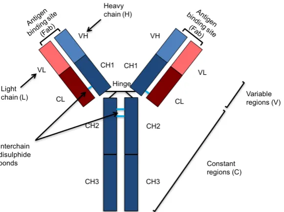
[image:24.595.177.468.447.664.2]Antibodies are immunoglobulins which bind to and neutralise the function of pathogens, while recruiting effector cells such as phagocytes and activating complement to promote pathogen clearance (Charles A Janeway et al., 2001). These glycoproteins form the basis of adaptive immunity and have a characteristic ‘Y’ shape, consisting of two light (λorκ) and two heavy (α,δ,γ,orµ) polypeptide chains linked by covalent interchain disulphide bonds. The five classes of human Abs share the same basic structure and are categorised depending on which type of heavy chain they possess. Heavy (H) and light (L) chains both consist of a combination of variable (V) and constant (C) regions. L chains have one variable (V) and one constant (C) regions while H chains have one V and 3-4 C regions, depending on the Ab class. Antigen specificity relies on subtle differences in the V region.
An antibody has two distinct functional regions. The two identical arms of the ‘Y’ are termed the fragment antigen binding (Fab) region because of their ability to bind antigens. These arms contain variable regions which define the specificity of the antibody for the target antigen. The other fragment participates in the recruitment of effector molecules and is termed the fragment crystalisable (Fc) region. A hinge region links the Fc and Fab portions and allows flexibility within the Ab molecule. The Fc region recruits effector cells by binding to glycoproteins on the surface called Fc receptors (FcRs). Each antibody isotype binds to a specific type of FcR which can be found on the surface of multiple effector cells (Table 1.1). Depending on the effector cell recruited, Abs have a wide range of functions such as phagocytosis of Ab-coated pathogens, antibody dependent cell-mediated cytotoxicity (ADCC), mast cell degranulation and complement activation.
Table 1.1: Fc receptors (FcRs) and their corresponding antibody ligands.
Receptor Ab Ligand Cells
FcγRI (CD64) IgG Macrophages, Neutrophils, Eosinophils, Dendritic
cells
FcγRIIA (CD32) IgG Macrophages, Neutrophils, Eosinophils, Platelets,
Langerhans cells
FcγRIIB (CD32) IgG B Cells, Mast cells, Macrophages, Neutrophils,
Eosinophils, Dendritic cells FcγRIIIA
(CD16a)
IgG NK cells, Macrophages
FcγRIIIB
(CD16b)
IgG Eosinophils, Macrophages, Neutrophils, Mast cells, Follicular dendritic cells
FcRn IgG Dendritic cells, Epithelial cells, Endothelial cells, Hepatocytes, Monocytes, Macrophages
FcRL5 IgG B cells
DC-SIGN IgG Macrophages, Dendritic cells FcμR IgM B cells, T cells, NK cells
Fcα/μR IgA, IgM B cells, Macrophages
pIg IgA, IgM Epithelial cells
FcRL4 IgA B cells
FcαRI (CD89) IgA Monocytes, Macrophages, Neutrophils, Eosinophils
FcεRI IgE Mast cells, Eosinophils, Basophils, Langerhans
cells, Monocytes, Dendritic cells, Platelets
Immunoglobulin-M
Immunoglobulin M (IgM) is the first Ab to be produced by B cells and has an important role in the initial stages of immunity (Klimovich, 2011). The membrane-bound monomeric form of IgM forms the BCR which controls B cell responses through interactions with endogenous and exogenous ligands (Ehrenstein and Notley, 2010). It also exists on mucous membranes in a secreted form and is found in the circulation at concentrations of 1 - 2 mg/ml in blood and, with a half-life of∼5 days, makes up about 15 % of Ig in the blood. The circulatory form can be divided into natural and immune IgM, both of which are crucial for Ab-mediated response to pathogens (Czajkowsky and Shao, 2009). The broadly reactive but low-affinity natural IgM (nIgM), produced by the B1 subset of B cells, occurs naturally in the blood of mice raised under germ-free conditions (Rapaka et al., 2010). In contrast, immune IgM is secreted by B-2 cells following exposure to pathogens and is mostly antigen-specific (Ehrenstein and Notley, 2010) although production levels fall upon development of IgG and other Abs. Immune IgM has been shown to be crucial for protection from viruses, bacteria, protozoa, fungi and helminths (Pleass et al., 2015).
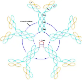
IgM molecules have a subunit with the characteristic ‘Y’ shape seen in other Ab isotypes, formed from two light chains and two heavy chains. Each heavy chain consists of one variable region (Vμ) and four constant domains (Cμ1 - Cμ4) as well as a tailpiece (tp)
located at the C terminus of the heavy chain (Sørensen et al., 1999). The tailpiece enables polymerisation and IgM exists both as pentamer with a five-subunit structure (mass∼970 kDa) and as a hexamer consisting of six subunits (∼1132 kDa). The pentameric form
contains a 15 kDa joining (J)-chain, attached to the tailpiece by disulphide bonds (Figure 1.2). The hexameric form lacks the J chain and is less common than the pentameric form, making up 20% of the total IgM. Hexameric IgM has increased avidity for both antigen and receptor, and is 15-20 times more efficient at activating complement than pentameric IgM (Pleass et al., 2015; Randall et al., 1990; Wiersma et al., 1998). Hexameric IgM without a J-chain is unable to bind to the polymeric Ig receptor (pIgR) which mediates transport of secreted pentameric IgM across epithelia, releasing them to mucosal surfaces (Johansen et al., 2000). This suggests that the role of hexameric IgM is solely in humoral immunity (Randall et al., 1990; Wiersma et al., 1998).
Figure 1.2: Simple schematic representation of a pentameric IgM molecule (five monomeric IgM molecules) showing the heavy and light polypeptide chains. The Fab domain contains one variable and one constant domain Cμ1. The Fc region contains three further constant
domains Cμ2 - Cμ4. Disulphide bonds cross-link adjacent Cμ3 and Cμ4 domains of
individual monomeric molecules and are shown in blue. The location of the J chain is given in red.
for structural integrity and stability of the Ab (Sola and Griebenow, 2009; Arnold et al., 2007).
Natural IgM is a vital part of the early innate immune response and is the first line of defence for protection against many pathogens. It is also able to recognise self-components. The multimeric structure gives the molecule poly-reactivity due to the presence of multiple antigen binding sites. This enables effective recognition of conserved structures on invading organisms. Although affinity for antigen is low, poly-reactivity also enables nIgM to bind multiple structures on the same antigen, thereby enhancing neutralisation by promoting it’s elimination through clearance by phagocytic cells (Ehrenstein and Notley, 2010). High poly-reactivity also gives nIgM a superior ability to detect transformed cells in malignancy where many tumour-related epitopes exist as repetitive carbohydrate motifs (Kaveri et al., 2012). In a study of tumour specific antibodies where thousands of Abs were isolated from patients, all were found to be IgMs (Br¨andlein et al., 2003). Most of these IgM Abs were nIgM which bound to modified tumour-specific receptors (Br¨andlein et al., 2003). Similarly, nIgM taken from healthy patients was found to have tumour specificity (Br¨andlein et al., 2003). Natural IgM therefore has significant importance in the immunogenic response to tumours and is routinely used in diagnosis and therapy of malignancies.
Although an ineffective opsonin by itself, nIgM is highly effective in facilitating clearance of small apoptotic particles, senescent erythrocytes and mis-folded proteins through complement-dependent mechanisms (Pleass et al., 2015). This crucial function is necessary for tissue homeostasis in order to prevent uncontrolled inflammation and to suppress autoimmunity. An accelerated development of IgG autoantibodies and autoimmune disease is seen in the absence of nIgM (Boes et al., 2000).
IgM-Fc receptors
The Fc region of an Ab mediates effector function through binding of host ligands and Fc receptors (FcRs). There are several known FcRs for IgM, the polymeric Ig receptor (pIgR), Fcα/μR, FcμR as well as a number of ligands known to bind IgM including
mannose-binding-lectin (MBL), CD22, TRIM21 and Sp alpha (McMullen et al., 2006; Adachi et al., 2012; Mallery et al., 2010; Tissot et al., 2002). Due to the multimeric structure discussed previously, affinity for IgM is high. The pIgR binds both secretory IgM and IgA on epithelia, as discussed previously. Another FcR shared with IgA is the Fcα/μR expressed on B cells and macrophages which is involved in the priming of helper T
lymphocytes and in defence against bacteria in peripheral organs (Sakamoto et al., 2001). Recently, the FcμR has been identified and is expressed in humans on B cells, T cells and
NK cells (Kubagawa et al., 2009; Shima et al., 2010). In mice, the FcμR is only expressed
on B cells, and studies of FcμR deficient mice have shown elevated non-immune IgM levels
in serum as well as natural autoantibody levels (Honjo et al., 2012; Ouchida et al., 2012). This suggests a role for FcμR binding in IgM homeostasis, autoimmune suppression and
regulation of humoral response (Honjo et al., 2012; Ouchida et al., 2012). The function of the human FcμR receptor remains unclear.
Immunoglobulin-G
Immunoglobulin G (IgG) is an Ab of molecular weight∼150 kDa consisting of a single ‘Y’ subunit formed from two light and two heavy chains. Each heavy chain consists of one variable region (Vγ) and three constant domains (Cγ1 - Cγ3). IgG is produced late in the
primary immune response and is the most abundant Ab found in serum and extracellular fluid (making up 75% of all Ig and found at 10 mg per ml in the circulation). IgG Abs have a wide range of functions and are involved in neutralisation of bacteria, viruses and toxins, as well as opsonising pathogens for engulfment by phagocytes and activating complement through C1q binding.
altering the conformation of the Ab and further influencing effector function. Although deficiency in a single IgG subclass can lead to susceptibility of an individual to specific pathogens, an IgG subclass deficiency is not usually detrimental (Vidarsson et al., 2014).
IgG contains a conserved N-linked glycosylation site at Asn-297 which is located between the Cγ2 and Cγ3 domains and creates an exposed docking site for FcγR binding
(Vidarsson et al., 2014). Fc glycans are known to play a role in Ab stability, and numerous studies have investigated the impact of glycosylation on structural integrity and effector function of IgG (Krapp et al., 2003; Zheng et al., 2014).
IgG-Fc receptors
IgGs are the only Abs to be passed from mother to fetus via the placenta by binding to the neonatal Fc receptor (FcRn) (Koch et al., 1967; Brambell, 1966). Binding to the FcRn also aids recycling of IgG by preventing lysosomal degradation. FcRn is unrelated to the classic FcγRs and is more similar in structure to MHC class I (Story et al., 1994; Roopenian et al.,
2003). Binding occurs in the Cγ2 - Cγ3 interface of the IgG Fc domain (Wines et al.,
2000). IgG binds to FcRns on epithelial cells in a variety of tissues, including the placenta where IgG are transported through vesicles and released by exocytosis into fetal blood (Alberts, 2008; Junghans and Anderson, 1996). FcRns are also located on white blood cells where they are important for IgG-mediated phagocytosis (Vidarsson et al., 2006). The half-life of IgG in the blood is greater compared to other Ab isotypes due to recycling mediated by the FcRn. The FcRn binds IgG at a slightly acidic pH, which is found in the intestinal lumen as well as in acidic endosomes. IgG internalised by pinocytosis binds the FcRn and is recycled to the cell surface and released at the basic pH of blood. This prevents lysosomal degradation and increases the half-life of IgG (Vidarsson et al., 2006; Ghetie et al., 1996).
There are six classic human FcγRs, four of which are activating Fc receptors while
two, FcγRIIB and FcγRIIIB (also known as CD16b), are inhibitory Fc receptors. The
inhibitory FcRIIB is a low affinity receptor and although homologous to the activating FcRs, contains a distinct ITIM sequence in the cytoplasmic domain (Ravetch and Bolland, 2001). The four activation FcRs (FcγRI, FcγRIIA, FcγRIIC and FcγRIIIA)
are co-expressed on the surface of effector cells and the ratio of their expression determines the effector response.
As well as FcγRIIB, another low-affinity inhibitory FcR is located on the surface
of dendritic cells, the dendritic cell-specific intercellular adhesion molecule-3-grabbing non-integrin (DC-SIGN) (Geijtenbeek et al., 2000). DC-SIGN is an important receptor in the efficacy of IVIG in controlling autoimmune disease, and binding to this receptor requires specific glycosylation patterns (Czajkowsky et al., 2015; Geijtenbeek et al., 2000).
The FcR-like protein FcRL5 expressed by B cells is homologous to FcRγI and has
recently been identified as an IgG receptor. FcRL5 is thought to play a role in the regulation of the B cell response (Wilson et al., 2012).
Immunoglobulin-E
Immunoglobulin E (IgE) is a monomeric Ab of low abundance (approximately 0.1 μg per
ml in circulation), consisting of two light and two heavy chains. Unlike IgG which has three, IgE has four constant domains (Cε1 - Cε4) meaning that it is heavier than IgG
at ∼190 kDa. The Cε3 and Cε4 domains of IgE are homologous to the Cγ2 and Cγ3
domains of IgG. The Cε2 domains of IgE are therefore their most distinguishing feature
and these replace the flexible hinge region of IgG. From the crystal structure of the IgE-Fc it is notable that the Cε2 domains bend back and make extensive contact with the Cε3
domains, causing an acute bend in the IgE molecule (Wan et al., 2002). This gives it a unique and more rigid structure than IgG.
IgE plays a role in both allergic and anti-parasitic response and is involved in the activation of mast cells and basophils leading to the consequential release of mediators such as histamine, which are associated with IgE-dependent allergic reactions (Kinet, 1999). When FcεRs are activated in the lungs, the release of toxic mediators are induced,
causing the symptoms of asthma (Gould and Sutton, 2008).
IgE is highly susceptible to cleavage and inactivation by proteases produced by parasitic helminths (Quinn et al., 2016). Several splice variants of IgE are known to exist, including one such variant called IgE-tailpiece (IgE-tp), which has eight novel residues including a cysteine in place of the two carboxy-terminal amino acids in classical IgE (Quinn et al., 2016). This variant has been shown to associate with α-1-antitrypsin (A1AT) in plasma
et al., 2016).
IgE-Fc receptors
Two IgE-Fc receptors are known, the high affinity FcεRI and the lower affinity FcεRII
which are present on most immune cells. The high affinity FcεRI captures both Cε3
domains of IgE and the subsequent and exceptionally slow rate of dissociation on mast cells and basophils is responsible for persistent sensitisation of these cells to allergic challenge (Stone et al., 2010).
Immunoglobulin-A
The highly hydrophilic immunoglobulin A (IgA) is the most abundant Ab in secretions at mucosal sites (making up 10 % of Ig in total). Consisting of the characteristic ‘Y’ structure common to most Ab molecules, IgA contains two heavy and two light chains. A molecule of IgA has three constant regions (Cα1 - Cα3) and, like IgM, contains a tailpiece
and J-chain at the heavy chain C terminus (Kerr, 1990). This also gives IgA the ability to polymerise, and although it is primarily found in monomeric form it can also take a dimeric form (Atkin et al., 1996). In serum, catabolic rates of IgA are five times faster than IgG which is synthesised at the same rate of IgA, therefore the concentration of IgA is about one-fifth the concentration of IgG (Kerr, 1990). In secretions, IgA synthesis rates are far higher than any other Ab class. IgA is therefore overall the most highly synthesised Ab isotype with more IgA produced in total than all of the other Ab classes combined (Kerr, 1990).
IgA-Fc receptors
Due to it’s ability to polymerise and the presence of the J-chain, secretory IgA binds the polymeric Ig receptor (pIg) through the Cα3 domain, leading to transport across
epithelial surfaces (Brandtzaeg and Prydz, 1984). Another receptor shared with IgM is the Fcα/μR expressed on B cells and macrophages. For this receptor interaction, the Cα2
- Cα3 domain interface is critical while the J-chain is not considered essential for binding
(Sakamoto et al., 2001). IgA has a high level of glycosylation although its glycans are not required for receptor interaction (Mattu et al., 1998).
A receptor unique to IgA, is the FcαRI (CD89), thought to play a role in mucosal
immune defence (Bakema and van Egmond, 2011). FcαRI has homology with FcγRs and
FcεRI, although the binding site at the Cα2 - Cα3 interface is distinct from the homologous
regions in IgG and IgE at the hinge proximal regions of Cγ2 and Cε3 respectively (Woof,
2002).
The FcR-like protein FcRL4 expressed by B cells has recently been identified as an IgA receptor. FcRL4, like the IgG receptor FcRL5, is thought to play a role in the regulation of the B cell response (Wilson et al., 2012).
Immunoglobulin-D
Co-expressed with IgM by mature B cells before isotype switching triggers expression of the other Ab classes, immunoglobulin D (IgD) is a monomeric Ab of relatively low abundance (approximately 30 μg per ml in circulation). The role of IgD is not fully understood. IgD
exists in both membrane-bound form and in serum and it consists of two light and two heavy chains. Although IgD has three constant domains (Cδ1 - Cδ3) similar to IgG, it has
a long hinge region which gives it a unique flexible ‘T’ shape compared to the traditional ‘Y’ shape of other Ab isotypes. This makes the molecule highly flexible thus enhancing antigen binding, although making it susceptible to proteolytic degradation (Chen and Cerutti, 2011; Vladutiu, 2000).
IgD-Fc receptors
The FcR for IgD remains elusive although there is evidence for the presence of FcδRs on
1.2 Fc Fusion Technology
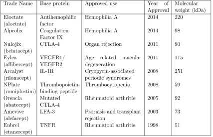
Fc fusion therapeutics are a fast growing field of bio-engineering. Monoclonal antibodies (mAbs) have dominated the field of therapeutics in recent years mainly due to the biological and pharmacological properties resulting from the effector function of the Fc region. A new generation of therapeutics whereby the Fc region of an IgG antibody is fused to an active protein drug, are proving successful. The IgG Fc allows interaction with FcRs on immune cells and gives the protein of interest antibody-like properties. The IgG Fc also binds to the neonatal Fc receptor (FcγRn) an action which facilitates recycling,
thus protecting the active protein from endosomal degradation. The addition of the IgG Fc therefore increases the serum half-life of the protein of interest, extending exposure to the target by limiting renal clearance, consequently improving pharmacological effect and its therapeutic potential. This technology has opened up therapeutic use and drug delivery of smaller active proteins in particular as clearance of such molecules (<60 kDa) is rapid with half-life ranging from between a few minutes to hours. Fc-fusion proteins also form homodimers which improves avidity of the active protein giving it further therapeutic advantage (Figure 1.3).
Other fusion proteins which are designed to improve the half-life of the fused protein include HSA-fusions and Transferrin-fusion proteins. The 66.5 kDa human serum albumin (HSA) is also known to bind the FcRn, with a slightly lower affinity than IgG and therefore shorter half-life (456 hours compared to 480 hours for IgG) (Strohl, 2015). HSA-fusion proteins are also currently in development and are showing some success in clinical trials with Tanzeum (GlaxoSmithKline) receiving FDA approval in April 2014 (Strohl, 2015). Novozymes have also shown that an engineered HSA is able to improve half-life by improving the affinity for FcRn 12-fold (Andersen et al., 2014). This has potential to improve the half-life of HSA-fusion therapeutics. Further fusion proteins designed to improve half-life include fusion of Transferrin or CTP although so far none have FDA approval (Strohl, 2015).
Figure 1.3: Schematic representation of a dimeric Fc-fusion protein. The fused peptide is covalently attached to the Cγ2 via a hinge region which forms a disulphide
bridge and produces a dimeric fusion protein. Adapted from Czajkowsky et al. (2012).
nine approved by the FDA for use in drug delivery (Table 1.2). Like many mAbs, most of these Fc-fusion proteins target receptor-ligand interactions acting either as antagonists or as agonists to manipulate immunity by either blocking or stimulating receptor function (Czajkowsky et al., 2012).
Table 1.2: FDA approved hIgG Fc-fusion proteins currently in the clinic.
Trade Name Base protein Approved use Year of Approval Molecular weight (kDa) Eloctate (aloctate) Antihemophilic factor
Hemophilia A 2014 220
Alprolix Coagulation Factor IX
Hemophilia A 2014 98
Nulojix (belatacept)
CTLA-4 Organ rejection 2011 90
Eylea (aflibercept)
VEGFR1/ VEGFR2
Age related macular degeneration 2011 115 Arcalyst (rilonacept) IL-1R Cryopyrin-associated periodic syndromes 2008 251 NPlate (romiplostim) Thrombopoietin-binding peptide
Thrombocytopenia 2008 59
Orencia (abatecept)
Mutated CTLA-4
Rheumatoid arthritis 2005 92
Amevive (alefacept)
LFA-3 Psoriasis and transplant rejection
2003 73
Enbrel (etanercept)
Currently, all FDA-approved Fc-fusion proteins use the Fc region from the IgG1 subclass. This is due to its increased half-life over other subtypes as well as its superior engagement of FcγRIIA which makes it the preferred subclass for use in delivery of antigen
to APCs (Czajkowsky et al., 2012). It has been suggested that IgG3 could be used to improve complement activation. However, the increased length of the IgG3 hinge region means that it is more susceptible to proteolytic cleavage than other subclasses and this technical difficulty would have to be overcome for its use in Fc-fusion proteins (Czajkowsky et al., 2012).
The recent success of Fc-fusion proteins has mostly been focussed on drug delivery, although direct targeting of FcRs by the Fc-fusion protein gives promise for Fc-fusion based vaccines (Czajkowsky et al., 2012). Once the target FcRs are identified, the need for receptor cross-linking in enhanced cell signalling highlights the importance of valency particularly when targeting low-affinity receptors. It has recently been shown that Fc-fusions can be modified to polymerize into a hexameric form with twelve fused partners (Mekhaiel et al., 2011). The Hexa-Fc scaffold has thus been developed to oligomerise monomeric IgG Fc into hexameric oligomers (Czajkowsky et al., 2015). This construct is able to enhance binding to low-affinity receptors with increased avidity, thus opening up the use of Fc-fusion proteins as vaccines (Czajkowsky et al., 2015).
1.3 Parasite Fc-Binding Proteins
We have seen that the function of the five Ab classes and the recruitment of effector cells relies on Ab Fc binding to glycoprotein FcRs which are present on immune cell surfaces. A successful field of bio-engineering takes advantage of these interactions to develop successful Fc-fusion based therapeutics and has further potential for the development of vaccines.
In some cases, an FcR-like pathogen is also able to take advantage of this system; hijacking the Ab by binding to the Fc portion of a non-immune antibody instead of the antigen-specific Fab region. This allows the pathogen to evade immunity as the Ab is prevented from interacting with host FcRs, the effector function is blocked and the pathogen is masked in host non-specific Ab. Parasites, which rely on remaining undetected within their host for survival, are particularly proficient in this immune evasion strategy. A large number of human parasites are able to bind the Fc portion of immunoglobulin non-specifically and prevent downstream Fc-mediated destruction. This strategy, mediated by Ig-binding molecules expressed on the surface of the parasite is also well documented in bacteria and viruses such asStaphylococcus aureuswhich expresses protein A, herpes simplex virus which expresses the glycoprotein complex gE-gI, a known Fcγ
receptor, as well as IgA-binding proteins identified in many strains of Streptococcus
(Watkins, 1964; Lubinski et al., 2011; King and Wilkinson, 1981; Pleass et al., 2001; Nezlin and Ghetie, 2004; Pleass and Woof, 2001). In parasites, Fc-binding proteins which play a role in evasion of the immune response may also add to the infectivity and persistence of the parasite in circulation.
IgM Fcμ-binding proteins are less well documented than for IgG and IgA. This may
be due to difficulties in distinguishing between low-affinity natural IgM Fab binding and Fc-receptor interactions (Czajkowsky et al., 2010). Several protozoa are known to utilise IgM-binding proteins, including Toxoplasma gondii and pathogenic species of
Trypanosomitidae (Vercammen et al., 1999; Vincendeau and Da¨eron, 1989; Czajkowsky
et al., 2010). More recently, proteins from the Plasmodium falciparum erythocyte membrane protein 1 (PfEMP1) family, expressed on the surface ofP. falciparum-infected erythrocytes (IEs) have been shown to bind natural IgM via the Fc. Only certain P.
falciparum strains have been shown to bind IgM, but binding phenotypes are linked to
the pathogenesis of malaria by mediating cytoadhesion (Smith et al., 2013). The role of IgM-binding in PfEMP1 is discussed further in Section 1.5.3.
1.4 Immunity and Malaria
With around half of the world’s population at risk, malaria is a major global health problem. Caused by the highly successful Plasmodium parasite, which is injected into a human host by the Anopheles mosquito upon taking a blood meal, in 2013 there were an estimated 198 million clinical cases of malaria worldwide, 584,000 of which resulted in death (WHO, 2015). Malaria immunity develops slowly and as a result 78% of malaria deaths occur in children under five years of age. Moreover, 90% of all malaria deaths occur in Africa where many mothers are unable to afford or access treatment, therefore prevention is critical (WHO, 2015; Miller et al., 2002). Despite some recent success in prevention and control measures (Yewhalaw et al., 2011; Greenwood and Targett, 2011), mosquito-insecticide resistance and drug-parasite resistance threaten to undermine these efforts. Antibody response is an important aspect in protective immunity toPlasmodium, particularly in the blood stages of infection where several parasite antigens that are known to cause clinical symptoms drive strong antibody responses. Antigenic variation means that immunity to a malarial infection is short-lived and leads only to partial immunity which is unable to protect an individual against a new infection. Malaria vaccine development is urgently needed but the complexity of the parasite lifecycle and intricacy of the host-parasite interaction means that it is a great scientific challenge to develop an effective vaccine.
There are more than 100 species ofPlasmodium, five of which cause malaria in humans
(Plasmodium falciparum, P. vivax, P. malariae, P. knowlesi and P. ovale). While P.
falciparum is the most virulent species and causes the highest rates of complications and
mortality,P. vivax is the most widely distributed species worldwide. In Africa, however, the risk of infection with P. vivax is low due to the absence of the Duffy gene in many populations, which is required for infection of red blood cells. Here, where the vast majority of malaria deaths occur, P. falciparum poses a huge burden on the poorest and most vulnerable communities.
1.4.1 Lifecycle of Plasmodium falciparum
within the midgut into ookinetes which burrow through the midgut wall and form oocysts on the other side. As oocysts grow and divide, thousands of active haploids called sporozoites are produced which burst into the body cavity of the mosquito where they travel to and invade the salivary glands. It is from the saliva of the infected mosquito that sporozoites are injected into the human host’s skin where they migrate into the blood stream.
Figure 1.4: Lifecycle ofPlasmodium falciparum within the mosquito and human hosts. Upon taking a blood meal, an anopheles gambiae mosquito injects sporozoites into the human host. These migrate to and invade liver cells where they differentiate and multiply, generating merozoites, the invasive form of the parasite. Merozoites subsequently infect and then multiply inside erythrocytes or differentiate into gametocyes which are injested by a mosquito upon taking a blood meal. The change of environment in the mosquito midgut induces gamete formation and the life cycle repeats.
approximately within 60 minutes of inoculation (Baron et al., 1996). A single sporozoite multiplies and re-differentiates within an infected hepatocyte over the course of 5 - 10 days to generate thousands of merozoites, which are the invasive form of the parasite (Maier et al., 2009). Subsequently, bulging hepatocytes burst and merozoites are released into the blood stream where they rapidly begin a cycle of invasion and infection of host erythrocytes.
Merozoites within an infected erythrocyte consume haemoglobin, giving them energy to develop into trophozoites. Upon gorging itself on haemoglobin, a trophozoite undergoes schizogony and develops into a schizont, consequently another round of asexual replication then produces multiple merozoites per schizont. A bulging schizont ruptures 48 hours after infection of the erythrocyte, releasing merozoites into the blood stream once again where they infect further erythrocytes. The cycle of infection, multiplication and bursting of erythrocytes continues, producing a high parasite burden on the host. The blood stage parasites are responsible for clinical manifestations of the disease and the cycle of fresh merozoites bursting into the blood stream responsible for the waves of parasitemia.
A few blood stage merozoites don’t develop into schizonts and multiply, instead they differentiate into gametocytes. Upon ingestion when a mosquito takes a blood meal from an infected human host, the change in environment in the mosquito midgut induces gamete formation and the life cycle repeats (Figure 1.4).
Plasmodium falciparum multiplies rapidly within a human host during cycles of
erythrocyte invasion by merozoites. The host first encounters the parasite during the symptomless pre-erythrocytic phase and the parasite is exposed to the immune system again in the form of the infective merozoite. The immune response to malaria is complex and stage-specific. Individuals exposed to repeat infection do eventually develop immunity, although as it is slow to develop children are of high risk.
1.4.2 Innate immunity and malaria
donors within 10 hours (Stevenson and Riley, 2004; Scragg et al., 1999). Recently, it has been suggested that innate immunity is triggered when parasite density crosses a threshold value (Stevenson and Riley, 2004). This means that blood parasite densities oscillate between a high level (where innate responses are triggered and parasites are partially cleared) and low level (where innate immune responses are not triggered) (Stevenson and Riley, 2004). This implies that innate responses are essential during the initial phase of infection and allow the host time to develop an adaptive response. From an evolutionary perspective, this is advantageous for the parasite as well as the host. A reduction in the virulence of the initial infection means a decreased incidence of early host death, the parasite therefore has more chance of being passed to the next mosquito host (Stevenson and Riley, 2004).
There have been relatively few investigations into the role of innate immunity in malaria. It has been suggested that regulatory cytokines produced by innate immune cells in response to malaria modulate the subsequent adaptive immune response (Stevenson and Riley, 2004). This may also help provide a first step in downstream T-cell activation (Stevenson and Riley, 2004). Macrophages, dendritic cells (DCs) and B cells have been implicated as the antigen-presenting cells (APC) involved in T cell activation in response to malaria (McCall and Sauerwein, 2010; Stevenson and Riley, 2004).
2004).
γδT cells bridge the responses between the innate and adaptive immune systems and
have been reported to increase in number in response to infection with P falciparum
(Hviid et al., 2001). The clinical relevance of this is unclear, however, they have been reported to have anti-parasitic properties (Jagannathan et al., 2014; Elloso et al., 1994). One suggestion is that the role ofγδT cells is to activate further cells such as T cells and
NK cells (Stevenson and Riley, 2004).
Natural Killer (NK) cells are often the first cells to respond to in vitro exposure of PBMCs to IEs with rapid IFN-γproduction (Artavanis-Tsakonas and Riley, 2002). They
are also known to be involved in controlling early parasitemia in murine models. The IFN-γ response is IL-12 dependent which suggests that the source is NK cells. NK cell
activation in response toP. falciparum infection produces IFN-γ24 - 48 hours beforeγδT
cells and NKT cells respond, and activation ofγδT cells and NKT cells correlates highly
with the NK response suggesting that this response initiates a further cascade of innate responses (Stevenson and Riley, 2004; Artavanis-Tsakonas and Riley, 2002). However, high levels of IFN-γ are associated with pathology and clinical immunity is associated
with decreased IFN-γ production (Riley, 1999).
The impact of the innate response on the outcome of infection by P. falciparum is unclear due to relatively few investigations. A rapid pro-inflammatory response might be able to control early parasitemia until the adaptive response is initiated. A potent pro-inflammatory response may also promote the development of severe malaria by overstimulating the adaptive response. In reality, it is possible that the innate response has the potential to be both initially beneficial and subsequently harmful unless modulated by the adaptive immune response (Stevenson and Riley, 2004).
Natural IgM and Plasmodium falciparum
The role of natural IgM in the innate immune response to malaria is unknown. However, given its exceptional ability to clear small apoptotic particles it has been suggested that natural IgM could be effective in the clearance of particle remnants produced upon erythrocyte rupture (Pleass et al., 2015).
benefit (Pleass et al., 2015). These will be discussed in Section (1.5) along with numerous other ingenious tactics.
1.4.3 Adaptive immunity and malaria
In malaria endemic regions, repeated exposure toP. falciparum infection over many years eventually results in relative immunity or “premunition”. However, because of the slow development time malaria deaths are highest in children who have not yet developed immunity. The adaptive response of B cells and antibodies forms the basis of naturally acquired immunity to malaria.
Antibodies use a variety of mechanisms to protect against malaria both directly and in collaboration with effector cells. By binding to surface-expressed antigens, protective antibodies are able to directly block invasion of erythrocytes as well as blocking the release of merozoites from schizonts. Antibodies are also able to bind phagocytes via the Fc receptor, limiting parasite growth. Antibodies further promote parasite clearance by stimulating the splenic removal of IEs as well as initiating opsonisation, therefore enhancing phagocytic activity and complement-mediated clearance.
Immune responses to pre-erythrocytic stages are thought to have little involvement in adaptive immunity (McCall and Sauerwein, 2010). When the liver is bypassed and blood-stage parasites directly injected, immune individuals are still able to reduce parasitemia and avoid symptoms. In contrast, immunisation with whole irradiated sporozoites or pre-erythrocytic antigens can induce immunity (Kumar et al., 2006). During erythrocytic stages, there is more exposure to the parasite and a more extensive immune response is established. Potential targets of an immune response at this stage are parasite-encoded surface-expressed proteins revealed by free merozoites in the bloodstream as well as intra-erythocytic parasites. Protective immunity is thought to be largely mediated by IgG1 and IgG3 subclasses, as treatment of patients with IgG extracted from immune individuals reduces parasitemia and improves clinical symptoms. Parasite-specific IgG molecules target clonally variant surface antigens (VSA) expressed by the parasite in the blood stages (Hviid, 2005).
IgM is a potent adjuvant (Couper et al., 2005; PG et al., 1982) while immune complexes containing IgM stimulate the development of acquired T-cell-mediated immunity (Pleass and Holder, 2005).
Transcriptional switching occurs between members of gene families such as the Var
gene family. Var genes code for expression of Plasmodium falciparum erythrocyte membrane protein 1 (PfEMP1), a family of proteins located on the surface of an infected erythrocyte. Switching is a tactic employed by the parasite to avoid immune recognition by parasite-specific antibodies. This process, known as antigenic variation, gives rise to a wide range of proteins causing parasites with different antigenic and phenotypic characteristics to appear at different times within a population (Recker et al., 2011). In order to develop immunity to the parasite, the host has to build a large repertoire of antibodies recognising the range of antigens expressed by the parasite, a process which takes years and involves repeat exposure. The immunity developed is also non-sterile and a state of relative immunity is reached instead, where low levels of parasitemia are unlikely to cause clinical symptoms.
1.5 Plasmodium falciparum Immune Evasion
The persistence of an infection by Plasmodium falciparum as well as the ability to cause recurrent infections is partly due to the parasite’s superior ability to evade the host immune response. The parasite is vulnerable whilst moving between cells within the host, however the parasite moves very rapidly within the bloodstream and swiftly infects cells, removing itself from the bloodstream and out of danger from antibodies. Upon invasion of erythrocytes, the parasite undergoes developmental changes and consequently the erythrocyte becomes more rigid, knob-like structures form on the membrane and parasite-expressed proteins appear on the membrane surface, hence an infected erythrocyte is readily distinguishable from an uninfected erythrocyte.
The parasite has itself developed mechanisms to avoid recognition by the immune system. Antigenic variation in P. falciparum infection is one reason that the parasite is able to proliferate so successfully and has the ability to reinfect time after time before the host can develop immunity. The wide range of protein variants expressed at critical stages in the lifecycle keeps the parasite one step ahead of the host immune response.
Plasmodium falciparum erythrocyte membrane protein 1 (PfEMP1) is a highly-variant
family of proteins encoded by thevar genes. Thevar gene family consists of 60var genes per parasite genome which code for PfEMP1. PfEMP1s are expressed on the surface of infected erythrocytes (IEs) following invasion by the merozoite. Transcriptional switching of this protein family slows immune recognition of the IE and allows the parasite time to develop before bursting out and infecting more erythrocytes. Susceptibility to malaria corresponds with gaps in an individual’s repertoire of PfEMP1-specific Abs (Hviid, 2010). The long acquisition time of immunity is partly due to the intraclonal and interclonal variation of PfEMP1 proteins, as well as the parasite’s ability to express multiple PfEMP1s at one time in a mutually exclusive manner and its ability to quickly switch expression among the different variants (Barfod et al., 2011). Moreover, a variant of PfEMP1 may only be expressed for a short period of time before this switching occurs, limiting its exposure to the immune system.
(known as rosetting) prevents clearance of the parasite by the spleen and leads to severe malaria pathogenesis. These surface proteins also aid the parasite in avoiding further clearance of the IE by binding to the Fc of natural IgM, preventing specific binding from more destructive IgG molecules. Protein variants which bind natural IgM are also present in the Merozoite Surface Protein (MSP) family located on the merozoite surface. The variant of surface protein expressed therefore determines pathogenesis and clinical disease. Antigenic variation of cytoadherent surface proteins is a deadly combination and the reason that the parasite thrives within the human host. Clinical disease depends on the cytoadherent property of the variant expressed and the pathogenesis that it produces.
1.5.1 Sequestration of parasites
One characteristic of the PfEMP1 protein family is its cytoadhesive property, an attribute linked to clinical malaria symptoms (Hviid, 2010). PfEMP1 variants act as ligands, adhering to host receptors such as chondroitin sulphate A (CSA), cluster of differentiation 36 (CD36) and intercellular adhesion molecule-1 ( ICAM-1) thus mediating tissue-specific sequestration of IEs (Barfod et al., 2011). Sequestration allows the parasite to avoid immune evasion by avoiding clearance by the spleen and leads to the accumulation of IEs in various tissues. Consequently, this pathogenesis leads to life-threatening inflammation and circulatory disturbances (Barfod et al., 2011). For example, the adherence of IEs to I-CAM1 has been linked to cerebral malaria, while adherence to CSA is associated with placental malaria.
1.5.2 Rosetting
1.5.3 Natural IgM Fc-binding