University of Warwick institutional repository: http://go.warwick.ac.uk/wrap
A Thesis Submitted for the Degree of PhD at the University of Warwick
http://go.warwick.ac.uk/wrap/51637
This thesis is made available online and is protected by original copyright.
Please scroll down to view the document itself.
A polyphasic approach to the study
of chitinolytic bacteria in soil
A thesis submitted by
Ashley Simon Johnson-Rollings
BSc (Hons)
to
The School of Life Sciences
for the degree of Doctor of Philosophy
University of Warwick
Contents
List of Figures vi
List of Tables ix
Acknowledgements x
Declaration xii
Abstract xiii
1 Introduction 1
1.1 Chitin . . . 1
1.1.1 Structure . . . 1
1.1.2 Presentation . . . 2
1.1.3 Uses of chitin and chitosan . . . 3
1.1.4 Degradation of chitin . . . 4
1.1.4.1 Two families of chitinases . . . 5
1.1.4.2 Two mechanisms, exo- and endo- acting . . . 7
1.1.5 Multiplicity in the chitinolytic system . . . 8
1.1.6 Chitin as a nitrogen source . . . 9
1.2 Molecular approach to studying microbial diversity . . . 11
1.3 Surveying the functionally dominant chitin degraders . . . 14
1.4 Hypotheses and aims . . . 15
2 Materials and General Methods 18 2.1 Reagents . . . 18
2.2 Materials and Equipment . . . 20
2.3 Strains and Media . . . 23
2.4 Field sites . . . 24
2.4.1 Sourhope, Scotland, UK . . . 24
2.4.2 Cayo Blanco, Cuba . . . 26
2.4.3 Test Soil . . . 29
2.4.4 Soil Properties . . . 29
2.4.5 Sampling methods for soil . . . 30
2.4.5.1 Sourhope . . . 30
2.4.5.2 Cayo Blanco . . . 30
2.4.5.3 Test Soil . . . 30
2.5 The preparation of the microcosms . . . 31
2.5.1 Calculating water content . . . 31
2.5.2 General microcosm preparation . . . 31
2.6 Preparation of a- and b- chitin for microcosms . . . 32
2.6.1 a-chitin from crab shells . . . 32
2.6.2 b-chitin from squid pen . . . 33
2.7 Preparation of Streptomyces coelicolor spores . . . 33
2.8 DNA extraction from soil . . . 34
2.8.1 Cayo Blanco and Test Soil . . . 34
2.8.2 Sourhope soil . . . 35
2.9 Polymerase chain reaction . . . 36
2.9.1 Primers . . . 36
2.9.2 GH18 Group A, Chitinases . . . 36
2.9.3 GH19 Actinobacterial Chitinases . . . 37
2.10 Creation of GH19 clone library . . . 38
2.11 Sanger sequencing and bioinformatics . . . 39
2.11.1 Sanger sequencing method . . . 39
2.11.2 Bioinformatics . . . 40
2.12 Pyrosequencing methods and bioinformatics . . . 40
2.12.1 Pyrosequencing method . . . 40
2.12.2 Quality control of sequences using in-house pyrosequencing bioin-formatic pipeline . . . 42
2.12.3 Identification of potential chimeras . . . 43
2.12.4 Implementation of Qiime pipeline . . . 44
2.12.4.1 Generation and validation of mapping file . . . 44
2.12.4.2 Similarity-based OTU classification and representative se-quence picking . . . 45
2.12.4.3 Assigning Taxonomy . . . 46
2.12.4.4 Aligning representative OTUs & creating phylogenetic trees. 47 2.12.4.5 Alpha & Beta diversity analysis . . . 48
2.13 Fluorometric chitooligosaccharide assay . . . 49
2.13.1 Reagent and Standard Curve Preparation . . . 49
2.13.2 Sample processing . . . 49
2.13.3 Assay procedure . . . 50
2.14 Extracting protein from soil . . . 50
2.14.1 Soil exoproteome extraction . . . 50
2.14.2 Soil total proteome extraction . . . 51
2.15 Monitoring cell lysis during exoproteome extraction . . . 52
2.16 Gel-based proteomics . . . 53
2.16.1 SDS-PAGE analysis . . . 53
2.16.1.1 Hand-cast gels . . . 54
2.16.1.2 Tris-Glycine extended gels . . . 54
2.16.3 Staining SDS-PAGE gels . . . 55
2.16.3.1 Coomassie stain . . . 55
2.16.3.2 Silver stain . . . 56
2.16.4 Photographing stained SDS-PAGE gels . . . 56
2.17 Mass spectrometry . . . 57
2.17.1 Gel-dependent HPLC-ESI-QToF analysis . . . 57
2.17.1.1 Band excision, destaining, digestion and peptide extraction 57 2.17.1.2 Peptide separation by in-line LC and ESI . . . 57
2.17.1.3 Database interrogation . . . 58
2.17.2 Gel-independent 2D-LC Velos LTQ-Orbitrap analysis . . . 59
2.17.2.1 TCA precipitation . . . 59
2.17.2.2 Pre-digestion clean-up . . . 59
2.17.2.3 Sample digestion . . . 60
2.17.2.4 Sample clean-up . . . 60
2.17.2.5 Column preparation . . . 61
2.17.2.6 Sample loading . . . 62
2.18 Creation of GH18 & GH19 databases from CAZy . . . 63
2.18.1 Initial scrape . . . 64
2.18.1.1 Proxy Server . . . 64
2.18.1.2 Scraping a page . . . 65
2.18.1.3 Outputting scraping sessions . . . 69
2.18.2 Scraping additional data . . . 70
2.18.2.1 Scraping initial GenBank page . . . 70
2.18.2.2 Scraping the amino acid Fasta information . . . 73
2.18.2.3 Scraping the CDS page . . . 74
2.18.2.4 Scraping the nucleotide base pair Fasta information . . . . 74
2.18.2.5 Retrieving organism name and taxonomy URL . . . 75
2.18.2.6 Scraping taxonomic information . . . 75
2.18.2.7 Quality control, orientation correction, and formatting . . . 76
3 Investigating the biogeography of chi gene diversity 79 3.1 Introduction . . . 79
3.2 Chitinolytic potential of Cayo Blanco and Sourhope soil . . . 80
3.3 Analysis of chitin degradation in Test Soil . . . 82
3.3.1 Gross amendment of Test Soil with carapace waste . . . 82
3.3.2 Retained chitinolytic activity of Test Soil post amendment . . . 83
3.4 Effect of chitin amendment on bacteria community structure . . . 84
3.4.1 Phylotypes dominating unamended but not amended soil . . . 88
3.4.2 Phylotypes dominating amended but not unamended soil . . . 89
3.5 Assessing dominant chitinolytic organisms by functional genomics . . . 90
3.5.2 Assessing dominant chitinolytic organisms by pyrosequencing . . . . 91
3.5.2.1 GH18chi gene diversity . . . 96
3.5.2.2 GH19chi gene diversity . . . 98
3.6 Discussion . . . 100
3.6.1 Microcosm setup . . . 100
3.6.2 Assaying chitinolytic potential of soils . . . 101
3.6.2.1 Retained chitinolytic activity of Test Soil post-amendment 102 3.6.2.2 Confidence in the result . . . 103
3.6.2.3 Alternative methods . . . 104
3.6.3 Justification of bioinformatic approach . . . 106
3.6.3.1 Applicability of method . . . 106
3.6.3.2 DNA extraction . . . 106
3.6.3.3 Choice of primers . . . 108
3.6.3.4 Choice of E-value cut-off when assigning identities . . . 109
3.6.4 Coverage of diversity with pyrosequencing . . . 109
3.6.4.1 16S rRNA . . . 109
3.6.4.2 GH18 and GH19 chi genes . . . 110
3.6.5 Structure of soil communities and their response to amendment . . . 110
3.6.6 Preliminary assessment of dominant chitinolytic bacteria by cloning 116 3.6.7 Assessment of dominant chitinolytic bacteria by pyrosequencing . . . 116
3.6.7.1 Beta diversity analysis . . . 116
3.6.7.2 Notable putative phylotypes expressing GH18 chi genes . . 117
3.6.7.3 Notable putative phylotypes expressing GH19 chi genes . . 121
3.6.8 Potential sources of bias . . . 123
3.6.9 Conclusions . . . 124
4 Extracting the metaexoproteome 128 4.1 Introduction . . . 128
4.1.1 History of metaproteomics . . . 129
4.1.2 Basis for the method . . . 132
4.2 Humic substances . . . 133
4.3 Agitation and Incubation . . . 134
4.3.1 Sample size . . . 134
4.3.2 Choice of extractant . . . 135
4.3.3 Effect of pH on protein yield . . . 137
4.3.4 Modifying agitation parameters . . . 139
4.4 Removal of detritus and cells . . . 142
4.5 Dialysis . . . 144
4.6 UF and concentration . . . 146
4.6.1 Membrane fouling . . . 146
4.6.1.2 Membrane material . . . 155
4.7 Effect of temperature on extraction . . . 160
4.8 Minimizing keratin contamination . . . 163
4.9 Chitin-protein complexing . . . 165
4.10 Validation of method . . . 167
4.10.1 Minimization of cell lysis . . . 167
4.10.2 Reproducibility of method . . . 169
4.10.3 Applicability to multiple soil types . . . 170
4.10.4 Retention of chitinolytic activity post-extraction . . . 172
4.11 Conclusions . . . 173
5 Determining the functionally dominant chitinolytic bacteria 176 5.1 Introduction . . . 176
5.2 Mass spectrometry analysis of a-chitin and b-chitin amended Cayo Blanco samples . . . 176
5.2.1 Taxonomy of the exoproteome . . . 179
5.2.2 Function and location of extracted proteins . . . 182
5.2.3 Identifying chitinases . . . 183
5.3 Comparison of the Test Soil exoproteome and total proteome . . . 185
5.3.1 Intrasample variability . . . 185
5.3.2 Intersample variation . . . 186
5.3.3 Relative taxonomy . . . 186
5.3.4 Protein functions . . . 191
5.3.5 Identifying putative chitinolytic enzymes . . . 196
5.4 Discussion of the efficacy of the metaexoproteome method . . . 196
5.5 Summary of the effects of amendment on the Test Soil . . . 199
6 General Discussion 203 6.1 Future work . . . 206
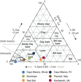
7 Appendix 209 7.1 Soil Texture Triangle . . . 209
7.2 Supplementary metaproteomic analysis information . . . 210
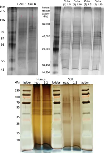
7.2.1 Sampling 1D SDS-PAGE gel . . . 210
7.2.2 Distribution of same-set hits . . . 210
7.2.3 Summary of pyrosequencing data . . . 211
7.2.4 Calculating the coefficient of determination . . . 211
7.2.5 Distribution of COGs between TS aTP and TSb TP . . . 212
7.2.6 Calculation of Spearman’s rank correlation coefficient . . . 212
7.3 Programmes and packages used . . . 213
List of Figures
1 Structure of chitin and its related, deacetylated product, chitosan . . . 1
2 Generalized structure of an exo- and endo- active site found in glycosyl hydrolases . . . 7
3 An incomplete summary of how chitin might be involved in the Nitrogen cycle . . . 11
4 Location of NERC Soil Biodiversity Field Experiment site . . . 24
5 An aerial view of the NERC Soil Biodiveristy Field Experiment . . . 25
6 Layout of NERC Soil Biodiversity Field Experiment site and plots . . . 26
7 A map of Cuba displaying its four largest cities . . . 27
8 Relative positions of Cayo Blanco and Hicacos Peninsula . . . 27
9 Locations of sample site on the island of Cayo Blanco . . . 28
10 Recent tourist developments on Cayo Blanco . . . 28
11 Calculating water content of soil by means of a desiccator . . . 31
12 Optimization of PCR conditions F19F2/F19R primers with eDNA . . . 38
13 Example sequence for a single clone provided by Macrogen . . . 40
14 Distribution of amplicon sequence lengths for 16S rDNA, GH18 and GH19 with chosen cut-offs . . . 43
15 An example of a sample mapping file for 16S rDNA data . . . 45
16 Schematic of soil metaexoproteome extraction . . . 51
17 Generalized construction of SCX-RP back column . . . 62
18 Retrieved HTML code from CAZy . . . 66
19 Relevant HTML highlighted and context menu displayed (Figure 19a), and code inserted into extractor patterns tab (Figure 19b). . . 66
20 Text liable to change with each iteration replaced with extractor tokens . . 67
21 A sub-extractor pattern window containing a sub-extractor pattern . . . . 68
22 Sample output from test pattern . . . 68
23 A screen capture of a script written in Interpreted Java. . . 69
24 Example datarecord on GenBank from which the sub-pattern extracts . . . 72
25 Example target page for scraping amino acid Fasta information . . . 73
26 Typical outputs from pre-Mafft alignment analysis . . . 77
27 Chitinolytic activity in Cayo Blanco and Sourhope samples in response to amendment . . . 81
28 Variation between biological duplicate microcosms and within the subsamples used for chitinase assay . . . 82
29 Degradation of surface and sub-surface shrimp carapaces in soil . . . 83
30 Chitinolytic activity of Test Soil 1, 6, and 12 months after last amendment with carapace . . . 84
32 Rarefaction curves indicating the observed number of OTUs at a genetic
distance of 3% for 16S samples . . . 86
33 Relative distribution of phyla detected from 16S rDNA pyrosequencing across soils . . . 87
34 2D unweighted and weighted discrete PCA plots for randomly sub-sampled bacterial communities . . . 88
35 Neighbour-joining phylogenetic tree for GH19 samples . . . 92
36 Distribution of sequences obtained for GH18 and GH19 pyrosequencing across soils and samples . . . 94
37 Rarefaction curves indicating the observed number of OTUs at a genetic distance of 3% for GH18 and GH19 samples . . . 95
38 Weighted and unweighted discrete PCA plots for GH18 and GH19 samples . 95 39 Relative abundances of phylogenetic groups at the genus level associated with GH18 chi genes . . . 98
40 Relative abundances of phylogenetic groups at the genus level associated with GH19 chi genes within samples . . . 99
41 Different coloured extracts from SH soil during the metaXP extraction . . . 104
42 Venn diagrams showing the distribution of unique OTUs across samples (irrespective of treatment) for GH18 and GH19 . . . 117
43 A representative chemical structure of a humic acid in soil. . . 134
44 A graph of the buffering capacity of the extraction solution . . . 137
45 Effect of pH on humic content of concentrated samples . . . 138
46 Effect of pH of sample on protein recovery from soil . . . 139
47 Modified Langmuir equation . . . 140
48 Repeat XP extraction from Cayo Blanco soil . . . 141
49 An equation for calculating adsorption equilibria using the Redlich-Peterson model . . . 149
50 Comparison of three UF membranes on extract quality . . . 152
51 Recovery of protein from gelatinous foulant cake layer resulting from mem-brane fouling . . . 153
52 Effect of allowing sample in stirred cell to ‘run dry’ . . . 153
53 Comparison of diffusate colour using 3 000 MWCO and 10 000 MWCO Vivaspin columns . . . 155
54 Molecular weight of HS in natural sediment samples . . . 158
55 Effect of temperature on protein recovery from metaexoproteome extraction 162 56 Co-extracted keratin visible on SDS-PAGE gels . . . 165
57 Effect of chitin on protein yield using metaexoproteme protocol . . . 167
58 Demonstrating minimal cell lysis during exoproteome extraction . . . 168
59 Demonstration of repeatability of method. Four extracts from the same soil run on the same SDS-PAGE gel . . . 170
61 Chitinolytic potential of soil extract post-extraction . . . 173
62 SDS-PAGE gel of Cayo Blanco exoproteome samples sent for mass spec-trometry analysis . . . 177
63 Distribution of quality-control passed peptide hits identified after a MAS-COT search . . . 177
64 A visual representation of the community present in a combined a-chitin and b-chitin amended Cayo Blanco exoproteome sample . . . 181
65 COGs associated with proteins identified in CB a+b sample . . . 183
66 Peptides recovered in exoproteome and their position within the chitinase ofNocardiopsis dassonvillei subsp. dassonvillei DSM 43111 . . . 184
67 A scatter plot of peptide spectral counts of soil proteins identified in the technical replicates . . . 187
68 Scatter plots of peptide spectral counts demonstrating intersample variabil-ity due to amendment and targeting the exoproteome . . . 188
69 Comparison of proteomic and genomic results for the Test Soil . . . 189
70 Relative distribution of COG categories between the exoproteome and total proteome ofb-chitin amended TS . . . 192
71 Comparison of relative abundance of COG functions across three samples: TSb XP, TSbTP, and TS aTP . . . 194
72 Graphical representation of soil texture for soils listed in Table 4 . . . 209
73 Illustration of the gel slices taken from Figure 62 . . . 210
74 Distribution of SSH in the CBa+bexoproteome . . . 210
75 Standard regression equations used by Excel . . . 211
76 Relative distribution of COG categories betweena-chitin andb-chitin amended TS . . . 212
List of Tables
1 List of reagents, media, and kits used in this thesis . . . 18
2 List of materials and equipment used in this thesis . . . 20
3 List of strains and media used in this thesis . . . 23
4 Summary of properties of soils used in this thesis . . . 29
5 PCR primers used in this thesis . . . 36
6 Reagents required to make 12% SDS-PAGE gel . . . 54
7 Solvent protocol for MS. . . 63
8 Chitinase primers previously used in environmental screens . . . 80
9 pH of various soils in water and with extract solution . . . 136
10 Comparison of UF membrane properties . . . 150
11 Summary of membranes used in other studies referenced in section 4.6 . . . 156
12 List of protein functions in addition to those listed in Figure 71 on page 194 (derived from COG assignments) that only occurred in a single sample . . 195
13 Chitinase and chitinase-like enzymes recovered from soil samples . . . 197
14 Abundance of three most common protein types in total proteome samples compared with their abundance in the exoproteome . . . 200
15 Summary of all pyrosequencing data. Coverage was calculated using the chao1 richness estimation metric. CB = Cayo Blanco, SH = Sourhope, TS = Test Soil, a= 1% a-chitin amended, b= 1% b-chitin amended, N = not amended . . . 211
Acknowledgements
Firstly I would like to thank my supervisor Professor Elizabeth Wellington, for
allowing me to tackle this PhD, use the latest techniques available, and for providing the opportunities for me to travel to Sydney, Cairns, Adelaide, Pisa, Florence, Havana, Varadero, Cayo Blanco, Santiago de Cuba, Keystone Colorado, New York, Knoxville, and yes, even Scotland.
I would like to thankRay Flintoft of Sourhope Farm, Kelso for permission to access
and sample from the formerNERC field site despite the lease on the land having expired
andMartin Krsekwho volunteered to drive the 8-hour-each-way journey so that I could
go sampling. Similarly, I would also like to thank Dr C Carlos Vallin Plou, the
Head of Development and Research Group in the Institute of Pharmacy and Food at
theUniversity of Havana, Cubafor collaborating and providing permits for sampling
Cayo Blanco in Cuba. For my amendments I wish to thankLoch Fyneseafood restaurant
in Kenilworth, Warwickshire and Clive Miller Fishmongers in Coventry Market for
providing the squid pen.
In the department I would to like to acknowledge and thank Cerith Harries, Anora
Johnson, and assorted members of the Prep’ Room and teaching labs. They were
always willing to provide otherwise unobtainable reagents and let me borrow all manner of weird and wonderful equipment that one doesn’t usually find in a microbiology lab. In
ad-dition, I would like to specially thankSue Sladefor providing complimentary sequencing
machine time during the initial development of the exoproteome extraction method.
In my lab I would like to thank Jane Green, my lab manager during my first 3 years,
whose breadth of knowledge of methods and the commercial aspects of science proved
invaluable. Leo Calvo-Bado, who took me under his wing when I was in my first year
and taught me most of the skills I came to rely on during my PhD. Victoria Hibberd
for all her help and for being a member of the ‘chitin bay’ helping to making it the noisiest
and most sociable bay in the lab. I’d like to give special thanks toHelena Wright, also
On the bioinformatics side, I want to thank Tanya Khera, Sam Mason and Brian
Oakleyfor their unfaltering patience, guidance, and problem-solving abilities during the
processing of my pyrosequencing data.
I would also like to thank Nathan Verberkmoes, Serena Doni, Cristina Macci,
and Grazia Masciandaro for allowing to collaborate with them and affording me the
opportunity to visit their labs to learn new skills. I would like to additionally thank to
Nathan, for finding time in his schedule at short notice for me to visit and for providing
complimentary mass spectrometry analysis of my soil samples.
I want to thank NERC for funding my studentship, SGM for providing travel money to
travel to Australia for ISME12, and Colorado State University who found funding
for me to attend the Inaugural International Workshop on Environmental Proteomics.
And finally I would like to thank Jim for his unwavering support through my PhD. He
Declaration
Abstract
Chitin is the most abundant nitrogen-containing polymer in nature, with >1⇥1010 tonnes
produced annually in terrestrial and marine habitats. Chitinolytic bacteria are able to degrade this recalcitrant substrate through a multiplicity of chitinases. A polyphasic approach was taken to studying these organisms within three diverse soil communities. Fluorometric assays employing 4-methylumbelliferyl-labelled chitinooligosaccharides were used to estimate basal soil chitinase activity as well as its chitinolytic potential in response
to a- andb-chitin amendment. A molecular approach was adopted to profile the bacterial
community and functional chi gene diversity within the soils. Finally, a method of
ex-ploring the metaexoproteome, enabling investigation of the dominant chitin degraders at a functional level, was developed and implemented. The metaexoproteome and metapro-teome, extracted with an existing method, were compared and used to infer the functional dominance of chitinolytic phyla.
The basal chitinase activity in all soils was found to be low, yet chitin amendment rapidly
induced chitinases in all soils although intersite differences were seen. b-chitin amendment
induced more chitinolytic activity in Cayo Blanco (CB) compared to Sourhope (SH). The Test Soil (TS), a site biannually amended with carapaces, retained higher chitinolytic potential many months after chitin had been consumed.
Next-generation pyrosequencing enabled >50% of the potential OTUs present in the soil
to be recovered. The 16S rRNA gene analysis of SH revealed dominant phyla to be
Proteo-bacteria, Actinobacteria, andAcidobacteria with little change between amendments. The
TS was dominated by the same phyla but saw a proliferation ofActinobacteria with chitin
amendment. CB experienced the inverse response to the Test Soil, initially dominated by
Actinobacteria only forProteobacteria to dominate with amendment. Firmicutes were also
prevalent with b-chitin amendment.
Functionalchi gene analysis foundStreptomyces-like GH19chi genes to dominate in both
SH and CB. A rare Actinomycete Planobispora dominated chitin-amended TS. This
analysis or the metaproteome; further analysis is required to confirm its presence. Strep-tomyces-like GH18chi genes only dominated CB with amendment and were absent in SH. A large number of OTUs were identified as uncultured organisms suggesting a large pool
of uncharacterized GH18chi genes.
Metaproteomics is the functional analysis of complex communities at a given point in time. The heterogeneity of soil, associated microbial communities, and presence of interfering compounds make the extraction of protein from soil a technical challenge. Chitinases are extracellular and so the metaexoproteome was targeted after development of a novel method that biased extraction towards exoproteins. The protocol successfully extracted the largest soil metaproteome to date. Actinobacterial chitinases were found to be functionally
dominant in the Test Soil, especially in response to b-chitin amendment.
Glossary
Abbreviations and Definitions
⇠ Approximately
4-MU 4-methylumbelliferyl
A Amperes
aa Amino acid
Abbr. Abbreviation
AcN Acetonitrile
APS Ammonium persulfate
ASCII American Standard Code for Information Interchange, a character-encoding
scheme originally based on the English alphabet
ASTM ASTM International, formerly known as the American Society for Testing
and Materials, an international standards organization
b-ME b-mercaptoethanol, a compound used to reduce disulfide bonds in
pro-teins, disrupting tertiary and quaternary structure
BLAST Basic Local Alignment Search Tool, an algorithm used in bioinformatics
bp Base pairs (nucleotide sequence)
BSA Bovine serum albumin, a large globular protein (approx. 66 kDa) dervied
from cow blood plasma
CA Cellulose acetate
CB Cayo Blanco soil. When followed bya,b, or N (e.g. CBa) it denotes that
the soil was amended with eithera-chitin, b-chitin, or left unamended.
CBPs Chitin-binding proteins, proteins containing chitin-binding domains and
lacking hydrolytic activity that mediate the interaction between chitinases and chitin
CDS Coding sequences
CE Cellulose ester
ChEBI Chemical Entities of Biological Interest, a dictionary of molecular entities provided by the EBI that focuses on ‘small’ chemical compounds
COG Clusters of Orthologous Groups of proteins
CSS Cascading style sheet(s), a language used to describe the presentation
semantics, the look and formatting, of a document written in a markup language, such as HTML [http://www.w3.org/]
CTAB Cetyl Trimethyl Ammonium Bromide
DDA Data dependent acquisitions
Dialysate That which is being dialysed against
Diffusate That which diffuses from the sample, across the semi-permeable
mem-brane, into the dialysate
DIN Deutsches Institut für Normung, the German Institute for
Standardiza-tion
DNA Deoxyribonucleic Acid
dNTPs Deoxyribonucleotide mixture, containg equal amounts of deoxyadenosine
monophosphate (aATP), thymidine monophosphate (dTMP), deoxyguanosine monophosphate (dGMP), and deoxycytidine monophosphate (dCMP)
EC Enzyme Classification
eDNA Environmental DNA, metagenomic DNA extracted from natural soil samples
EDTA Diaminoethanetetra-acetic acid disodium salt
EMBL-EBI European Bioinformatics Institute, a centre for bioinformatic research and
services based at the Wellcome Trust Genome Campus, Hinxton, UK. The EBI is part of the European Molecular Biology Laboratory and hosts the nucleotide sequence database EMBL-Bank and UniProt, the combined Swiss-Prot–TrEMBL protein sequence database
ESI Electron spray ionisation
Exoprotein Protein present in the extracellular milieu (Desvauxet al., 2009)
Export Active transport from the cytoplasm (Desvaux et al., 2009)
Fr. Formerly
GB GenBank, an open-access annotated sequence database maintained by
NCBI as part of the INSDC, collecting all publicly available nucleotide sequences and their protein translations
GFP [glu1]-fibrinopeptide B, a peptide derived from amino acid residues 1-14
GH Glycoside hydrolases, enzymes that hydrolyse glycosidic linkages in sub-strates to release smaller sugars
(GlcNAc) N-acetyl-b-D-glucosamine
(GlcNAc)2 N,N�-diacetylchitobiose
(GlcNAc)3 N,N�,N��-triacetylchitotriose
GUI Graphical User Interface
HA Humic acids
HPLC High-performance liquid chromatography
HS Humic Substances, a fraction of soil organic matter containing a complex
mixture of carboxyl and phenolic acids formed as a by-product of micro-bial degradation of plant material. Can be sub-divided into fulvic acids (FA), humic acids (HA), and humins
HTML Hypertext markup language, the main markup language for web pages
[http://www.w3.org/]
HTTP Hypertext transfer protocol, an application protocol for distributed
col-laborative hypermedia information systems. The foundation of data com-munication for the World Wide Web
IANA Internet Assigned Numbers Authority, the entity that oversees, amongst
other areas, Internet Protocol-related symbols and numbers
ICT Intracellular trafficking
InChIKey IUPAC International Chemical Identifier Key, a textual identifier for
chem-ical substances, designed to provide a standard and human-readable way to encode molecular information and to facilitate the searching of data-bases on the Internet
INSDC International Nucleotide Sequence Database Collaboration, a
collabora-tion between DNA Data Bank of Japan, GenBank (USA) and EMBL (European Molecular Biology Laboratory, Germany) to collect and dis-seminate databases containing DNA and RNA sequences
in silicio Literally, in silicon, performed on computer or by computer simulation.
Equivalent to the more prevalent and erroneously derived, in silico
IPTG Isopropylb-D-1-thiogalactopyranoside
ISE-CNR Istituto per lo Studio degli Ecosistemi, Consiglio Nazionale delle
ISO International Organization for Standardization
iTOL Interactive Tree of Life, an online tool for the display and manipulation
of phylogenetic trees
IUPAC International Union of Pure and Applied Chemistry
JGI Joint Genome Institute, a centre for bioenergy and environmental research
LB Lysogeny Broth (Bertani, 2004)
LC Liquid chromatography
LFH Laminar Flow Hood, an enclosed bench where air is drawn through a
HEPA (High-Efficiency Particulate Air) filter and blown in a laminar flow
towards the user
LTQ Linear Trap Quadrupole, a technique that radially confines ions using a
set of linear quadrupole rods and axially confines using static electrical potential on-end electrodes
Mafft Multiple Alignment using Fast Fourier Transform
Maldi Matrix-Assisted Laser Desorption/Ionization
MetaXP Metaexoproteome, the subset of proteins present in the extracellular
mi-lieu within an environmental sample including protein lysed cells and protein adhered to the soil matrix
Mimarks Minimum Information about a marker gene sequence (Yilmazet al., 2011)
MS Mass spectrometry
Muscle Multiple Sequence Comparison by Log-Expectation, a multiple sequence
alignment program for amino acid and nucleotide sequences
NAST Nearest Alignment Space Termination, an algorithm for creating multiple
sequence alignments
NCBI National Center for Biotechnology Information, part of the United States
National Library of Medicine (NLM), a branch of the National Institutes of Health (NIH). NCBI provides, via Entrez, the Global Query Cross-Database Search System, genome sequencing data in GenBank, an index of biomedical research articles in PubMed, and other information relevant to biotechnology
NNI Nearest Neighbour Interchanges, a tree topology strategy that reroots
internal branches or subtrees to obtain new topographical configurations until a maximum-likelihood is achieved
№ Number
OTU Operational Taxonomic Unit, similar taxa grouped for phylogenetic
ana-lysis
Page Polyacrylamide Gel Electrophoresis, as in SDS-Page
PBS Phosphate buffered saline
PCA Principle Component Analysis, a method of multivariate statistical
ana-lysis that aims to reduce the dimensionality of a data set whilst retaining representation of variation within the dataset
PCR Polymerase chain reaction, a molecular biology technique for amplifing
DNA
PDI Polydispersity Index, a measure of the distribution of molecular mass in
a given polymer sample
per. comms. Personal communications, unpublished
PES Polyethersulfone, a hyrophobic, low-protein-binding, non-crystalline,
heat-resistant engineering plastic
PKL(s) Peak list file(s), a QToF output file containing peak list data
PriA Phosphoribosyl isomerase A, a novel bifunctional enzyme from
Strepto-myces coelicolor involved in both histidine and tryptophan biosynthesis
PTM Post-translational modification
PVDF Polyvinylidene fluoride
PVP Polyvinylpyrrolidone
PVPP Polyvinylpolypyrrolidone, a highly cross-linked water-insoluble
modifica-tion of polyvinylpyrrolidone
pyNAST Python Nearest Alignment Space Termination, a Python
reimplementa-tion of the NAST sequence aligner
Qiime Quantitative Insights Into Microbial Ecology, an open source software
package for comparison and analysis of microbial communities based on
high-throughput amplicon sequencing data, pronounced [tSaIm]
QToF Quadrupole mass filtered time-of-flight
RAM Random access memory, a buffer for temporary storage of information
during calculations in computers
Retentate That which remains within the semi-permeable membrane envelope after dialysis
rpm Revolutions per minute
rRNA Ribosomal ribonucleic acid
RT Room temperature (approximately 20°C)
SCX-RP Strong Cation Exchange - Reverse Phase
SDS Sodium Dodecyl Sulfate
sdw Sterile distilled water
Secreted protein Protein actively transported via a secretion system (Desvauxet al., 2009)
Secretion Active transport from the interior to the exterior of a cell (Desvauxet al.,
2009)
Secretome Components of the translocation systems and their substrates (Desvaux
et al., 2009)
SH Sourhope soil. When followed by a,b, or N (e.g. SH b) it denotes that
the soil was amended with eithera-chitin, b-chitin, or left unamended.
sv Standard deviation
Smiles Simplified molecular-input line-entry specification, a line notation for
de-scribing the structure of chemical molecules using short ASCII strings
Sonicated Disrupted or resuspended through the use of ultrasonic vibrations
sp. / spp. Species / Species pluralis
SPR Subtree-Pruning-Regrafting, a tree topology strategy that removes
sub-trees and reinserts them onto other branches to form new sub-trees until maximum-likelihood is achieved
SSH Same set hit, where identified peptides have an equal probablility of
be-longing to two or more different proteins or organisms
STD Standard
TBE Tris/Borate/EDTA buffer
TCA Trichloroacetic acid
TE Tris/EDTA buffer
TEMED Tetramethylethylenediamine
TGX Tris-Glycine eXtended gels, a type of precast SDS-Page gel which employs
TOC Total orgaic carbon
TON Total organic nitrogen
TP Total Proteome, all proteins in an environment
Tris Tris(hydroxymethyl)aminomethane
TS Test Soil soil. When followed by a, b, or N (e.g. TS N) it denotes that
the soil was amended with eithera-chitin, b-chitin, or left unamended.
TSV Tab-separated variables, a delimiter-separated values format allowing a
database table to be formatted in simple text by separating each field value of a record from the next using a tab stop character
UB Urea dilution buffer
UF Ultrafiltration, filtration through membranes with MWCO less than
⇠1 000 000 or pores smaller than 0.1mm
UHMW Ultra-High Molecular Weight
UNIX Originally, Unics (UNIpleXed Information and Computing System), a
multi-tasking, multi-user computer operating system controlled by The Open Group
UPGMA Unweighted Pair Group Method with Arithmetic Mean, an agglomerative
clustering method that assumes a constant rate of evolution
URL Uniform/Universal resource locator, a specific character string that
con-stitutes a reference to an Internet resource
UV Ultraviolet, 10–400 nm electromagnetic radiation
Vortex To mix vigorously with a vortex mixer
w/v weight (g) / volume (ml)
w/w weight (g) / weight (g)
¯
x Arithimetic mean
X-gal 5-bromo-4-chloro-indolyl-b-D-galactopyranoside, or galactose linked to a
substituted indole
XP Exoproteome, the subset of proteins present in the extracellular milieu
(Desvaux et al., 2009)
Units
° � � ° (degrees),1/360of a full rotation or 17.45 mrad
� (arcminutes),1/60of a degree or 290.9 mrad
� (arcseconds),1/60of an arcminute or 4.848 mrad
°C Degree(s) Celsius
Da Daltons, the unified atomic mass unit defined as 1/12th of the rest mass
of an unbound neutral atom of 12C in its nuclear and electronic ground
state
dS m 1 deciSiemens / metre, the SI derived unit of electric conductance and
elec-tric admittance
g Gram(s)
h Hour(s)
l Litre(s), a unit of volume equal to 1 dm3
lm 2h 1MPa 1 Normalized water flux, a unit of achievable membrane flux per unit
trans-membrane pressure
M Molar concentration
m Metre(s), (e.g. km, kilometre, cm, centimetre, mm, millimetre, mm,
mi-crometre)
m/z Mass to charge ratio
min Minute(s)
MWCO Molecular weight cut-off
N Normality, defined as the molar concentration, ci divided by an
equival-ence factor,feq
NMWL Nominal molecular weight limit
OD600 Optical Density at 600 nanometres
Pa Pascal(s), the SI derived unit of pressure defined as one newton per square
metre (e.g. MPa, megapascals, kPa, kilopascals)
pKa Logarithmic acid dissociation constant, equal to−log10Ka, a quantitative
measure of the strength of an acid in solution
ppm Parts per million
S Svedberg, a unit of sedimentation coefficient. e.g. 16S, 18S
V Volt(s), the SI derived unit for electric potential, defined as the difference in electric potential across a wire when an electric current of one ampere dissipates one watt of power
1 Introduction
1.1 Chitin
1.1.1 Structure
Chitin is the most abundant nitrogen-containing polymer and, after cellulose, is the second
most abundant polymer in nature, with >1⇥1010 tonnes produced annually in terrestrial
and marine habitats (Gooday, 1990; Chateret al., 2010). It has a similar structure to
cellu-lose but with the C-2 amino polysaccharide having acetamide groups rather than hydroxyl
groups. It is often described as “a simple polymer of b(1,4)-linked N-actylglucosamine
residues” (Sinnott, 1990), but in nature its presentation is more complicated. It is better
de-scribed as a versatile, linear unbranched fibrous biopolymer ofb-1,4-linkedN
-acetylglucosa-mine and glucosa-acetylglucosa-mine. Pure chitin is completely acetylated; when deacetylated, its de-rivative is known as chitosan (Figure 1). There is no set definition in the literature for
the acetylation/deacetylation cut-off between chitin and chitosan but it is generally
accep-ted that chitin must be at least 30–40% acetylaaccep-ted, though natural samples are typically 85–95% acetylated (Ravi Kuma, 2000; Kurita, 2001).
Figure 1: Structure of chitin and its related, deacetylated product, chitosan
Chitin polymers can be arranged into three naturally occurring allomorphs, in order of
abundance in the environment they are: a-chitin, b-chitin, andg-chitin. a-chitin is a dense
and hard structure with anti-parallel polymer chains. In aquatic systems it is commonly found in the cuticles of crustacea such as barnacles, crab, lobster, crayfish and shrimp
(Rhaziet al., 2000); in terrestrial systems the common sources include fungal cell walls and
protistan and invertebrate exoskeletons (Gooday, 1990), and there are other sources such as
in pogonophoran and vestimentiferan worms, the monocrystalline spines of the marine
diatomThalassiosira fluviatilis,and in squid pen, or gladii, the hard internal feather-shaped
vestige of the ancestral mollusc shell formed by secretions from the shell sac in which it
resides (Chandumpaiet al., 2004; Fanet al., 2009; Muzzarelli, 2011). It has parallel chains
which pack less densely, allowing for greater hydration that produces a more flexible, softer structure. Chitosan can be found in nature in some fungi, including Mucorales and a few
Basidiomycetes (Mario et al., 2008), the majority however is produced industrially (Alves
and Mano, 2008). The preference of chitosan in industry is in part due to its solubility, which increases with the degree of deacetylation within the molecule (Ravi Kuma, 2000).
g-chitin is a hybrid of a-chitin and b-chitin, where two chains run parallel with a third
lying anti-parallel (Jang et al., 2004).
1.1.2 Presentation
Environmental chitin, with the exception of diatom spines, is always presented in conjunc-tion with other substances such as glucans, lipids or proteins (Gooday, 1990; Schrempf, 2001). As part of an expanded nitrogen cycle, figure 3 on page 11 shows a typical example of a complex presentation of chitin in nature, that of the hierarchical structure within the exoskeleton of a crab. Chitin fibrils (3 nm Ø) are clustered and wrapped with proteins to form fibres (60 nm Ø), which are assembled into bundles that form horizontal planes, and are superimposed in a helicoid stack to create a twisted Bouligand structure. This forms the endocuticle and denser exocuticle which are then coated with a waxy waterproof epicuticle. In fungi, chitin fibrils fold to form anti-parallel nascent chitin chains (a-chitin structure), these then form inter-chain hydrogen bonds to create strong fibrous microfib-rils. The microfibrils form a lattice which is then covalently bonded the major constituents of the fungal cell wall, glucans and mannans, to provide additional structural support
(Bulawa, 1993; Lenardon et al., 2010).
utilizes substrate analogues such as colloidal chitin, and small chitinooligosaccharides. This can be misleading as measures of hydrolase activity against such substrates can be up to 1 000-fold greater than that observed with the natural substrate (Keyhani and Roseman, 1999). For this reason, organisms identified as having highly chitinolytic enzymes in the literature may not dominate under natural conditions where there is a diverse presentation of chitin and environmental conditions are sub-optimal.
1.1.3 Uses of chitin and chitosan
Chitin and chitosan are biocompatible, biodegradable, nontoxic, anti-microbial and
hy-drating agents (Jayakumar et al., 2010, 2011) that are readily available from inexpensive
biological material obtained from invertebrate skeletons, fungal cell walls, and squid pen
(Merzendorfer, 2003; Sagheer et al., 2009; Jayakumar et al., 2010, 2011). The polymer
structure lends itself to modification allowing the creation of many forms. The novel prop-erties of some of these can be exploited for medical purposes: hydrogels; in wound dressings
to coagulate blood and prevent infection (Tamuraet al., 2011); membranes, as
antimicro-bial layers in food packaging (Abdou et al., 2008); sponges, as scaffolds for chondrocyte
grow in cartilage replacement (Suzuki et al., 2008); beads, for the delivery of anti-cancer
drugs (Yusof et al., 2001); nanoparticles, for the delivery of drugs (Huang et al., 2009);
nanofibres, for anti-microbial wound dressings that promote healing (Fan et al., 2009;
Homayoni et al., 2009; Cai et al., 2010); and nano-fibrous scaffolds, onto which human
mesenchymal stem cells can adhere and proliferate (Shalumon et al., 2009).
Crude chitin and chitosan also have myriad uses in industry where their polycationic properties are exploited for recovering suspended solids, proteins, lipids and other organic compounds during processing (Bough, 1975; Fernández and Fox, 1997; No and Meyers,
1989; Shahidiet al., 1999), in the removal of metal ions from industrial effluent (Ravi Kuma,
2000), and in the purification of water by flocculation and aggregation of organic material
(Zemmouriet al., 2012).
The Norway lobster, Nephrops norvegicus, has had global landings of over 60 000 tonnes
(In-stitute of Marine Research), 2011). The inedible fraction accounts for 60% of the organism
by mass. The status quo for disposal is burial in landfill, this is costly, anti-social due to
the smell, and non-productive as many chitinolytic organisms are aerobes (Healey et al.,
2003). The bioprocessing of shell waste has been investigated by many as an alternative to
disposal at landfill but there are few reports of industrial scale processing (Healey et al.,
2003; Oh et al., 2007; Jo et al., 2008; Wang et al., 2008b; Xu et al., 2008; Wang et al.,
2009a; Wang, 2012) despite the introduction of EU Council Directive 1999/31/EC requir-ing a 75% reduction in biodegradable municipal waste over 1995 levels by 2020 (Healey
et al., 2003). It is unclear whether this is a result of the biology not yet being refined,
or resistance to move away from the status quo. Products from chitin need not be for
high-technology applications. Chitinooligosaccharides are easily recovered and have been
shown to have anti-tumor, anti-oxidant, and anti-microbial actions in vitro (Lian et al.,
2007; Wang et al., 2008a). The requirements of purity and safety of these compounds, if
they go on to be used medically, makes bioprocessing a viable option rather than using
chemical means (Chandumpaiet al., 2004; Chaussard and Domard, 2004).
1.1.4 Degradation of chitin
The crystalline networks of chitin, stabilized by hydrogen bonding and van der Waals’
interactions, can have a molecular weight up to several MDa (Chateret al., 2010). In this
state chitin is unable to enter cells. Induction of the chitinolytic system and the uptake
of chitin must therefore be mediated by breakdown products of chitin. (GlcNAc)2 is the
smallest substrate that induces chitinases and appears to be the main inducer (Tsujibo
et al., 1999; Miyashitaet al., 2000).
The main route for degradation of chitin in soil is via extracellular chitinases (EC 3.2.1.14)
that hydrolyse the bonds between GlcNAc residues releasing oligomeric (GlcNAc)n, dimeric
(GlcNAc)2or monomeric chitooligosaccharide (GlcNAc) products (Seidlet al., 2005).
Chit-inases have been isolated from many sources including plants, fungi, yeast, bacteria, insects
and vertebrates (Bhattacharya et al., 2007; Karlsson and Stenlid, 2009). The role of
are responsible for morphogenesis, e.g. hyphal tip extension or daughter cell separation; in plants, which do not contain chitin, they are pathogenesis-related proteins that are in-duced by stress or pathogenic attack (Kasprezewska, 2003). In bacteria, chitinases are used to degrade chitin-containing substrates into carbon and nitrogen sources; they may also
have an antifungal role in some species (Bormann et al., 1999; Wang et al., 2002; Kawase
et al., 2006). A second route involves the deacetylation of chitin to chitosan by chitin deacetylase, hydrolysis of chitosan by chitosanase (EC 3.2.1.99), and a final hydrolysis step by glucosaminidase to glucosamine (Gooday, 1990). The distinction between chitinases and chitosanases is not sharp. Both classes of enzyme have an ability to degrade chitin and
chitosan with different degrees of deacetylation, depending on the enzyme, but chitinases
will preferentially hydrolyseN-acetylated regions and chitosanasesN-deacetylated regions
(Somashekar and Joseph, 1996). Chitosanases will be not investigated in this thesis.
1.1.4.1 Two families of chitinases Based on amino acid sequence similarity within the catalytic domain as well as structural and mechanistic characteristics, chitinolytic enzymes are grouped into family 18 and 19 glycosyl hydrolases (GH18 and GH19) (Henrissat, 1991; Henrissat and Bairoch, 1993; Davies and Henrissat, 1995; Henrissat and Bairoch, 1996).
Glycosyl hydrolase family 18 GH18 chitinases account for the majority of microbial chitinases (Karlsson and Stenlid, 2009). Several systems have been employed by bacteri-ologists, mycbacteri-ologists, and phytologists to categorize chitinases found in their respective fields. Svitil and Kirchman (1998) grouped bacterial chitinases into groups I, II, III and IV based on conservation of amino acids within the catalytic domains, with an additional
group V for chitinases that did not fall in to I-IV. After a study of partialchi genes from
aquatic habitats, LeCleir et al. (2004) further subdivided group I into A, B, C, and D,
based on conserved residues found within diverse proteobacteria. A second method of
cat-egorizing GH18 bacterial chitinases was employed by Suzuki et al. (1999) who separated
similar to that of group A but lack the cysteine-rich domain at the N-terminus; and group
C has no homology with either group A or B (Shinshiet al., 1990; Watanabeet al., 1993).
Fungal chitinases have been categorized into three groups, A, B, and C by Seidl et al.
(2005). Plant GH18 chitinases are traditionally divided into classes III and V based on their amino acid sequence and structure, with classes I, II, IV, VI, and VII being GH19 chitinases (Passarinho and de Vries, 2002; Kasprezewska, 2003).
With the growing number of chitinase sequences available from genome projects,
Karls-son and Stenlid (2009) more finely divided the GH18 groups, as set out by Suzuki et al.
(1999). Group A was split into subgroupI containing bacterial and viral sequences,II–VIII
containing only bacterial sequences, A-II–A-VI and C-I–C-II containing only fungal
se-quences, Class V containing plant chitinases, and three additional groups for Archaea,
Caenorhabditis elegans, and Drosophila melanogaster. Group B was split into subgroups
IVa–IVb containing bacterial chitinases, B-I–B-V containing fungal chitinases, and Class
III plant chitinases. Group C retained its function as a ‘not A or B’ group
contain-ing sequences with no obvious pattern of domain structure. Bacterial, fungal, and plant
chitinases were represented in both groups A and B suggesting that the differentiation
of these clusters preceded the appearance of eukaryotes. Bacterial and fungal chitinases did not form monophyletic groups, suggesting that previous methods of categorizing these chitinases were based on incomplete coverage of available diversity.
Glycosyl hydrolase family 19 GH19 chitinases differ from GH18 in their amino acid sequence, three-dimensional structure and in the molecular mechanism of their catalytic
reactions (Kawaseet al., 2004). Until the discovery of chitinase C-1 inStreptomyces griseus
HUT 6037 (Ohnoet al., 1996), GH19 chitinases were thought to only occur in higher plants;
since then they have been discovered in many other bacteria including, Burkholderia
gla-dioli, Vibrio cholerae, Haemophilus influenza and Pseudomonas aeruginosa (Itoh et al.,
2002), and a few other organisms including nematodes (The C. elegans Sequencing
Con-sortium, 1998). GH19 chitinases can be separated into five clusters. It is interesting to note that all actinomycete GH19 chitinases are grouped with plant class IV chitinases in cluster
chit-Exochitinase Endochitinase
Figure 2: Generalized structure of an exo- and endo- active site found in glycosyl hydro-lases. Adapted from Davies and Henrissat (1995)
inase, CHB101, belong to cluster III which is most distantly related to the rest of GH19 chitinases. This suggests that these chitinases are a recent acquisition by the
actinomy-cetes from higher plants (Kawase et al., 2004). However, a pair-wise distance comparison
betweenchiF and housekeeping genes demonstrated that the acquisition is an ancient one
that has remained highly conserved, raising questions of their purpose (Ul-Hassan, 2006).
GH19 chitinases still remain poorly understood (Ubhayasekera, 2011). In the literature
8 crystal structures exist, only 2 of which are from bacterial chitinases: Streptomyces
coelicolor A3(2) andS. griseus HUT 6037 (Kezukaet al., 2005; Hoell et al., 2006).
1.1.4.2 Two mechanisms, exo- and endo- acting Chitinases from both families are fur-ther classified by their enzymatic method of action into endo- and exo-chitinases based
on their structure and the method by which they act upon (GlcNAc)n (Figure 2).
1.1.5 Multiplicity in the chitinolytic system
Organisms that degrade chitin often have complex co-regulated chitinolytic systems, ex-hibiting a high multiplicity of chitinases and chitosanases that work synergistically with
accessory chitin-binding proteins (CBPs), this enables them to efficiently degrade
recal-citrant crystalline environmental sources of chitin for carbon and nitrogen. Streptomyces
coelicolor A3(2) has 11 GH18, and 2 GH19 chitinases (Kolbeet al., 1998; Saitoet al., 1999,
2000; Svergunet al., 2000; Saitoet al., 2001; Schrempf, 2001; Bentleyet al., 2002; Kawase
et al., 2006). Other bacteria with high multiplicity includePhotobacterium sp. andVibrio angustum with 8 chitinases each (Karlsson and Stenlid, 2009).
Complex chitinolytic systems are not necessarily required for highly chitinolytic organisms.
A demonstration of synergy in a chitinolytic system can be seen in S. coelicolor A3(2)
and Aeromonas sp. O-7. Both organisms have individual chitinases that exhibit higher
activity against certain presentations of chitin. In the case of Aeromonas sp. O-7, it has
4 chitinases, 3 of which exhibit their activity against powdered, glycol, or colloidal chitin, and one chitinase which is cold adapted. In combination, chitinolytic activity is 2-fold higher than the combined activity of the individual enzymes against the same substrate (Orikoshi et al., 2005; Kawaseet al., 2006).
Chitin binding protein (CBPs) are small extracellular proteins that contain chitin-binding domains. They are thought to mediate interactions between the chitinolytic organism and
various chitin-containing substrates such as a-chitin in fungal cell walls and crab shell,
b-chitin, chitosan, or cellulose (Schnellmann et al., 1994; Saito et al., 2001; Schrempf,
2001). The category contains proteins with different functions and the precise
mechan-isms by which CPBs improve the efficiency of chitin degradation are not known. The first
chitin-binding protein discovered was CHB1 from Streptomyces olivaceoviridis
(Schnell-mann et al., 1994). Later, a related a-chitin-binding protein from Streptomyces reticuli, CHB2, was characterized microscopically and immunologically, and found to act like a
glue, mediating the contact between the fungal and the Streptomyces hyphae (Kolbeet al.,
1998). In Streptomyces tendae Tü901 an anti-fungal protein (AFP1) was found to target
potentially by allowing chitin synthetase inhibitor nikkomycin to enter (Bormann et al., 1999). CHB1 and CHB2 are responsible for bacterial proliferation and retarded develop-ment of fungi (Schrempf, 2001; Siemieniewicz and Schrempf, 2007). Although AFP1 also has anti-fungal activity, it does not show sequence similarity with CHB1 or CHB2 and is of smaller size.
When studying the chitinolytic system ofSerratia marcescens2170, a 21 kDa protein
lack-ing chitinase activity was identified in the culture supernatant, this was termed
‘chitin-binding protein 21’ (CBP21) (Watanabeet al., 1997). Further analysis of CBP21 found it
to share 45% amino acid identity with CHB1 from Streptomyces olivaceoviridis, although
unlike CHB1, which specifically binds a-chitin, CBP21 had its highest activity against b
-chitin (Suzukiet al., 1998). The degradation ofb-chitin is biphasic, with a fast phase where
easily accessible amorphous substrate is degraded followed by a slower second phase where
recalcitrant crystalline regions are degraded. CBP21 from Serratia marcescens has been
shown to aid chitinases during the second phase by interfering with the crystalline structure
ofb-chitin in a non-specific way (Vaaje-Kolstad, 2005b). The structure of CBP21 revealed
a pyramidal molecule with conserved tryptophan residues, previously hypothesized to be involved in chitin binding, on the interior of the molecule. A conserved flat surface of solvent-exposed polar side chains were instead found to mediate binding (Vaaje-Kolstad, 2005a). CBP21 is classified as a class 33 ‘carbohydrate-binding module’ (CBM33). Further work revealed CBP21 to be a hydrolytic, metal ion dependent ‘chitin oxidohydrolase’,
sim-ilar to family 61 glycosyl hydrolases in fungi (Vaaje-Kolstadet al., 2010). The mechanism
of action for both CBM33 and GH61 is unknown.
1.1.6 Chitin as a nitrogen source
Being an involved and energy-consuming process the chitinolytic system is totally repressed
in the presence of more readily available carbon sources such as glucose (Miyashitaet al.,
1991, 2000). As well as chitinases, the chitin oligomers can be hydrolysed by related
enzymes such as chitodextrinases and b-N-acetylglucosaminidases to produce monomeric
GlcNAc and (GlcNAc)2. The molecules can be taken up by the cell (Chapin III et al.,
Various catabolic extracellular enzymes degrade the macrostructure of environmental chitin. The depolymerization and degradation is mediated by exo- and endo-chitinases and CBPs
that act upon the (GlcNAc)n, along with chitodextrinases andb-N-acetylglucosaminidases,
to eventually produce monomeric and dimeric chitinooligosaccharides (GlcNAc and
(Glc-NAc)2) which can be transported into the cell. GlcNAc is taken up by the
phosphoenolpyr-uvate:glucose phosphotransferase system, eventually yielding NH3, acetate, and
fructose-6-phosphate (F-6-P), for metabolism. (GlcNAc)2 is acted upon by N,N�-diacetylchitobiose
phosphorylases converting it to N-acetylglucosamine-1-phosphate which enters the same
metabolic pathway as GlcNAc. The exoenzyme-driven demineralization and depolymeriz-ation of N-containing substrates is the rate-limiting step in the generdepolymeriz-ation of bioavailable
ON ON ɅN &QJDVUJDMF &OEPDVUJDMF &YPDVUJDMF Organic N
Richardson, David J. and Watmough, Nicholas J. Current Opinion in Chemical Biology, 1999, 3:207-219
NO2⁻(+3)
NH2OH (-1) N2O (+1)
NO (+2)
NO3⁻(+5)
N2(O)
NH2+(-3) ANAMOX nitrification fixation denitrification denitrification denitrification assimilation dissimilation detoxification nitrification heterotrophic nitrification NH3 NH4+
Plants Monomers Microbes (GlcNAc)2 (GlcNAc) GlcNAc-6-P GlcNH2-6-P
GlcNAc-1-P F-6-P Metabolism respiration dissimilation assimilation nitrification Nitrog en C
ycle
Bacteria
Chitin (GlcNAc)n
Figure 3: An incomplete summary of how chitin might be involved in the Nitrogen cycle, created from figures and information from Richardson and Watmough (1999);
Schimel and Bennett (2004); Chen et al. (2008); Jackson et al. (2008); Jung
et al. (2008)
1.2 Molecular approach to studying microbial diversity
It has long been known that the vast majority (>99.9%) of soil bacteria are not culturable
using standard cultivation techniques (Handelsman et al., 1998; Vogel et al., 2009). The
percentage of phyla recovered in classical taxonomic studies varies greatly reflecting either
type differences between soils or biases in the techniques adopted (Bakken, 1997). The
historical difficulties associated with bacterial taxonomy in soil made the field a small
(Dini-Andreoteet al., 2012).
The term ‘metagenomics’ was coined by Handelsmanet al.(1998) to describe the approach
of treating the combined DNA of microorganisms in soil as analogous to that of a genome from a single species. True metagenomics does not include studies that employ PCR, either using primers specifically towards genes that are representative of phylogeny such as 16S rRNA, or random primers. Instead it is an umbrella term for techniques that sequence DNA obtained directly from the environment such as environmental DNA (eDNA) libraries
and whole genome shotgun sequencing (Riesenfeld et al., 2004).
Twenty years ago Torsviket al.(1990) used DNA-DNA reassociation techniques to estimate
that 1 g soil contained ⇠1.5⇥1010 bacteria containing ⇠7 000–10 000 different genomes.
Such a large diversity was surprising at the time, and remained the upper estimate for
soil microbial diversity until the turn of the millennium (Handelsman et al., 1998) when
modern sequencing techniques and statistical modelling revealed the number to be in excess
of 1⇥106 genomes per 1 g of soil (Curtis and Sloan, 2005; Gans, 2005). Historically, the
bacterial genomes represented in repositories such as NCBI, have been selected based on
their applicability to industry or medicine (Dini-Andreote et al., 2012). The first major
contribution of environmental information was 1 Gbp of non-redundant sequences from
microorganisms collected from the Sargasso Sea near Bermuda (Venter et al., 2004). To
date only a single soil metagenome has been made available (Tringeet al., 2005) but other
large projects such as the Terragenome (Vogelet al., 2009) are under way.
As sequencing technologies continue to improve, data acquisition becomes increasingly easy
and more affordable producing a glut of phylogenetic data. A situation is developing where
the data cannot be manually curated and previous molecular biology methods for compu-tationally processing data are no longer possible. A result of this is the increased number of ‘uncultured bacterium’ hits when trying to search for alignments of sequences. Slightly
more useful is the ever increasing list of ‘Candidatus’ putative taxa1. These are organisms,
discovered by molecular approaches, that cannot be sustained in culture for more than a few serial passages, but for which more information than a mere sequence is available (Murray and Schleifer, 1994). Some have questioned the purpose of deeply sequencing
small quantities of soil to access the rare biosphere given the lack of understanding of the relationships between these unculturable organisms and inability to handle such data in a meaningful way (Baveye, 2009).
A different approach is to pose a question and target sequencing effort towards a gene
that can answer it. The 16S rRNA gene is the ‘gold standard’ for estimating microbial diversity. It is universally present and contains both highly conserved regions suitable for
primer design and variable regions that allow for the discrimination of different microbial
taxa. Because of the near ubiquity of this approach, large curated datasets exist that allow
the comparison of obtained data across myriad habitats and samples (Vos et al., 2012).
Current high-throughput sequencing methods do not provide reads long enough to span the entire 16S rRNA gene, so shorter hypervariable regions are chosen to be representative of the whole. There is healthy debate in the literature over the the relative merits of
various regions (Kimet al., 2011). Using the 16S rRNA approach targeting V1–3, bacterial
communities were profiled from three soils and the effects of different chitin amendments
on the relative abundance of bacteria within the soil communities investigated.
Previous studies have investigated the diversity of chitinases in the soils used in this thesis
at the infancy of functional screening of chi genes (Williamson et al., 2000; Williamson,
2001; Metcalfe, 2002; Metcalfe et al., 2002, 2003). At the time, the efficient recovery of
eDNA from soil was in its infancy and these two studies represented the first attempts
at recovering chi genes from the environment. For CB, a library of 100 clones was
cre-ated. These were established to be streptomyces/not streptomyces using specific 16S rRNA primers (Williamson, 2001). In SH the bacterial community was investigated using the re-striction fragment length polymorphism analysis (RFLP) technique which identified tens of types (Metcalfe, 2002). The ability to return to the sites and exploit advancements in molecular biology to survey the microbial communities provides an opportunity to reeval-uate previous research at these two sites and the conclusions drawn.
1.3 Surveying the functionally dominant chitin degraders
Both Williamson (2001) and Metcalfe (2002) concluded that mere detection of functional
genes from uncultured bacteria in the environment was insufficient, and that a polyphasic
approach was necessary to integrate knowledge of the community structure with functional information and investigate chitinases at the protein level in the soil environment. At the time, methods for extracting protein from soil were not at the level where they could provide access to less abundant proteins in soil (Ogunseitan, 1993; Singleton, 2003).
The soil enzyme pool theory suggests that the total activity of an enzymatic process in soil is a composite of many biotic and abotic components. From an enzyme’s perspective the soil is a very inhospitable environment; physical conditions such as pH and temperature may be prohibitive and other factors such as non-biological denaturation, adsorption and
inactivation, and proteolytic degradation will also reduce enzymatic efficiency. Increased
longevity of intracellular enzymes from lysed cells and secreted extracellular enzymes may be achieved by adsorption onto external surfaces, or within the lattices, of silicates or through association with humic colloids by adsorption, entrapment, or co-polymerisation (Burns, 1982). The functional significance of the stabilized enzyme pool when compared to the active enzyme population has been questioned as bound enzymes have reduced
enzymatic efficiency (Allison, 2006). Research into phosphatase activity in 1 g soil has
shown activity to be equivalent to that produced by 1010 bacteria or 1 g fungal mycelia,
more than the amount contained in the soil. This strongly suggested that a portion of the phosphatase activity was no longer associated with the organisms in the soil (Tabatabai and Dick, 2002).
Soil enzymes have in the past been been studied indirectly by enzymatic assaying of soil solutions and soil extract, but little work has focussed on extracellular enzymes themselves
(Murase et al., 2003). Metagenomics can reveal the rare biome and provide information
on the potential interactions between organisms in the soil. There is, however, a need to correlate the data obtained from genome characterization with that of soil functionality.
The “one gene-one enzyme” hypothesis, proposed by Beadle and Tantum (1941), and the
Several events can occur between gene expression and protein function, generating protein isoforms. These can include post-translational modifications, proteolytic regulation, and
compartmentalization, and affect the influence of a protein on the environment (Nannipieri,
2006). This makes nucleic acid based approaches of investigating microbial communities, that rely on the coordination of protein function and gene amplification less representative of the environment.
Extracellular proteins play an essential role in bacterial lifestyles, mediating the interface between the cell and its environment. They are involved in processes such as nutrient uptake, secretion of cell waste and the secretion of compounds such as catabolic enzymes, antibiotics, toxins, virulence factors, pheromones and quorum-sensing molecules (Belasco,
2010; Rahman et al., 2011). The study of proteins in soil has a long complicated history
that grew from the interest of biogeochemists in the nitrogen cycle. These techniques have traditionally focussed on developing methods optimized for rapidly assaying specific enzymes to monitor changes in soil in response to treatment (Ogunseitan, 2006).
Historically the terminology describing the exoproteome (XP) has been vague and confus-ing, with crucial terms such as ‘secretome’, ‘secreted protein’, ‘exo protein’, and
‘extra-cellular protein’ ill defined. This thesis will use the definitions set out by Desvaux et al.
(2009). In brief, the term XP is the broadest term, referring to the subset of proteins local-ized in the extracellular milieu. This includes proteins that are actively secreted, and those that find themselves outside of cells by other methods, such as cellular lysis. A detailed discussion of the background behind metaproteomics is provided in Chapters 4 and 5.
1.4 Hypotheses and aims
There is a strong biogeography ofchi genes in bacterial populations, and in environments
where chitin is thought a major organic nitrogen input there will be a greater diversity of chitinolytic genes.
• Compare the bacterial populations in the Test Soil, Sourhope soil, and Cayo Blanco
• Investigate functionalchi gene diversity using existing, established primers targeting the dominant GH18 A chitinase and GH19 Actinobacterial chitinases
Actinobacteria are the most dominant chitinolytic bacteria in the soil environment and have a key role in the release of nitrogen from chitin.
• Analyse the diversity of chitinolytic bacteria in soil using functional high-throughput
sequencing employing chi gene primers
• Develop a method of exploring the metaexoproteome to enable investigation of the
2 Materials and General Methods
2.1 Reagents
Table 1: List of reagents, media, and kits used in this thesis
Name Description Manufacturer Cat. №
a-chitin
Practical grade, coarse flake from crab shells (product now listed as sourced from shrimp shells)
Sigma-Aldrich,
MO, USA C7170
Acetic Acid Glacial acetic acid, analytical reagent grade Fisher Scientific,
NH, USA A/0400/PB17
Acrylamide/Bis Solution
30% Acrylamide/Bis solution, 37:5:1, BioReagent, suitable for electrophoresis
Sigma-Aldrich,
MO, USA A3699
Ammonia 35% Ammonia solution, 0.88 specific gravity Fisher Scientific,
NH, USA A/3280/PB17
Chitinase
Assay Kit Fluorometric, 200 multiwell tests
Sigma-Aldrich,
MO, USA CS1030
Citric Acid 99.8 % citric acid HPLC grade Fisher Scientific,
NH, USA C/6230/53
DTT DL-Dithiothreitol, BioUltra, 99.0% (RT) Sigma-Aldrich,
MO, USA 43817
EDTA Diaminoethanetetra-acetic acid disodium
salt, analytical grade
Fisher Scientific,
NH, USA D/0700 /53
Ethanol 99.8 % AnalaR Normapur absolute ethanol VWR, PA, USA 20821.330
FastDNA Spin Kit for soil
An eDNA extraction kit with ‘GENECLEAN’ purification
MP Biomedicals, OH, USA (Fr. Qbiogene)
6560-200
Formaldehyde Molecular biology grade formaldehyde
solution, 36.5–38% in H2O
Sigma-Aldrich,
MO, USA F8775
GeneJET Gel
Extraction Kit Gel extraction kit
Thermo Fisher Scientific NH, USA (Fr. Fermentas)
K0691
Glutaraldehyde 25%, electron microscopy grade Agar Scientific,
Stansted, UK R1012
HotStar HiFidelity Polymerase Kit
HotStar HiFidelity DNA Polymerase kit designed for highly sensitive high-fidelity PCR Qiagen, Venlo, Netherlands 202602 HotStar Taq Plus Master Mix Kit
HotStarTaq Plus DNA Polymerase kit that minimizes the need for optimization, and dNTPs
Qiagen, Venlo,
Netherlands 203642
InstantBlue Ready-to-use, mass spec compatible
Coomassie stain
Expedeon (Fr. Generon), Harston, UK