Original Article
Effects of weightlessness on expression of TLR4/CD14
and chemotactic factors in gingival tissue
of rhesus macaques
Chuhua Tang1*, Yanhua Zheng2*, Tongfei Wang1,3, Yuhui Chen1, Qingping Jing4, Guanghan Kan5,
Shaoyan Si2, Jianzhong Zhang4, Zhongying Niu1
Departments of 1Stomatology, 2Center for Special Medicine and Experimental Research, 4Pathology, 306 Hospital of PLA, Beijing, China; 3Department of Stomatology, Liaoning Medical University, Jinzhou, China; 5China Astronaut Research and Training Center, Beijing, China. *Equal contributors.
Received May 30, 2016; Accepted June 25, 2016; Epub August 1, 2016; Published August 15, 2016
Abstract: Background and objective: Healthy stomatognathic system is a guarantee for nutrient intake of astronauts during long-term spaceflight, but few researches have been carried out to investigate the influence of weightless-ness on oral health. This study was aimed to explore the effects of simulated weightlessweightless-ness on the histology and immune status of gingival tissues of rhesus macaques using a 6w ground-based weightlessness simulation model. Methods: An internationally recognized ground-based microgravity simulation model (-10° Head-down tilted bed rest, HDBR) on rhesus macaques was employed. Fifteen healthy male rhesus macaques weighing 4 to 8 kg were divided into three groups: control group (Group A), 6w -10° HDBR group (Group B) and 6w -10° HDBR followed by 4w recovery group (Group C), according to stratified random grouping design. After the macaques were sacrificed with humanistic care, oral gingival tissues were sampled immediately. The pathological changes of gingival tissues were investigated by hematoxylin and eosin (HE) staining. Histopathochemical method and real-time PCR were per-formed for the determination of protein and mRNA expression of TLR-4, CD14, MCP-1, MIP-1α and CCL20. Results: Inflammatory cells infiltration was found in the free marginal gingiva of all of the three groups, but it was more se-vere in group B than in the other two groups. In addition, the cornified envelope of gum in group B was also thinner than that in group A and group C. The expression of TLR-4, CD14, MCP-1, MIP-1α and CCL20 was all slightly reduced by 6w -10° HDBR at both protein and mRNA levels, but only the CD14 IHC scores showed statistically significant dif-ference between Group A and Group B (P < 0.05). We also found that all of the reduced expression of TLR-4, CD14, MCP-1, MIP-1α and CCL20 were restored by 4w recovery, however, only MCP-1 IHC scores showed significant differ-ence between Group B and Group C (P < 0.05). Conclusion: -10° HDBR for 6 weeks could promote the inflammatory cells infiltration by weakening the local immunity of oral gingiva. This study fills a gap in space medical researches and provides some experimental basis for oral healthcare of astronauts during their spaceflight.
Keywords: Simulated weightlessness, immune response, gingival, rhesus macaque, TLR-4, CD14, MCP-1, MIP-1α, CCL20
Introduction
According to the third national oral health epi-demiological investigation, periodontal disease is the primary oral health problems boring the adult population in China, which has a preva-lence rate higher than 80% [1]. Such a high prevalence rate attracts our attentions to the oral health of astronauts on spaceflight, espe-cially on their oral healthcare of long-term spaceflight.
effects of weightlessness on oral health, espe-cially on oral immunity that is pivotal for oral health [7, 8], remain to be clarified.
Head-down tilted bed rest (HDBR) is an interna-tionally recognized ground-based microgravity simulation model for studying the effects of microgravity on human systemic functions, especially on the loss of minerals in bone [9, 10]. The similarity of major physiological func-tions between human and monkey makes non-human primates a suitable model especially for the experiments in weightlessness [11]. The rhesus monkey has been proposed as an ideal model for the effects of space flight on immu-nity [12]. Thus, in this study, we used rhesus macaques as the subjects to explore the effects of weightlessness on the oral immunity, especially on the gingival tissue due to its sen-sitivity to the whole oral immune status. This experimental model has been widely used [3, 11, 13].
Our previous studies showed that 30d -6° HDBR can induce inflammation in free and attached gingival tissues [14]. Then we expect-ed to investigate whether the HDBR-inducexpect-ed inflammation was mediated by Toll-like recep-tor - 4 (TLR4)/CD14 axis or chemotactic fac-tors, such as monocyte chemoattractant protein-1 (MCP-1), macrophage inflammatory protein-1α (MIP-1α) and CC chemokine ligand 20 (CCL20). Thus, we used rhesus macaques ground-based weightlessness simulation mo- del to investigate the in situ effect of weight-lessness on the gingival tissues of rhesus macaques by exploring the histopathological changes of the gingival tissues and the chang-es in the exprchang-ession of TLR-4/CD14, MCP-1, MIP-1α and CCL20, which play crucial roles in oral immunity, so as to investigate the changes of oral immunity under weightless-ness condition.
Materials and methods
Animals and grouping
Fifteen healthy male rhesus macaques, aged 4 to 8 years and weighing 4 to 8 kg, were from Beijing Institute of Xie’erxin Biology Resource (Beijing, China). All rhesus macaques were sole-ly fed in stainless steel mesh cages at the Laboratory Animal Center of China Astronaut
Research and Training Center, with a standard primate diet and had free access to water. Animals were housed in rooms with controlled room temperature (22 ± 2°C) and humidity (55 ± 5%), with 12-h artificial day/night light cycle. All procedures were performed in accordance with the guidelines for the use of experimental animals established by the National Institutes of Health (NIH, USA) and approved by the Institutional Animal Care and Use Committee of China Astronaut Research and Training Center (ACC-IACUC-2014-001).
Animals were divided into three groups: control group without any treatment (Group A, n=5), group treated with head-down tilt for 6 weeks (Group B, n=5) and group treated with head-down tilt for 6 weeks and then recovered for 4 weeks (Group C, n=5), according to stratified random grouping design.
Treatment and sampling
Simulated weightlessness was realized by -10° HDBR. Briefly, macaques in Group B and Group C were fixed on special devices and kept in -10° head-down-tilt for 6 weeks. Before HDBR, macaques received appropriate domestication to ensure they could adapt the restrictive living environment of 6w HDBR. Macaques in Group A were still bred in the cages for 6 weeks with free movement. Macaques in Group C subject-ed to free movement in the cages for another 4 weeks after 6w head-down tilting.
After the corresponding treatment, macaques in each group were sacrificed under deep anes-thesia with hydration ketamine. All macaques received humanistic care before they were sac-rificed. Then tissues were sampled immediately from the attached gingiva at buccal side of lower teeth of each macaque. Each sample was divided into two parts: one was fixed in 10% neutral buffered formalin for histopathological or immunohistochemical examinations and the other stored in liquid nitrogen for real time PCR.
Pathological examination
Interpretation of IHC results
TLR4 and CD14 protein expressions are mainly located in cellular membrane and cytoplasm, while MCP-1, MIP-1α and CCL20 are mainly expressed in cytoplasm, characterized by brown granules. According to the cell staining degree and the proportion of positive cells, slides were scoring as follows: 0, 1, 2 and 3 for staining intensity of no staining, yellow staining, tan staining and brown staining, respectively; while according to the proportion of positive cells, 0, 1, 2, 3 and 4 for positive rate < 10%, 10%-25%, 26%-50%, 51%-75% and > 75%, respectively. Five high visions (400×) of each case were selected for the comprehensive score by two experienced pathologists in dou-ble-blinded conditions. The total score of each slide was semi-quantified by the formula: total score = intensity score × positive proportion score.
RNA extraction and real-time PCR
The fresh gingival mucosa tissues of each macaque kept in liquid nitrogen were used for total RNA extraction which was performed using Trizol reagent (Invitrogen, Carlsbad, CA, USA) according to the manufacturer’s instruc-tion. Then, 2 μg of total RNA was reversely tran-scribed to the first-strand cDNA using AMV First Strand cDNA Synthesis Kit (Thermo Fisher Scientific, Foster City, CA). For each sample, 1 μg of cDNA was subjected to real-time PCR amplification with a SG Fast qPCR Master Mix kit (BBI, Shanghai, China). The specific primer pairs were shown in Table 1. The real-time PCR reactions were run on a LightCycler® 480
Real-Time PCR System (Roche, Basel, Switzerland) under the following conditions: a holding step at 95°C for 3 min, and 40 cycles of 95°C for 7 s, 57°C for 10 s, and 72°C for 15 s. The data were processed using LightCycler® 480
Software System and the relative expressions of target genes were calculated by 2 meth-subjected to hematoxylin and eosin (HE)
stain-ing for morphological evaluation.
Immunohistochemical detection (IHC)
[image:3.612.90.524.86.177.2]Slides with 5 μm-thickness of paraffin-embed-ded gingival mucosa tissues of rhesus macaques were treated conventionally to be hydrated and then incubated with 3% hydrogen peroxide (H2O2) at room temperature for 20 min to eliminate the endogenous peroxidase. In order to fully expose the antigen sites, slides were immersed in 0.01 mol/L citrate buffer (pH 6.0, Beijing ZSGB Bio-tech. Co., China) and heated in a pressure-cooker for 2 min at the highest pressure and then cooled naturally at room temperature for 30 min. After blocked with 5% BSA in PBS for 30 min at room tem-perature, slides were respectively incubated with mouse monoclonal antibody to TLR4 (dilut-ed 1:100, ab8376, Abcam, San Francisco, USA), rabbit polyclonal to MCP1 (diluted 1:300, ab73680, Abcam, San Francisco, USA), mouse monoclonal antibody to CD14 (diluted 1:300, NB100-77758, Novus Biological, Littleton, CO, USA), goat polyclonal antibody to CCL20 (dilut-ed 1:1000, AF360, R&D, MN, USA) and rabbit polyclonal to MIP-1α (diluted 1:1000, ab32609, Abcam, San Francisco, USA) at 4°C overnight, followed by sequential incubation with poly pelper reagent and polyer peroxidase-anti-mouse/rabbit or anti-goat IgG (PV-9000/ PV-9003 Polymer Detection System, Beijing ZSGB Bio-tech. Co., China) at room tempera-ture for 20 min, respectively. After stained with diaminobenzidine (Beijing ZSGB Bio-tech. Co., China) and counterstained with hematoxylin, slides were observed under a Leica microscope (Leica Microsystems, Wetzlar, Germany). For the negative control, the specific primary anti-body was omitted and replaced by phosphate-buffered saline. One human Lymph node tissue served as positive control. Positive and nega-tive controls were performed with each batch of slides.
Table 1. Specific gene primers used in this study
Gene Forward Reverse Product size (bp)
TLR-4 5’-TTTAGACCTGTCCCTGAACCC-3’ 5’-CCAGAACCAAACGATGGACTT-3’ 161
MCP-1 5’-AGGCTGGCGAGCTATAGAAGA-3’ 5’-AGGCTTCGGAGTTTGGATTT-3’ 159
CD14 5’-CAACTTCTCCGAACCTCATCC-3’ 5’-AGCCTTGATCGTGTCAGCATA-3’ 157 CCL20 5’-TACAGACCGTATCCTTCATCCTAA -3’ 5’- CGACGTACAATAAGTTTCACCCA -3’ 156
Figure 1. Histological changes in gingival tissues of rhesus macaques determined by HE staining (Original magnifi-cation, × 100). The gingival tissues were harvested from Rhesus monkeys in each group, fixed with neutral buffered formalin and paraffin-embedded. Then the 5 μm sections were stained with hematoxylin and eosin. A. Representa-tive of control group; B. RepresentaRepresenta-tive of weightlessness group; C. RepresentaRepresenta-tive of recovery group.
[image:4.612.95.521.337.632.2]od. PCR reaction of each sample was run in triplicate.
Statistical analysis
Statistical analysis was performed using SPSS (version 18.0, SPSS, Chicago, IL, USA). The quantitative data were expressed as mean ± standard error (SE) if in accordance with nor-mal distribution or they were shown as median (range). Intergroup differences were compared using one-way analysis of variance. In case of homogeneity of variance, LSD test was per-formed. In case of heterogeneity of variance, Dunnett’s T3 test was employed. If the data were in abnormal distribution, the differences were compared by rank sum test. Differences
with P < 0.05 were considered statistically significant.
Results
Histological changes in oral gingival tissue of rhesus macaque induced by weightlessness
[image:5.612.94.521.75.429.2]in the other two groups (Figure 1A and 1C). The inflammatory cells infiltration could be com-monly seen in the free marginal gingiva of macaques. In addition, we also found that the cornified envelope of gum in group B was thin-ner than that in group A and group B.
Simulated weightlessness reduced the expres-sions of TLR4 and CD14 in oral gingival tissue of rhesus macaques
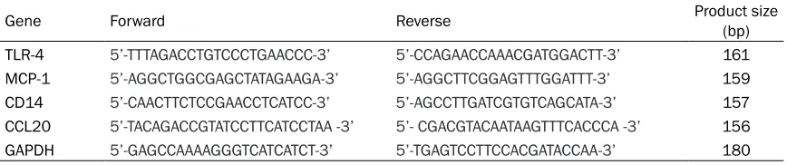
Due to the inflammatory cells infiltration in gin-gival tissues of rhesus macaques and the histo-pathological changes induced by simulated weightlessness, we expected if TLR4/CD14 axis, a crucial pathway in the recognition of microorganisms, could be changed by simulat-ed weightlessness. Thus, we detectsimulat-ed the expression and distribution of TLR4 and CD14 in the gingival tissues of rhesus macaques by using IHC method. Results showed that TLR4 mainly expressed in the infiltrated
inflammato-ry cells, and there were no marked difference for TLR4 expression among the three groups (Figure 2). CD14 was highly expressed in the base layer of the gingival mucosa of control ani-mals (Group A). After 6w -10° HDBR (Group B), the expressions of CD14 were significantly reduced as compared with that in Group A. However, with the 4w recovery (Group C), the expression of CD14 increased comparable to the level in Group A (Figure 2).
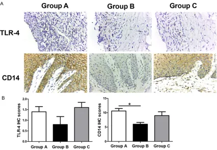
Simulated weightlessness had mild effects on the expression of chemotactic factors MCP-1, MIP-1α and CCL20 in oral gingival tissues of rhesus macaques
[image:6.612.96.522.76.367.2]mucosal epithelial cells. MCP-1 was mildly expressed in Group A and C, but almost not expressed in Group B. Statistical difference was found between Group B and Group C but not between Group A and Group B or between Group A and Group C. MIP-1α and CCL20 were highly expressed in all of the three groups, although they were decreased slightly in Group B, no statistical difference was found (Figure 3).
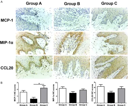
Simulated weightlessness slightly reduced the mRNA expressions of TLR4, CD14, MCP-1 and CCL20 in oral gingival tissue of rhesus macaques
In order to verify if weightlessness could affect the immune response of gingival mucosa at mRNA level, we detected the mRNA expression of TLR4, CD14, MCP-1 and CCL20 by using real-time PCR. Due to lack of an optimal specific primer pair for MIP-1α, no determination of
MIP-1α mRNA was carried out. The results revealed that all of the mRNA expression of
TLR-4, CD14 and MCP-1 were slightly reduced by 6w -10° HDBR, but they would be restored after 4w recovery. Nevertheless, all of the ferences showed no statistically significant dif-ference (Figure 4).
Discussion
Weightlessness has a variety of effects on the human physiological functions. It has been reported that about half of the astronauts exe-cuting Apollo missions had minor bacterial or viral infections within a week after their return, although the effects only lasted for short time [15]. The reactivation of latent herpes viruses has also been observed in crew during flight and within 1 week of return, indicating the downregulation of cellular immunity [16, 17]. In addition, Mehta et al. recorded subclinical acti-vation of Epstein-Barr virus, varicella-zoster virus and cytomegalovirus in 14 of the 17 astro-nauts who undertook short-duration flights on Space Shuttle [18]. All these evidences urged us to explore the effects of weightlessness on the oral immunity due to the large number of oral flora existing in oral cavity.
We employed a ground-based simulated weightlessness model on rhesus macaques in this study to explore the histopathological change of gingival tissues under 6w -10° HDBR
condition and if it can be restored after 4w recovery. The pathological examination re- vealed that inflammatory cells infiltration could be seen in the gingival tissues of all of the three groups, especially in the HDBR group. Because no oral care had been taken for the macaques during the experiment, all of the macaques had periodontal diseases in differ-ent degrees, and the HDBR group was in the most severity, suggesting that 6w -10° HDBR could aggravate the inflammation in gingival tissues, which was consistent with the results from human test [14]. However, after 4w recov-ery, the inflammatory cells infiltration was obvi-ously alleviated, indicating that the effect of weightlessness on the oral health was reversible.
In the current study, we found that the cornified envelope of rhesus macaque gum was attenu-ated as compared with that in the control group and the recovery group. Cornified envelope is the outermost layer of gum, formed by cross-linked proteins and lipids, which is the main composition of the epithelial barrier. It pos-sesses high resistance to dissolve and strong flexibility, so as to protect the epithelial cells in the inner layer. Kim W et al. demonstrated that spaceflight can promote the number of viable cells, biofilm biomass, and thickness of biofilm by Pseudomonas aeruginosa [19]. Oral micro-bial community is a typical biofilm, the bacteria in which have some special properties [20, 21]. When bacteria in biofilm-forming state, they have greater resistance to antibiotics. Due to the multiple species characteristics of dental plaque biofilm, oral microbial interactions may influence the whole human community. Thus, we speculated that the thinning cornified enve-lope may be induced by the accumulation of biofilm biomass.
immune responses against microbial infec-tions. In this study, we found that both the pro-tein and mRNA expressions of TLR-4 and CD14 in the macaque gingival tissues were reduced by -10° HDBR compared with those in control group, but the downregulation could be restored after 4w recovery. Unfortunately, the changes could only show an obvious trend, because only the difference of CD14 IHC score showed sta-tistical significance between the control group and HDBR group. Our pathological results showed that HDBR promoted inflammatory cells infiltration in the gingival tissues, but the expressions of TLR-4 and CD14 in the HDBR group was reduced at both protein and mRNA levels, instead. This is maybe a result of impaired innate immune induced by weight-lessness, which has been verified by several studies [23, 24]. Marcu, O et al. applied Drosophila (fruit fly), which shares many simi-larities with human innate immunity at the level of molecular and genetic pathways, to investi-gate the effect of spaceflight on innate immu-nity, and found that the phagocytosis of plas-matocytes was attenuated following space-flight, and in parallel, the constitutive expres-sion of pattern recognition receptors and opso-nins that specifically recognize bacteria, antimi-crobial peptide (AMP) pathway and immune stress genes were also reduced [23]. Kaur I et al. also reported that the astronauts’ mono-cytes exhibited reductions in ability to phagocy-tose Escherichia coli following 5-11 days of spaceflight [24].
Monocyte chemoattractant protein 1 (MCP-1), macrophage inflammatory protein-1α (MIP-1α) and CC chemokine ligand 20 (CCL20) are all cytokines that belong to the CC chemokine family, which play vital roles in various inflam-matory diseases including periodontitis [25-27]. Our results showed that MCP-1 could be slightly decreased following 6w -10° HDBR, but then return to normal after 4w recovery. MCP-1 can recruit monocytes, memory T cells, and dendritic cells to the sites of inflammation pro-duced by either tissue injury or infection [28]. CCL20 (also refers as macrophage inflamma-tory protein (MIP)-3alpha) and MIP-1α can attract activated T and B lymphocytes and immature dendritic cells in host responses to bacterial infection. However, although MCP-1, MIP-1α and CCL20 were decreased to some extent, the inflammatory cells infiltration was still severe in the HDBR group. This may be another evidence for the loss of function of
immune cells induced by HDBR. Recently, one report on Science revealed that IAV-infected monocytes from older humans have intact inflammasome responses but the antiviral resistance is impaired [29], which may partly explain the phenomenon in our study. Stowe RP
et al. also showed that neutrophils increase in circulating leukocyte subsets but their chemo-tactic ability decrease significantly after short-term spaceflight [30], indicating that not only the cytokines production of immune cells but also the chemotactic ability of them could be reduced by weightlessness. Thus, we may attri-bute the aggregation of inflammatory cells in oral gingival tissues of HDBR group to some else mechanisms, such as the redistribution of blood and others, but not the homing environ-ment in the gingival tissue of rhesus macaque. Our results can only show a change trend of the effects of stimulated weightlessness on the structure of oral gingival tissue and its immune status, because the differences were not very statistically significant between the groups. As we known, rhesus macaques have complicated genetic background, just like human. Thus, 5 rhesus macaques in each group may be a fatal weakness of this study. This may be comple-mented in the following studies. Another limita-tion of this study is all of the rhesus macaques having some degree of gingivitis due to lack of oral care for them. But it is perhaps a model more suitable to the real conditions owing to the high prevalence of periodontal disease in China.
In conclusion, our results revealed that 6w HDBR could promote the inflammatory cells infiltration in oral gingival tissues of rhesus macaques. The thinning cornified envelope of macaque gum indicated a reducing protection of the epithelial cells against the oral microbial community. The decreased expression of TLR-4/CD14 axis, MCP-1, MIP-1α and CCL20 induced by HDBR suggested an inhibitory effect of 6w HDBR on the local immunity of oral gum. This study filled a gap in space medical researches and provides some experimental basis for oral healthcare of astronauts during their spaceflight.
Acknowledgements
Laboratory Animal Center of China Astronaut Research and Training Center for their kindly supports during the animal experiment.
Disclosure of conflict of interest
None.
Address correspondence to: Zhongying Niu, De- partment of Stomatology, 306 Hospital of PLA, 9 Anxiang North Road, Chaoyang District, Beijing 100101, China. Tel: +86-10-66355041; E-mail: [email protected]
References
[1] Qi XQ. Report on the third national oral health epidemiological survey. People’s medical pub-lishing house 2008.
[2] Astakhov DA, Baranov MV and Panchenkov DN. [Physiological effects of microgravity as risk factors of diseases during space flight]. Patol Fiziol Eksp Ter 2012; 70-76.
[3] Convertino VA, Koenig SC, Krotov VP, Fanton JW, Korolkov VI, Trambovetsky EV, Ewert DL, Truzhennikov A and Latham RD. Effects of 12 days exposure to simulated microgravity on central circulatory hemodynamics in the rhe-sus monkey. Acta Astronaut 1998; 42: 255-263.
[4] Zhu H, Wang H and Liu Z. Effects of real and simulated weightlessness on the cardiac and peripheral vascular functions of humans: A re-view. Int J Occup Med Environ Health 2015; 28: 793-802.
[5] Oganov VS, Bogomolov VV, Bakulin AV, Novikov VE, Kabitskaia OE, Murashko LM, Morgun VV and Kasparskii RR. [Comparative analysis of cosmonauts skeleton changes after space flights on orbital station Mir and international space station and possibilities of prognosis for interplanetary missions]. Fiziol Cheloveka 2010; 36: 39-47.
[6] Zayzafoon M, Meyers VE and McDonald JM. Microgravity: the immune response and bone. Immunol Rev 2005; 208: 267-280.
[7] Hajishengallis G. Periodontitis: from microbial immune subversion to systemic inflammation. Nat Rev Immunol 2014; 15: 30-44.
[8] Darveau RP. Periodontitis: a polymicrobial dis-ruption of host homeostasis. Nat Rev Microbiol 2010; 8: 481-490.
[9] Gmunder FK, Baisch F, Bechler B, Cogoli A, Cogoli M, Joller PW, Maass H, Muller J and Ziegler WH. Effect of head-down tilt bedrest (10 days) on lymphocyte reactivity. Acta Physiol Scand Suppl 1992; 604: 131-141.
[10] Voogel AJ, Stok WJ, Pretorius PJ, Van Montfrans GA, Langewouters GJ and Karemaker JM.
Circadian blood pressure and systemic hae-modynamics during 42 days of 6 degrees head-down tilt. Acta Physiol Scand 1997; 161: 71-80.
[11] Nogues C and Milhaud C. A new technique for iliac crest biopsy in rhesus monkeys for use in weightlessness experiments: some results of ground studies. Aviat Space Environ Med 1988; 59: 374-378.
[12] Sonnenfeld G, Schaffar L, Schmitt DA, Peres C and Miller ES. The Rhesus monkey as a model for testing the immunological effects of space flight. Adv Space Res 1994; 14: 395-397. [13] Zwart SR, Crawford GE, Gillman PL, Kala G,
Rodgers AS, Rogers A, Inniss AM, Rice BL, Ericson K, Coburn S, Bourbeau Y, Hudson E, Mathew G, Dekerlegand DE, Sams CF, Heer MA, Paloski WH and Smith SM. Effects of 21 days of bed rest, with or without artificial grav-ity, on nutritional status of humans. J Appl Physiol (1985) 2009; 107: 54-62.
[14] Shi TP, Niu ZY, Shi SG, Bao B, Tang CH and Chen YH. Effects of 30 d Head-down Bed Rest on Gingiva Color in Men. Space Medicine & Medical Engineering 2012; 25: 135-137. [15] Taylor GR. Recovery of medically important
mi-croorganisms from Apollo astronauts. Aerosp Med 1974; 45: 824-828.
[16] Stowe RP, Mehta SK, Ferrando AA, Feeback DL and Pierson DL. Immune responses and latent herpesvirus reactivation in spaceflight. Aviat Space Environ Med 2001; 72: 884-891. [17] Sonnenfeld G and Shearer WT. Immune
func-tion during space flight. Nutrifunc-tion 2002; 18: 899-903.
[18] Mehta SK, Laudenslager ML, Stowe RP, Crucian BE, Sams CF and Pierson DL. Multiple latent viruses reactivate in astronauts during Space Shuttle missions. Brain Behav Immun 2014; 41: 210-217.
[19] Kim W, Tengra FK, Young Z, Shong J, Marchand N, Chan HK, Pangule RC, Parra M, Dordick JS, Plawsky JL and Collins CH. Spaceflight pro-motes biofilm formation by Pseudomonas ae-ruginosa. PLoS One 2013; 8: e62437.
[20] Kara D, Luppens SB, van Marle J, Ozok R and ten Cate JM. Microstructural differences be-tween single-species and dual-species biofilms of Streptococcus mutans and Veillonella par-vula, before and after exposure to chlorhexi-dine. FEMS Microbiol Lett 2007; 271: 90-97. [21] Kara D, Luppens SB and Cate JM. Differences
between single- and dual-species biofilms of Streptococcus mutans and Veillonella par-vula in growth, acidogenicity and susceptibility to chlorhexidine. Eur J Oral Sci 2006; 114: 58-63.
[23] Marcu O, Lera MP, Sanchez ME, Levic E, Higgins LA, Shmygelska A, Fahlen TF, Nichol H and Bhattacharya S. Innate immune respons-es of Drosophila melanogaster are altered by spaceflight. PLoS One 2011; 6: e15361. [24] Kaur I, Simons ER, Castro VA, Ott CM and
Pierson DL. Changes in monocyte functions of astronauts. Brain Behav Immun 2005; 19: 547-554.
[25] Zhu H, Lin X, Zheng P and Chen H. Inflammatory cytokine levels in patients with periodontitis and/or coronary heart disease. Int J Clin Exp Pathol 2015; 8: 2214-2220.
[26] Dommisch H, Chung WO, Jepsen S, Hacker BM and Dale BA. Phospholipase C, p38/MAPK, and NF-kappaB-mediated induction of MIP-3alpha/CCL20 by Porphyromonas gingivalis. Innate Immun 2010; 16: 226-234.
[27] Shaddox LM, Spencer WP, Velsko IM, Al-Kassab H, Huang H, Calderon N, Aukhil I and Wallet SM. Localized Aggressive Periodontitis Immune Response to Healthy and Diseased Subgingival Plaque. J Clin Periodontol 2016; 43: 746-53.
[28] Gerard C and Rollins BJ. Chemokines and dis-ease. Nat Immunol 2001; 2: 108-115.
[29] Pillai PS, Molony RD, Martinod K, Dong H, Pang IK, Tal MC, Solis AG, Bielecki P, Mohanty S, Trentalange M, Homer RJ, Flavell RA, Wagner DD, Montgomery RR, Shaw AC, Staeheli P and Iwasaki A. Mx1 reveals innate pathways to an-tiviral resistance and lethal influenza disease. Science 2016; 352: 463-466.