Published online March 4, 2015 (http://www.sciencepublishinggroup.com/j/ajbio) doi: 10.11648/j.ajbio.s.2015030203.11
ISSN: 2330-0159 (Print); ISSN: 2330-0167 (Online)
Undifferentiated Catecholaminergic and NO-producing
cells of forebrain matrix zones and intercellular
relationships in periventricular diencephalon of juvenile
Oncorhynchus masou
Pushchina E. V.
1, *, Varaksin A. A.
1, Shukla S.
2, Obukhov D. K.
31
Laboratory of Cytophysiology, A.V. Zhirmunsky Institute of Marine Biology Far Eastern Branch of Russian Academy of Sciences, Vladivostok, Russia
2
Faculty, Prof. Brien Holden Eye Research Centre, L.V. Prasad Eye Institute, Hyderabad, India
3
St. Petersburg State University, Universitetskaya nab. 7/9, St. Petersburg, Russia
Email address:
[email protected] (Pushchina E. V.)
To cite this article:
Pushchina E. V., Varaksin A. A., Shukla S., Obukhov D. K.. Undifferentiated Catecholaminergic and NO-Producing Cells of Forebrain Matrix Zones and Intercellular Relationships in Periventricular Diencephalon of Juvenile Oncorhynchus masou. American Journal of BioScience. Special Issue: Adult and Reparative Neurogenesis: Actual Questions. Vol. 3, No. 2-3, 2015, pp. 1-11.
doi: 10.11648/j.ajbio.s.2015030203.11
Abstract:
Localization of TH- and NO-producing systems in the diencephalon of juvenile masu salmon Oncorhynchus masou was investigated by using histofluorescence labeling of glyoxylic acid (GA), and ICH labeling of tyrosine hydroxylase (TH) and neuronal nitric oxide synthase (nNOS). High density distribution of catecholaminergic and NO-ergic cells has been found in preoptic, posterior tuberal and hypothalamic areas. Cells revealed in above mentioned diencephalic areas were assigned to three main types: pear-shaped, bipolar and rounded. Most of the TH- and NO-producing cells had the phenotype of undifferentiated elements localized on territory of diencephalic matrix zones. Paracrine and autocrine relationships between TH- and NO-producing cells in periventricular diencephalon of juvenile O. masou before formation of blood-brain barrier have been hypothesized. The lack of differentiated cells and the presence of a large variety of size groups of cells indicate a possible heterochrony in growth and differentiation of O. masou diencephalic areas. Location of catecholaminergic and NO-producing cells in the territory of diencephalic matrix zones suggests that catecholamines and nitric oxide are involved in the regulation of post-embryonic neurogenesis in diencephalon of O. masou.Keywords:
Catecholaminergic Cells, Proliferation, Adult Neurogenesis, Cell Migration, NO, Paracrine Regulation, Cerebrospinal Fluid Contacting Cells, Development of Hypophysotropic Areas1. Introduction
Hypophysotropic nuclei of diencephalon in teleost´s brain contain periventricular nitroxidergic neurons [1-3]. The presence of NO-synthesizing cell groups in the periventricular diencephalic nuclei, despite for interspecies differences in teleosts is the most consistent [4-7]. Interestingly, this region of teleost´s diencephalon also contains catecholaminergic (dopaminergic) cell groups [8-10]. It has been shown that dopaminergic neurons in diencephalon of Danio rerio are the source of ascending afferents to the ventral telencephalon, considered now as a functional analog of the mammalian striatum [11, 12]. Similar results have also
been revealed in other specimen of cyprinoid Rhodeus cericeus, where in the periventricular and posterior tuberal regions are identified TH-immunopositive (TH-ip) cells projected to the ventral telencephalon [13].
amniotes [18]. Meanwhile, a CA-producing and NO-producing systems in vertebrates brain, particularly mammals are often seen as systems antagonists, which usually have no spatial co-localization. However, in our studies on Salmoniformes fish large projection neurons in periventricular diencephalon were not found, but the presence of catecholamine-producing cells was observed [19].
The aim of our study was to investigate the spatial relationships of catecholamine-synthesizing and NO-producing systems in periventricular diencephalon of juvenile masu salmon Oncorhynchus masou in the context of continued adult neurogenesis and cell migration in this area retaining the features of segment structural organization.
2. Materials and Methods
As the study objects, we used 6 month (10 sp.) and 1 year old (10 sp.) juvenils of masu salmon Oncorhynchus masou
obtained from Ryazan′ experimental-manufacturing fish-breeding hatchery in 2014. Experimental procedures were conducted in accordance with European Community guidelines on animal care and experimentation. The fish were kept in aquaria with aerated fresh water at 17-180 C and anesthetized in 0.1% solution of tricaine methanesulfonate (MS-222, Sigma, United States) for 10-15 min.
2.1. Histofluorescence Labeling of Sugar-Phosphate Glyoxylic Acid (SPGA)
For identification of cateholamine-containing neurons we used a histofluorescence labeling of sugar-phosphate glyoxylic acid [20]. Fishes were anesthetized in a cuvette with 0.1% solution of MS-222, the skull was opened and the brain was removed. Fresh brains were frozen in a cryostat, and serial sections of 25-30 µm were cut. Sections were mounted on gelatinized slides. In a 100 ml SPGA prepared in distilled water containing sucrose - 10.2%, KH2PO4 - 4.2%,
and glyoxylic acid - 1.5% (pH 7.2). This solution was adjusted to a volume of 150 ml with distilled water. In the final solution, SPGA (pH 7.4), sections were incubated for 3-5 seconds and then dried. A drop of mineral fluorescent immersion oil was applied onto sections and placed in an oven at a temperature of 95°C for 2.5 min. Preparations were viewed under fluorescent microscope Leica DM (235-250 nm).
2.2. Immunohistochemistry of Tyrosine Hydroxylase
IHC of tyrosine hydroxylase (ТН) was conducted, using a standard avidin-biotin peroxidase labeling on free-floating slices. The slices were incubated with mouse monoclonal antibodies against TH (Vector Laboratories, Burlingame, USA), (1:10000) at 4°С temperature for two days. For visualization of the IHC labeling, a standard kit (Vectastain Elite АВС Kit, Burlingame, USA) was used. For identification of the reaction products of TH, the substrates of red colors (VIP Substrate Kit, Vector Labs, Burlingame, USA) was used. The labeling process was controlled with
microscope; the slices were rinsed in water, mounted on slides, and dehydrated according to a standard method and incorporated into medium Bio Optica (Italy). The measurements were performed with the use of an Axiovert Apotome microscope 200M.
2.3. nNOS Immunohistochemistry
The sections for nNOS-immunohistochemistry were incubated with methanol containing 0.3% hydrogen peroxide for 30 min to block endogenous peroxidase, followed by 30 min in 5% normal goat serum in PBS containing 1% bovine serum albumin (BSA), at room temperature to block nonspecific binding. Thereafter, the sections were reacted for 2 days at 4˚C with the polyclonal primary anti-nitric oxide synthase antibody (Biomedicals, Germany) diluted 1:500 in 0.01 M PBS containing 1% goat serum and 0.1% BSA. Subsequently, the sections were incubated with biotinylated goat anti-rabbit IgG (1:200, MP Biomedicals, Inc) for 1 hour and then streptavidin peroxidase (1:100) for 1 hour at room temperature. The washes between treatments were done using PBS. The sections were then incubated in the HRP substrate kit (Vector VIP Substrate kit for peroxidase, SK-4600, Vector Laboratories, Burlingame, CA), at room temperature for 30 min. Finally, the sections were washed in tap water and mounted into medium Bio Optica (Italy).
To evaluate the specificity of the immunohistochemical reaction, we used a negative control. Brain slices were incubated for 1 day with 1% nonimmune horse serum instead of with primary antibodies and then were stained as described above. In all control experiments the immunopositive reaction did not occur.
2.4. Statistical Analysis
For statistical and morphometric analysis we used software of microscope research class Axiovert with module Apotome. For the quantitative analyses, all cells stained for TH or nNOS in all diencephalic nuclei were counted under a 40x objective lens using an Axiovert Apotom 200 M (Carl Zeiss, Germany). Densitometric investigation of the optical density of immunolabeled cells was performed using software Axiovert Apotome 200 M. The optical density of immunoprecipitate in labeled cells was studied on the samples from 50-100 cells. On the basis of morphometric analysis we allocated 3 morphological types of cells in accordance with generally accepted neurohistological classification [4]. Morphometric parameters and studied correlations between some of the parameters were analyzed by Microsoft Excel 2010.
Data are expressed as mean ± S.E.M. and were analyzed with an ANOVA followed by post hoc Tukey's tests unless otherwise stated. P-values < 0.05 were considered to be statistically significant.
3. Results
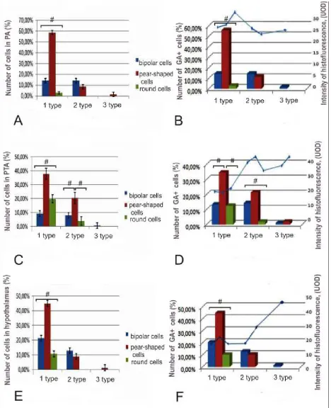
posterior tuberal and hypothalamic nuclei showed that 1 year-old O. masou diencephalon contain several types of cells (Figs. 1A, C, E). Three types of cells were identified in preoptic, posterior tuberal and hypothalamic areas. Small cells with diameter less than 8 µm form 1st type of cells; the cells of 2nd type have average diameter of 8-14 µm, and 3rd type was represented by large cells with diameter greater than 14 µm (Figs. 1A, C, E). All cells in diencephalic nuclei were located in periventricular and subventricular areas. Histofluorescence was determined in all types of glyoxylic acid (GA)-labeled cells in preoptical area of O. masou (Fig. 1B). Small GA-labeled cells of 1st type were presented by unipolar pear-shaped cells, which also were TH-ip (Fig. 2A); the proportion of such cells was 55.7% (Fig. 1B). Other subtype was presented by bipolar (fusiform) and TH-immunonegative (TH-in) cells; their proportion was 14.3%. We believe that this subtype of cells does not contain enzymes of catecholamines synthesis and may belong to a population of serotonergic cerebrospinal fluid contacting cells (CFC). Among this subtype, 2.8% of cells had no outgrowths, had round shape and were located in periventricular area. The maximal level of histofluorescence GA (Fig 1B) and TH-immunoreactivity (Fig. 2A) have been registered in these cells. Large bipolar cells were founded in subvetricular area; they were GA-labeled/TH-in (Fig. 1B). The proportion of large neurons was 1.5% (Fig. 1B).
The morphological composition of cells from posterior tuberal area (PTA) of O. masou similar with preoptic one, but predominant type of cells was small, pear-shaped cells (55%). Small cells in PTA were also presented by bipolar and round subtypes; their proportion was much lower to compare with pear-shaped cells (Fig. 1C). In PTA of 1-year-old O. masou
were discovered pear-shaped and spindle-shaped cells of medium size, as well as a small number (1.3%) were large, pear-shaped cells with a diameter of 14 to 20 µm (Fig. 1C). In PTA the histofluorescence of GA was detected in 65% of neurons. Most of GA-labeled elements were small pear-shaped cells localized in periventricular area (Fig. 1D), which were TH-ip too (Fig. 2E, G). In larger cells of similar morphological structure, histofluorescence of GA and TH-immunoreactivity were also registered (Fig. 1D, 2G). We believe that these populations of cells are early and later stages of development of the same type of cells; larger cells have also more intense histofluorescence of GA (Fig. 1D), which confirms our assumption. Among neurons labeled by GA we recorded only few small rounded cells (10%). These cells usually have periventricular localization and TH-expression always has revealed. Rounded cells of larger size presented small population of TH-ip neurons in periventricular area. We believe that processes of these cells may be cut or arranged in a different plane; but it is possible that these cells also presented a later stage of development of small round cells not forming processes. Cells of bipolar morphology did not express TH, but they were GA-labeled. The proportion of bipolar small cells was 20.5%; and bipolar medium-sized cells were 12.8%. Most of the GA-labeled and TH-ip cells (44.8%) were small, pear-shaped neurons;
number of larger cells of same type in O. masou was 10%. In
O. masou PTA were identified cells in size from 14 to 20 µm (1.3%). Processes of such cells ramify in the subventricular region, forming a sparse network. This pattern was observed after impregnation by Cajal. The intensity of histofluorescence after GA-labeling in cells of PTA was varied (Fig. 1D). Most small cells (about 60%) have a low level of histofluorescence (20 UOD); more intense labeling (more than 35 UOD) was founded in medium and large pear-shaped neurons similar to those in preoptic area (Fig. 1B, D). In hypothalamus, which occupies a considerable part of O. masou diencephalon, lateral, central and ventral areas according to common classification were examined [21]. In the lateral hypothalamus same types of cells as in other areas of diencephalon were identified (Fig. 3A). Identified cells were assigned to two types: small cells (diameter 8 µm) and the medium one (their diameter was from 8 to 14 µm). In the lateral hypothalamus, the quantity of small cells was 82% of the total cell´s number. Rounded elements located in periventricular area constitute 30% of the cells (Fig. 3A). In
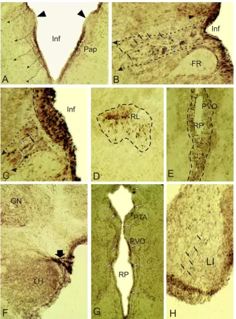
O. masou brain during 1st year of development, majority of small cells expresses TH and localized in periventricular area (Fig. 2G). Part of TH-ip cells located around the lateral ventricle has a single process (Fig. 2D). Cells with process usually are pear-shaped or oval-shaped, others cells are rounded. The number of small pear-shaped cells in the lateral hypothalamus is 44%, and rounded cells more than 32% (Fig. 3A). The proportion of larger pear-shaped cells in lateral hypothalamus of O. masou is 8%. The same number of cells has a bipolar morphology and probably such cells are cerebrospinal fluid contacting elements (Fig. 3A). Only a small part (2%) of round cells has a size greater than 8 µm (Fig. 3A). Thus, in the lateral hypothalamus of O. masou 1 year-old pear-shaped neurons that make up 44% of the total number of cells are the predominant cell type; 16% of the cells are spindle-shaped and are likely cerebrospinal fluid contacting elements.
In the central hypothalamus, majority of cells localized in periventricular area, intercellular distance does not exceed the size of single cell´s body; the density of cells distribution is significant. In the subventricular area we founded larger cells, their location was diffuse. In central region, the cells with structure similar to that of the lateral part were observed; the ratio of cell´s types is shown in Fig. 3B. The predominant type of cells is small, pear-shaped neurons, constituting 42% of the total number of cells in central area of hypothalamus; the number of rounded cells is 20% and bipolar cells - 18% (Fig. 3B). Among the larger neurons also identified all types of cells as described above, but their ratio is markedly different; the group of pear-shaped neurons constitute 16%, the round cells constitute only 4% of total number of neurons (Fig. 3B). Thus, in central hypothalamus of 1 year-old O. masou, 78.5% of the cells have a size of up to 8 µm.
of the cells, located in this region were pear-shaped, their processes directed toward mesencephalo-hypothalamic tract (Fig. 1H). In the ventral hypothalamus were divided three size groups of cells, similar to those in the preoptic area and PTA (Fig. 3C). Among different neurons in ventral hypothalamus were identified small pear-shaped and rounded cells that have periventricular localization; bipolar cells in this region were not detected (Fig. 3C). Most of cells (60%) in ventral hypothalamus had dimensions of 8 to 14 µm (Fig. 3C). In this size group the number of pear-shaped cells was 38%, bipolar cells - 16% and rounded cells - 6% of total number of cells (Fig. 3C). In O. masou ventral hypothalamus large fusiform cells, accounted 2% have been identified.
Summary data about ratio in different types of cells in hypothalamus O. masou are shown in Fig. 1E. In hypothalamus GA marks about 40% of all cells. Total histofluorescence activity of GA in 1 year-old O. masou
hypothalamus is shown in Fig. 1F. Most of the GA-labeled cells (60.2%) refer to populations of small periventricular cells of hypothalamus. Larger cells make up 37% of the total number of GA-marked cells. The level of histofluorescence of these cells is twice higher than in small cells (Fig. 1F). Most identified cells in hypothalamus except the bipolar one also were TH-ip. Finally, in O. masou hypothalamus 2% of cells were size of 14 to 20 µm (Fig. 1E). These large cells had a high level histofluorescence of GA coinciding with that in medium-size cells (Fig. 1F).
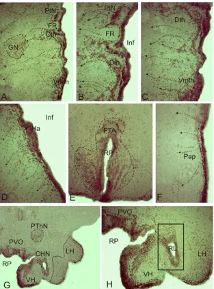
Study of TH- and NOS-immunolocalization in 6-month-old juveniles of O. masou showed that in hypophysotropic zones of diencephalon (preoptic area, paraventricular organ, anterior and posterior tuberal areas, hypothalamus) immunopositive cells were also found in subventricular regions, additionally with periventricular one (Fig. 1A, B, 4A-C, D, F). We believe that these cells are neuroblasts, migrating from diencephalic proliferative zones. Migration trajectory and direction of cell´s migration can be traced to guide their radial fibers (Fig. 2 B, C), or on the specifics of cell´s groups to form «chains» (Fig. 4C, D). Figures (2A-C, 4A-D, F) show the direction of cell´s movement in deeper layers of the brain. Most of TH-ip migrating cells at the brain sections of O. masou can be easily identified, however, on the borders of prosencephalic neuromeres in 6 month-old fishes TH-labeling is absent (Fig. 2A), as in other age groups. Clusters of intensively labeled TH-ip cells migrating along
Figure 2. Immunolocalization of tyrosine hydroxylase (TH) in diencephalon of Oncorhynchus masou. A - immunopositive cells in 6-month-old juvenile in preoptic area pars parvocellular (Pap), small dashed arrows indicate the direction of cells migration from periventricular region, immunonegative border between neuromeres marked with triangular black arrow, Inf - infundibulum; B – cells migrating along radial fibers (indicated by black arrows), TH-ip cells (indicated by white arrows) in the pretectal area of 6-month-old O. masou, FR – fasciculus retroflexus; C - migrating cells from the dorsal thalamic neuromer of 1 year-old O. masou; D – TH-ip cells around the lateral ventricle (RL) of 6-month-old juveniles; E - TH-ip cells around paraventricular organ (PVO) limited by dotted line, RP - posterior ventricle; F - catecholamine-contained fibers (shown in black broad arrow) within the lateral hypothalamic tract of 1-year-old juvenile, GN - glomerular nucleus; G - paraventricular and posterior tuberal (PTA) area of 1-1-year-old juvenile, white arrows indicate TH-ip cells; H – TH-ip fibers (black arrows) within the lobus inferior (LI) of 1 year-old juvenile. Magnification: A, F, G - ob. x20, oc. x10; B-E, H - ob. x40, oc. x10.
Figure 3. Morphological parameters of cellular composition in hypothalamus of juvenile O. masou. Number of cells in the lateral (A), central (B), and ventral
Figure 4. Immunolocalization of neuronal nitric oxide synthase (nNOS) in the diencephalon of 6 month old juvenile O. masou. A - a general view of thalamus, a large dotted line marked pretectal cluster of cells (PtN), dorsal (Dth) and ventromedial (Vmth) cells clusters, dotted with small arrows indicate the direction of cells migration from the periventricular areas to glomerular nucleus (GN) and in deep zone of thalamus, FR - fasciculus retroflexus; B and C micrographs are with higher magnification of A; the proposed trajectory of cell´s migration are shown in small dotted line, Inf - infundibulum; D - nNOS immunolocalization in the dorsal preoptic area, Ha - habenula; E - posterior tuberal area (PTA) in caudal hypothalamus limited by dotted line, RP - posterior ventricle; F - parvocellular part of preoptic area (PAp); G - general view of nNOS immunolocalization in anterior hypothalamus, posterior thalamic nucleus (PThN), lateral hypothalamus (LH) and diffuse nucleus of the lateral ventricle limited dotted CHN - central hypothalamic nucleus; H – fragment of paraventricular organ (PVO limited by dotted line) at higher magnification, lateral hypothalamic ventricle (RL) in the rectangle with adjacent cells clusters. Magnification: A, E, H - ob. x20, oc. x10; B-D, F - ob. x40, oc. x10; G - ob. x12, oc. x10.
4. Discussion
According to our data, hypothalamic area of O. masou
includes periventricular accumulation of CE neurons, located with high density of cells distribution. In subventricular hypothalamic zone, cell clusters with more diffuse structure were dominated. Such morphology and presence of poorly differentiated forms of catecholaminergic cells, in our opinion, indicate that in hypothalamus of O. masou
differentiation and migration of cells are continued. Minor intercellular distance and high density distribution of cells, and the absence of developed network of processes, suggests that among these cells is carried out not synaptic
assumption in discharge a hypothetical one. In hypothalamus of O. masou, the majority of cells are represented by small pear-shaped neurons, processes of which are directed towards the opposite course from the lumen of hypothalamic ventricle. The population of small pear-shaped cells is 66.9% of all cells in hypothalamus. Neurons of same morphological type, but a larger size contain 33%, most of these cells are localized in the ventral hypothalamus. Based on these data, we assume that postembryonic morphogenesis in the hypothalamus of O. masou is characterized by a certain heterochrony. As a result, in ventral hypothalamic region, the cells during 1st year of development have some phenotypic traits acquired specialization expressed to a greater extent than in the central and lateral areas. Among these cells, pear-shaped neurons constitute 58.3%. We believe that this type of cells found in all size groups of cells in preoptic area, PTA and hypothalamic regions is the main type of diencephalic periventricular cells. According to our data in O. masou during 1st year of development the cells of this type are available in different size groups, but the number of these cells in different areas of the hypothalamus varies.
Cells with bipolar morphology, in our opinion, can be attributed to population of CFC cells, but morphologically they are more heterogeneous than pear-shaped cells. Rounded cells, we believe, can be attributed to a population of undifferentiated neuroblasts, because most of them are localized in periventricular region of lateral and central hypothalamus. In these areas of hypothalamus morphological differentiation of cells and the presence of larger cell forms with processes is less than in the ventral hypothalamus. We suppose that the round cells represent the earliest stages of cellular differentiation after exiting from mitosis in a matrix area of brain. Larger rounded cells are apparently at further stage of differentiation prior to the formation of processes. Expression of some specific substances (TH, NO) in cells at stages of development in which the morphological differentiation of cells weakly expressed or absent, indicate that these substances in O. masou brain can act as morphogenetic factors controlling further development of brain. An additional argument is the presence of several morphologically similar sized groups of cells, which in our opinion indicates that the data size groups are simultaneously gradually existing populations of differentiating cells produced in proliferative (matrix) zones of diencephalon. We also believe that pear-shaped cells with processes of varying lengths expressed TH and marked by GA are the source of dopamine and serotonin in diencephalon of O. masou. These neurotransmitter substances may excrete into the extracellular space and render regulatory effect to differentiation and migration of target-cells located in same area. Lack of developed processes in this cells and small intercellular distance largely determine the effect of dopamine and nitric oxide as a paracrine factors that have a regulatory effect on the differentiation of the cells microenvironment. Undifferentiated TH-ip cells in subvetricular area have distinct signs of fetal organization: they often lack or have ill-defined processes and large
centrally located nucleus. Patterns of distribution of TH-ip cells in the subventricular layers of O. masou diencephalon allow us to suggest that these conglomerates of cells can be attributed to migrate from proliferative zones of diencephalon. Cells from proliferative periventricular zone and from deeper subventricular region (where cells are in the initial stage of differentiation) express a number of specific syntheses, in particular TH and NO. We believe that the substances synthesized during adult brain morphogenesis of
O. masou, in particular dopamine and NO are signaling molecules that possibly have a paracrine and autocrine impact on the target-cells to the formation of interneuronal connections and synaptogenesis. Undifferentiated cells expressing specific syntheses were localized in the territory of rostral and caudal vascular plexuses. Signal molecules synthesized in these regions can be received into the portal circulation system and then into the system of systemic circulation, exerting effects on peripheral endocrine organs. We assume that dopamine and NO in hypophizotropic areas of diencephalon of juvenile O. masou are inducers of development (morphogenetic factors).
Our observations are consistent with the results of recent studies, obtained by Ugrumov on fetus of mammals in natal and early postnatal periods of development prior to the formation of the blood-brain barrier (BBB) [23, 24]. Undifferentiated neurons of rat’s hypothalamus in these periods of fetal development have not formed synaptic structure yet and cells expressed a number of specific syntheses, including TH. In neurons from rat fetus, reuptake of dopamine and its release in response to depolarization of neurons were recorded. However, the formation processes designed to target-cells and growth of processes from afferent receptors in such cells took place much later.
system before closing BBB sufficient to provide the physiological effect on target-cells? Experiments with microsurgical removal portion of hypothalamus with dopamine synthesis, as well as the use of a specific model, which causes the suppression of dopamine synthesis in the brain by 50% resulted in the decrease of dopamine levels by 75% in the portal blood shows that indeed dopamine in the brain has a portal blood origin [26]. Do the signaling molecules synthesized in the brain, affect peripheral organs? To show the influence of dopamine on prolactin secretion in rat’s lactotrophes administered to intact fetus blocker of dopamine D2 receptors - haloperidol, thereby obtaining an
increase in the concentration of dopamine in plasma and a decrease concentration in the pituitary. In microsurgical removal of dopamine synthesized areas of the brain in fetuses of rats, similar response to haloperidol has not been received. This indicates that the dopamine affect the synthesis of prolactin in rats before BBB formation.
The results of our observations in different age groups of masu salmon showed that in the periventricular area of diencephalic, mesencephalic, medullary and spinal parts of brain are localized morphologically and neurochemically heterogeneous population of cells, some of which have processes and produce TH, GABA and NADPH-d/nNOS [27]. Study has shown that O. masou brain in different age periods contains proliferative zones (PZ) [28]. Cells expressing TH and NADPH-d/NOS were identified in PZ of diencephalon and medulla oblongata. We believe that the cells expressing TH and NADPH-d/NOS are adult born neurons. It is possible that proliferative activity of cells in PZ of O. masou ensure a growth of diencephalic, mesencephalic and medullary brain structures and their physiological regeneration. It is shown that the molecule of neurotransmitters have a significant impact on the development of cells during embryogenesis [29], as well as during adult neurogenesis in subgranular zone of dentate gyrus and the medial surface of brain hemisphere [30, 31]. Recently, there are details that neurotransmitters regulate adult neurogenesis in the subventricular region of brain in different vertebrates [32]. Signaling mechanisms, including receptors, sources of release and molecule transporters were installed for some neurotransmitters [31, 32]. Our results suggest that TH and NO may act as signaling molecules that regulate processes of adult neurogenesis in the diencephalon and other regions of brain of O. masou [28]. In diencephalon,
tectum, medulla oblongata and spinal cord of three-, six-month and one-year-old juvenile of masu salmon in the periventricular cells synthesis of TH is defined [19, 27]. In adults O. masou, TH marks periventricular cells with radially oriented outgrowths in diencephalon and medulla oblongata departments that have not previously been shown to fish. In all age groups of O. masou, TH-labeling elicited neuromeric structure of brain.
In adults O. masou, TH-labeling was found at the boundary separating pallial part of telencephalic hemispheres from subpallial one [27]. This is consistent with the data obtained for D. rerio, where it showed the presence of
TH-immunoreactive descendants of neuroblasts, migrated in the rostral migratory flow [33]. TH-ip elements resembling to radial glia were localized in rostral areas of spinal cord. In this part of O. masou CNS main neuromorphological features of radial glia cells corresponding with classification of Rakic were presented most clearly [34]. In studies on D. rerio
participation of dopaminergic signaling in development of larval brain was shown [35]. It has been demonstrated that TH may act as morphogenetic or transcription factor in certain periods of ontogenesis, providing long-term impact on the differentiation of non catecholaminergic neurons and expression of their phenotype [24]. The proposed mechanism of such effects of dopamine is the presence of dopaminergic D1-D3 receptors in target-cells [36]. Subventricular zone (SVZ) in embryos and adult animals contained high levels of D3 receptor that was shown by methods of autoradiography and hybridization in situ [37]. Type D2 receptors have been identified in cells of transit-amplifying type, and D1 and D2 receptors were found in neuroblasts [38]. Despite the fact that some neuroblasts of SVZ differentiate into dopaminergic interneurons of olfactory bulbs, they do not express TH in mammalian brain [39]. TH was not detected in other types of cells in mammalian SVZ. Nevertheless, it has been shown that dopamine is released from the afferents of substantia nigra, contacted with the transit-amplifying cells in SVZ [38]. Data about distribution of dopamine receptors in fishes brain are limited and information about D1 receptors exist in only one species of teleost Anguilla anguilla [40] and TH-immunolocalization in forebrain of fish is quite different from that of mammals [8, 11, 41]; it is possible that high levels of TH in the periventricular cells with radial outgrowths in O. masou, is an example of an alternative adaptation mechanism, the functional role of which is not known yet. We assume that dopamine in periventricular TH-ip cells in O. masou carries morphogenetic regulation, influencing paracrine to neighboring target-cells or excreted into the circulation system and providing neuroendocrine regulation through a common system of blood flow.
Other neurochemical marker in cells of the periventricular zone of fishes brain is NADPH-d/NOS. It was firstly described in periventricular zone of sunfish’s brain [42]. In diencephalon of 6-month and one year-old juveniles O. masou similar IHC patterns of nNOS activity were registered, which suggests the participation of NO as a factor regulating cell’s migration. We also found clusters of nNOS-ip undifferentiated cells localized in periventricular, subventricular and deeper regions of the diencephalon. This data confirms participation of NO in the regulation of cell proliferation and neuronal differentiation during the post-embryonic ontogenesis [43, 44]. Features topography of subventricular cell clusters as immunolabeled by nNOS, as TH-labeled allow us to trace the path of their migration from the proliferative region of diencephalon brain. Analysis of the migration routes of preoptic and thalamic areas of O. masou
amount of migrating cells in preglomerular and pretectal area of thalamus, which presented the major sensory centers of diencephalon [45]. In rostral and caudal parts of hypothalamus we also found large accumulations of undifferentiated nNOS-ip cells in the subventricular areas. We believe that in these cells NO can regulate synthesis of hypothalamic hormones, acting by paracrine way. During embryogenesis of zebrafish posterior tuberal area is the caudal border of cell migration in diencephalon [46], we believe that in the same region of juvenile O. masou brain during all ages continue the processes of cells migration and formation of a differentiated structure of hypothalamic nuclei.
At the levels of forebrain and diencephalon of zebrafish, rapidly and slowly proliferating subpopulations of RG co-exist along all ventricular lumen; RG cells differ for BrdU labeling [33]. PCNA-labeling in the O. masou brain showed that yearling and adult individuals in the periventricular region of diencephalon and central gray layer of medulla oblongata show a population of proliferating cells [27]. At the level of forebrain the distribution of TH-ip cells corresponds to neuromeric structure of brain, which is confirmed by PCNA labeling of matrix zones. On the border between the dorsal prosomeres P2-P3 the immunolabeling of TH and PCNA is absent [27, 28]. In the diencephalon, localization of proliferative PCNA-ip regions correspond to prosomeric design of forebrain. Thus, in the CNS of O. masou revealed several PCNA-ip zones with proliferation, contained cells expressing TH and NO. We assume that these cells are newly formed neurons, which is consistent with the data obtained in the zebrafish [33]. CA and NO-producing cells are located in areas with PCNA-immunoreactivity. Perhaps TH- and NO-producing cells participate in the regulation of cell differentiation and migration, as well as the growth and physiological regeneration of diencephalic structures.
This work were supported by Program of fundamental investigation FEB RAS «Far East» 2015-2017 (grant number № 15-I-6-116) and DST-INSPIRE Faculty Grant (IFA14-LSBM-104).
References
[1] Holmqvist B., Östholm T., Alm P. (1994). Nitric oxide synthase in the brain of a teleost. Neurosci. Lett., 171:205-208.
[2] Brüning G., Katzbach R., Mayer B. (1995). Histochemical and immunocytochemical localization of nitric oxide synthase in the central nervous system of the goldfish, Carassius auratus.
J. Comp. Neurol., 358:353-382.
[3] Bordieri L., Persichini T., Venturini G. et al. (2003). Expression of nitric oxide synthase in the preoptic-hypothalamo-hypophyseal system of the teleost Oreochromis niloticus. Brain Behav. Evol., 62:43-55.
[4] Arevalo R., Alonso J. R., Garcia-Ojeda E. et al. (1995). NADPH-diaphorase in the central nervous system of the tench (Tinca tinca L., 1758). J. Comp. Neurol., 352:398-420.
[5] Villani L., Guarnieri T. (1995). Localization of NADPH-diaphorase in the goldfish brain. Brain Res., 679:261-266.
[6] Anken R. H., Rahmann H. (1996). An atlas of the distribution of NADPH-diaphorase in the brain of the highly derived swordtail fish Xiphophorus helleri (Atherinoformes: Teleostei). J. Brain Res., 37:421-449.
[7] Jadaho A.G., Wilke I., Meyer D.L. (1999). NADPH-diaphorase expression in the hypothalamo-hypophysial system of different catfish. Brain Res., 39:513-523.
[8] Meek J. (1994). Catecholamines in the brains of osteichtyes (bony fishes). Phylogeny and development of catecholamine systems in the CNS of vertebrates. Cambridge: Cambridge Univ. Press, pp. 49-76.
[9] Björklund A., Dunnett S.B. (2007). Dopamine neuron systems in the brain: an update. Trends Neurosci., 30:194-202.
[10] Yamamoto K., Vernier P. (2011). The evolution of dopamine systems in chordates. Front Neuroanat., 5:21, doi: 10.3389/fnana.2011.00021.
[11] Rink E., Wullimann M.F. (2001). The teleostean (zebrafish) dopaminergic system ascending to the subpallium (striatum) is located in the basal diencephalon (posterior tuberculum). Brain Res., 889:316-330.
[12] Tay T.L., Ronneberger O., Ryu S., Nitschke R., Driever W. (2011). Comprehensive catecholaminergic projectome analysis reveals single- neuron integration of zebrafish ascending and descending dopaminergic systems. Nat. Commun., 2:171.
[13] Pushchina E.V. (2012). Morphofunctional and the hodological characteristics of the ascending dopaminergic system of the Amur bitterling (Rhodeus sericeus, Cyprinidae). Neurosci. Behav. Physiol., 42:521-525.
[14] Smeets W., Gonzalez A. (2000). Catecholamine systems in the brain of vertebrates: new perspectives through a comparative approach. Brain Res. Rev., 33:308-379.
[15] Klejbor I., Domaradzka-Pytel B., Ludkewich B. (2004). The relationships between neurons containing dopamine and nitric oxide synthase in the ventral tegmental area. Folia Histochem. Cytobiol., 42:83-87.
[16] Le Moal M., Simon H. (1991). Mesolimbic dopaminergic network: functional and regulatory roles. Physiol. Rev., 71:155-234.
[17] Kalivas P.W. (1993). Neurotransmitter regulation of dopamine neurons in the ventral tegmental area. Brain Res Rev., 18:75-113.
[18] Dawson T.M., Zhang J., Dawson V.L. et al. 1994. Nitric oxide: cellular regulation and neuronal injury. Prog. Brain Res., 103:365-369.
[19] Pushchina E.V., Obukhov D.K., Varaksin A.A. (2013). Features of adult neurogenesis and neurochemical signaling in the Cherry salmon Oncorhynchus masou brain. Neuron. Regen. Res., 8:13-23.
[20] Bures J., Bureshova O., D. Houston. (1991). Techniques and basic experiments on the brain and behavior. Mosсow: Higher School. (In Russian).
[22] Wullimann M.F., Vernier P. (2007). Evolution of the nervous system in fishes. Evolution of nervous systems in non-mammalian vertebrates. Vol. 3. Ed. J. Kaas. Amsterdam: Elsevier, pp. 39-60.
[23] Ugrumov M.V. (2009). Non-dopaminergic neurons partly expressing dopaminergic phenotype: distribution in the brain, development and functional significance. J. Chem. Neuroanat., 3:241-256.
[24] Ugrumov M.V. (2010). Developing brain as an endocrine organ: a paradoxical reality Neurochem. Res., 35: 837-850.
[25] Ugrumov M.V. (1997). Hypothalamic monoaminergic systems in ontogenesis: development and functional significance. Int. J. Dev. Biol., 41: 809-816.
[26] Ugrumov M.V., Sapronova A.Y., Melnikova V.I., Proshlyakova E.V., Adamskaya E.I., Lavrentieva A.V., Nasirova D.I., Babichev V.N. (2005). Brain is an important source of GnRH in general circulation in the rat during prenatal and early postnatal ontogenesis. Comp. Biochem. Physiol. A Mol. Integr. Physiol., 141:271-279.
[27] Pushchina Y.V., Obukhov D.K., Varaksin A.A. (2012). Neurochemical markers of cells of the periventricular brain area in the masu salmon Oncorhynchus masou (Salmonidae). Rus. J. Develop. Biol., 43:39-53.
[28] Pushchina E.V., Varaksin A.A., Obukhov D.K. (2014). Participation of neurochemical signaling in adult neurogenesis and differentiation. Neurochemistry. Ed. T. Heinbockel. Rijeka: Intech, pp. 225-255.
[29] Platel J.C., Laca B., Bordey A. (2007). GABA and glutamate signaling: homeostatic control of adult forebrain neurogenesis. J. Mol. Histol., 38:602-610.
[30] Cameron H.A., McEwen B.S., Gould E. (1995). Regulation of adult neurogenesis by excitatory input and NMDA receptor activation in the dentate gyrus. J. Neurosci., 15:4687-4692.
[31] Aniol V.A., Stepanichev M.Yu. (2007). Nitric oxide and gamma-aminobutyric acid as regulators of neurogenesis in the brain of adult mammals: models of seizure activity. Neurochem. J., 1:265-274.
[32] Platel J.C., Stamboulian S., Nguyen I. et al. (2010). Neurotransmitter signaling in postnatal neurogenesis: the first leg. Brain Res. Rev., 63:60-71.
[33] Adolf B., Chapouton P., Lam C.S. et al. (2006). Conserved and acquired features of adult neurogenesis in the zebrafish telencephalon. Dev. Biol., 295:278-293.
[34] Rakic P. (2007). The radial edifice of cortical architecture: from neuronal silhouttes to genetic engineering. Brain Res. Rev., 55:204-219.
[35] Souza B.R., Tropepe V. (2011). The role of dopaminergic signalling during larval zebrafish brain development: a tool for investigating the developmental basis of neuropsychiatric disorders. Rev Neurosci., 22:107-119.
[36] Callier S., Snapyan M., Le Crom S. et al. (2003). Evolution and cell biology of dopamine receptors in vertebrates. Biol. Cell, 95:489-502.
[37] Diaz J., Ridray S., Mignon V. et al. (1997). Selective expression of dopamine D3 receptor mRNA in proliferative
zones during embryonic development of the rat brain. J. Neurosci., 17:4282-4292.
[38] Hoglinger G.U., Rizk P., Muriel M.P. et al. (2004). Dopamine depletion impairs precursor cell proliferation in Parkinson disease. Nat. Neurosci., 7:726-735.
[39] Baker H., Liu N., Chun H.S. et al. (2001). Phenotypic differentiation during migration of dopaminergic progenitor cells to the olfactory bulb. J. Neurosci., 21:8505-8513.
[40] Kapsimali M., Vidal B., Gonzalez A., Dufour S., Vernier P. (2000). Distribution of the mRNA encoding the four dopamine D(1) receptor subtypes in the brain of the european eel (Anguilla anguilla): comparative approach to the function of D(1) receptors in vertebrates. J. Comp. Neurol., 419:320-343.
[41] Yamamoto K., Vernier P. (2011). The evolution of dopamine systems in chordates. Front Neuroanat., 5:21.
[42] Ma P.M. (1993). Tanycytes in the sunfish brain: NADPH-diaphorase histochemistry and regional distribution. J. Comp. Neurol., 336:77-95.
[43] Moreno-López B., Noval J.A., González-Bonet L.G. et al. (2000). Morphological bases for a role of nitric oxide in adult neurogenesis. Brain Res., 869:244-250.
[44] Gibbs S.M. (2003). Regulation of neuronal proliferation and differentiation by nitric oxide. Mol. Neurobiol., 27:107-20.
[45] Ishikawa Y., Yamamoto N., Yoshimoto M. et al. (2007). Developmental origin of diencephalic sensory relay nuclei in teleosts. Brain Behav. Evol., 69:87-95.