Case Report
Prenatal Diagnosis and Postnatal Management of Meconium Peritonitis
Diagnosis Prenatal dan Tatalaksana Postnatal Mekonium Peritonitis
Mila Maidarti, Aria Wibawa, Eva Roria
Department of Obstetrics and Gynecology Faculty of Medicine University of Indonesia/
Dr Cipto Mangunkusumo Hospital Jakarta
INTRODUCTION
Meconium peritonitis is defined by sterile chemical peritonitis due to prenatal small bowel perforation in utero.1 Meconium leakage into the abdominal cavity induces an inflammatory response by stimulating peri-toneal macrophages followed by intestinal obstruction in several cases.2 The incidence of intrauterine bowel perforation is 3 in 100.000 births. The underlying cause of perforation is unknown in the vast majority of cases.3 Meconium periorchitis is a rare clinical en-tity with a reported incidence of 1 per 35,000 live births.4 Most of cases can be diagnosed by fetal ul-trasound. The prenatal sonographic appearance of me-conium peritonitis varies according to the underlying anatomical finding that clear definition of postnatal treatment options and prognosis is difficult.1,2
Management of meconium peritonitis is controver-sial and varies between definitive surgery in the early
neonatal period and staged surgery consisting of in-itial drainage followed by elective definitive surgery.5 Early recognition of the underlying etiology, patho-physiology as well as a specific perinatal management are prerequisites for optimizing postnatal outcome.1,2 In 1982, survival was; 70%.5 Due to advances in sur-gical technique and postoperative care, recent litera-ture indicates that survival is now near 100%.6
Most large neonatal units see only one or two cases of meconium peritonitis each year. Most cases are di-agnosed postnatally. When diagnosis is made ante-natally, the main decisions that have to be made are when and how to deliver the baby.7 Two cases in which bowel and abdominal distension progressively worsened were present. The literature was reviewed and the widely varying clinical features and manage-ment options were discussed.
Abstract
Objective: To present two cases of fetal meconium peritonitis with perforated ileum and without a definite intestinal obstructive lesion.
Case report: Two patients who presented prenatally with ultra-sound findings of meconium peritonitis and postnatally were found to have perforation of the terminal ileum and meconium peritonitis. In both cases, the diagnosis of meconium peritonitis was suspected prenatally based on the ultrasound findings which were hyperecho-genic bowel and abdominal free fluid with increased echohyperecho-genicity. Both babies were delivered by c-section due to obstetrical indica-tion. After delivery the babies were admitted to the intensive care unit because of a distended abdomen and respiratory distress. An ex-plorative laparotomy revealed perforations of the ileum, and the pe-diatric surgeon performed adhesiolysis and ileostomy. The infant re-covered well after the operation and was fed uneventfully.
Conclusions: Echogenic intraabdominal free fluid was the most common ultrasound findings in meconium peritonitis. Early detec-tion of meconium peritonitis was not indicative of poor neonatal outcomes, and selective termination was not necessary, unless indi-cated for other reasons.
[Indones J Obstet Gynecol 2011; 35-4:191-8]
Keywords: prenatal diagnosis, postnatal management, me-conium peritonitis
Abstrak
Tujuan: Menyajikan dua kasus fetal peritonitis mekonial yang dengan perforasi ileum, tanpa obstruksi usus.
Laporan kasus: Dua pasien yang datang pada masa prenatal dengan temuan pemeriksaan ultrasonografi sesuai dengan peritoni-tis mekonial dan postnatal, ditemukan perforasi pada ileum termi-nal dan peritonitis mekonial. Pada kedua kasus, diagnosis peritoni-tis mekonial ditegakkan pada masa prenatal berdasarkan temuan ultrasonografi, yaitu: kalsifikasi intraabdomen, gambaran usus hi-perekogenik, dan cairan bebas intraabdomen dengan peningkatan ekogenisiti. Kedua bayi dilahirkan dengan seksio sesarea atas indi-kasi obstetrik. Setelah persalinan kedua bayi dirawat di unit pe-rawatan intensif karena distensi abdomen dan distress pernapasan. Laparotomi eksplorasi menunjukkan perforasi ileum, dan bedah anak melakukan adesiolisis dan ileostomi. Penyembuhan post ope-ratif berjalan baik dan tidak ada masalah dengan diet.
Kesimpulan: Cairan bebas intra abdomen dengan internal eko dan kalsifikasi merupakan temuan yang umumnya didapat pada pe-meriksaan ultrasonografi peritonitis mekonial. Peritonitis mekonial yang ditemukan sejak awal kehamilan tidak mengindikasikan luaran neonatal yang buruk, dan terminasi kehamilan tidak harus dilaku-kan kecuali atas indikasi lain.
[Maj Obstet Ginekol Indones 2011; 35-4:191-8]
Kata kunci: diagnosis prenatal, manajemen postnatal, peritoni-tis mekonial
Correspondence: Mila Maidarti. Department of Obstetrics and Gynecology Faculty of Medicine University of Indonesia/ Dr. Cipto Mangunkusumo Hospital, Jakarta.Telephone: 0811.19679592; Email: [email protected]
CASE ILLUSTRATION
CASE 1
A 27-year-old primigravida was presented to Fetoma-ternal ultrasound clinic Dr. Cipto Mangunkusumo Hospital at 36 weeks of gestation. Ultrasound
exami-nation showed a viable fetus in cephalic presentation, and the presence of intra abdominal free fluid with marked echogenicity and multiple highly echogenic foci were seen scattered throughout the visceral peri-toneal surface of liver and the parietal peritoneum. (Figure 1).
This assessment confirmed the presence of the coarse hyperechogenic free fluid in other parts of the fetal abdomen. Fetal biometry was corresponded to 36 weeks of gestation with head circumference 329, abdominal circumference 360 mm, femur length 61 mm. The estimated fetal weight was 3500 g. Both kidneys and urinary bladder were normal. There was no evidence of fluid accumulation in one or more body cavity. Fetal lungs and heart were unremarkable. No bowel dilatation or other anomalies were present. There was no significant past medical or family his-tory, including no history of cystic fibrosis. Other findings revealed that although all the long bones were of normal shape, femur length measured was below the 5th percentile for gestation. There were no other anatomical abnormalities. Based on these find-ings a diagnosis of meconium peritonitis or ruptured of cystic teratoma were suspected. The mother was offered serial ultrasound scans and Doppler studies in a tertiary fetal medicine unit for further follow up.
The fetal condition was managed expectantly. No therapeutic prenatal intervention was necessary. As ultrasonographic improvement was sustained, the preg-nancy was allowed to continue until the 38th gesta-tional week and the initial plan was vaginal delivery. Unfortunately, patient came in 39 weeks gestation with spontaneous rupture of the amnionic membrane. The pregnancy was complicated by pregnancy in-duced hypertension. Repeated ultrasonography exami-nation in emergency service revealed increased ab-dominal circumference, which was 422 mm. Am-nionic fluid index was decreased with the level of 9. It was decided to perform emergency caesarean sec-tion. Advice was taken from the pediatric team re-garding neonatal management after delivery. At this stage, opinion was left open pending further fetal evaluations.
B
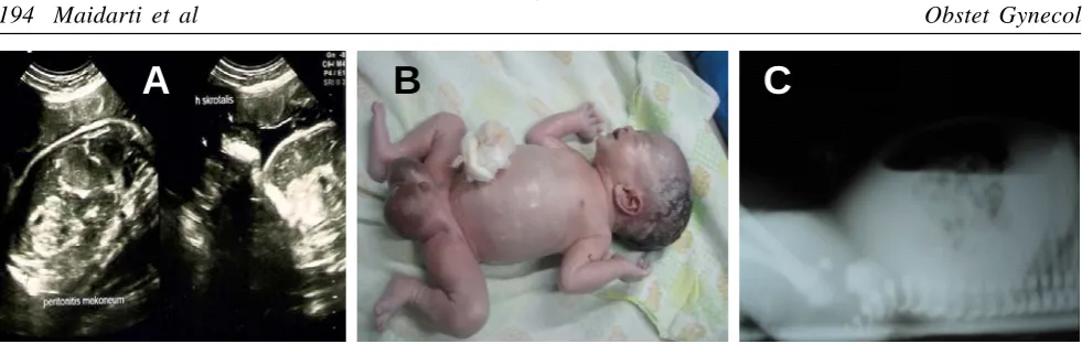
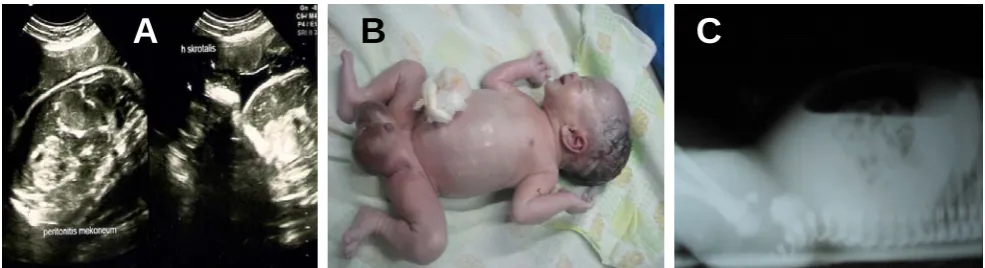
Figure 2: A. Prenatal ultrasound at 36 weeks of gestation revealed that the abdominal circumference was smaller than the head circumference, and calcium deposits around liver capsule.
B. An abdominal X-ray showing speckled calcification owing to meconium peritonitis. Arrows show the areas of calcification.
C. After birth, the child was noted to have marked abdominal distention such a degree as to cause decreased respiratory function. The abdomen was described as "shiny," with no bowel sounds heard and no visible bowel loops
over the abdomen.
A
C
A
Figure 1: A. Intra abdominal free fluid with increased genicity (meconium ascites), and intra-abdominal calcium deposition. B. Fetal surveillance revealed a reactive non stress test and systolic diastolic umbilical artery ratio was still normal.
B
By caesarean section delivered a live female baby with a birth weight of 3500 g and apgar scores 6 and 9 at 1 and 5 minutes, respectively. The child was noted at birth to have marked abdominal distention such a degree as to cause decreased respiratory func-tion (Figure 2). After immediate resuscitafunc-tion the baby was transferred to neonatal intensive care unit (NICU) for further evaluation. The abdomen was de-scribed as "shiny," with no bowel sounds heard and no visible bowel loops over the abdomen. An abdomi-nal x-ray showed free intestiabdomi-nal air that highly sug-gested viscous perforation (Figure 2). The baby un-derwent surgery at 24 hours of life and a persistent distal ileal perforation was discovered. The child was found to have a large meconium pseudocyst in the lower abdomen. A copious amount of meconium was removed, and the pseudocyst was resected, and an ileostomy was performed. After surgery, the baby’s severe respiratory difficulty quickly improved, but she was being continued to have a small supplemental oxygen requirement for several days. The immediate post-operative period was difficult due to bacterial colonization, signs of sepsis and complicated by feed-ing difficulties includfeed-ing a requirement for intrave-nous hyperalimentation. The child gained weight on a combination of total parenteral nutrition and enteral feeding. Finally the intestine was reversed 4 weeks later and evolution was without major complications. The baby continued to do well and was discharged on day 61 of life. At the age of 26 months, the baby has continued to thrive and is developmentally appro-priate. She is gaining weight normally and has normal bowel habit.
CASE 2
The patient was a 35-year-old woman in her fourth pregnancy, presented to Dr. Cipto Mangunkusumo ul-trasound clinic for routine antenatal ulul-trasound exami-nation. Ultrasound scan showed echogenic bowels with no other fetal abnormalities were noted, fetal biometry was consistent with 25 weeks gestation. Be-cause there were no other ultrasound abnormalities
including sign of bowel dilatation, hyperechogenic free fluid, so it was thought as a sign of intrauterine infection.
The family history was unremarkable, including no history of cystic fibrosis. After being advised of the possible causes, the patient was followed closely by serial fetal ultrasounds. The follow-up ultrasonogra-phy at 29 weeks of gestation revealed normal growth velocity, and echogenic material outside the bowel. The fetus was found to have an enlarged scrotum measuring 30x30 mm and containing a large fluid col-lection with densities of echogenicity, highly sugges-tive of calcifications. Scrotalis hernia was suspected (Figure 3). All these findings suggested intrauterine intestinal perforation. We suggested that the patient should be referred to the hospital with complete neo-natal facility to support the baby after delivery since regarding the neonate might need complete resuscita-tion. After 1 week the patient was presented to our policlinic and the pregnancy was complicated by se-vere preeclampsia. The patient was managed
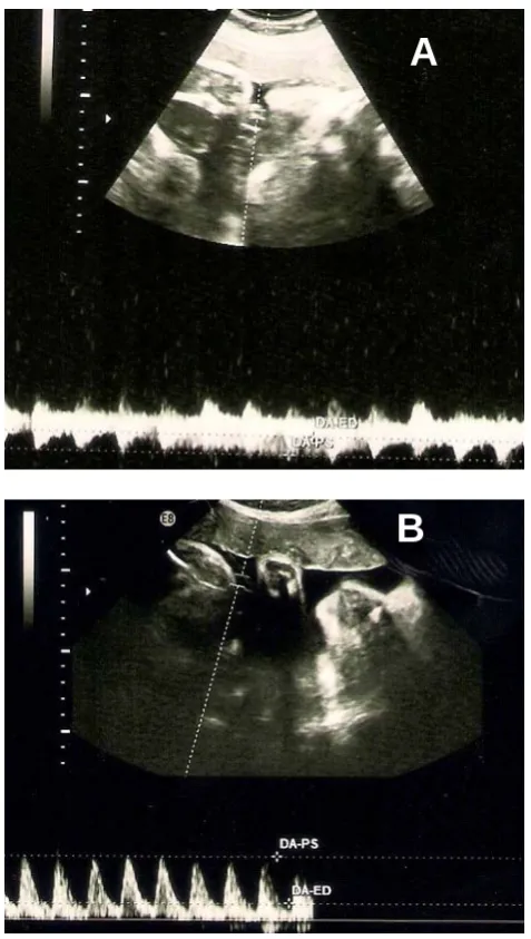
expec-Figure 4: Doppler measurements of the umbilical artery on followed up ultrasound on weekly outpatient visits at 34 weeks of gestation (Figure A) which was 3.4 and markedly increased the S/D ratio (3.9) at 35 weeks of gestation (Figure B)
B
Figure 3: Trans abdominal ultrasound image at 29 weeks’ gestation showing homogeneous appearance of the meconium filling the scrotum (open arrow) and scrotalis hernia (small arrow)
A
tantly with close monitoring of maternal and fetal conditions. Because of umbilical arterial flow deterio-ration (Figure 4), the pregnancy was terminated with the neonatology intensive care preparation.
The neonate weighing 2050 g was delivered by c-section on June 11th 2010. Apgar scores of 5 and 8 after 1 and 5 minutes, respectively. The neonate was admitted to the neonatal intensive care unit for further study. Plain abdominal radiographs showed right up-per quadrant calcification, an echogenic bowel, scat-tered peritoneal calcifications consistent with meco-nium peritonitis. No other fetal abnormalities were de-tected. A physical examination revealed abdominal distension and moderate scrotal swelling.
Since there was no vomiting or any other sign of intestinal obstruction, the baby was managed conser-vatively. He was kept nil orally and on intravenous (IV) fluids. Within 24 hours of expectant treatment, the baby was noted to have marked abdominal dis-tention resulting in severe respiratory distress. A peri-toneal drainage was inserted leading to evacuation of 400 ml of meconium. After improvement in his over-all status, the infant underwent a transverse laparo-tomy on the third day following delivery. The entire intestine was contained in a pseudo capsule and a large quantity of meconium and fibrin filled the
peri-toneal cavity. Meconium peritonitis was confirmed. When pseudo capsule was dissected from the intes-tine, a perforation in the terminal ileum, 5 cm proxi-mal to Bauhini valve was noted. A primary ileostomy was performed at the site of the perforation for fecal diversion.
His post-operative course was complicated by ane-mia and intestinal continuity was restored by end-to-end anastomosis 14 days after the first surgical pro-cedure. The baby was given pack cells transfusion. The baby was started to be given oral feeds seven days after the second operation. During this period, he received total parenteral nutrition. Scrotalis hernia was managed conservatively, no specific surgical pro-cedure was done. The boy is now 16 months old and subsequent follow-up revealed growth and develop-ment within normal range.
DISCUSSION
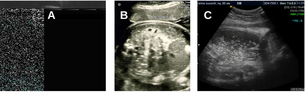
Meconium peritonitis results from in utero bowel per-foration and almost always involves small bowel which frequently complicates congenital bowel ob-structions. Approximately 20% of cases are a result of meconium inspissations secondary to cystic fibro-sis and 30% result from intestinal atresia or obstruc-Figure 5: A: Ultrasound followed up at 35 weeks gestation, abdominal free fluid with markedly increased echogenicity and
scrotalis hernia which was more apparent.
B. Within 24 hours of life the infant showed no sign of viscous obstruction nor perforation. The fetus was found to have an enlarged scrotum containing a large fluid collection with densities of echogenicity, highly suggestive of calcifications. C. Plain abdominal radiographs supine position of an 2-hours-old girl with air-fluid level (left image), the peripherally
displaced bowel loops with no remarkable distension.
C
B
A
Table 1. Etiology of meconium peritonitis.11
Common causes Uncommon causes
Meconium ileus (secondary to cystic fibrosis) Meckel’s diverticulum
Intestinal atresia Colonic atresia
Intestinal stenosis Torsion of a fallopian tube cyst
Intestinal hernia Perforated duplications
Hirschsprung’s disease Foetus in fetu
Volvulus Cytomegalovirus, hepatitis A and Parvovirus B19 infection
Intussusception Hydrometrocolpos
Extrinsic bands and adhesions Appendicitis
Duplication Rectal strictures Imperforate anus
Prenatal anoxic events causing bowel inchemia Vascular insufficiency including thrombosis Idiopathic
tion.5 Although half of all cases of prenatal bowel perforations and meconium peritonitis have no known etiology, most are probably secondary to a vascular event causing intestinal wall ischemia and eventual perforation.8 In addition, bowel ischemia secondary to prenatal anoxic events may cause perforation with subsequent meconium peritonitis.6
Prenatal bowel perforation usually occurs proximal to some form of obstruction, although this cannot al-ways be demonstrated.9 Lloyd suggested that intesti-nal perforation is associated with diminished mesen-teric blood flow due to perinatal asphyxia. Tibboel et al hypothesized that a temporary reduction in mesen-teric blood flow may lead to necrosis of the mucosa, which may in turn cause complete obstruction of the intestinal lumen, resulting in atresia of the small bowel.10 Persistent reduction of mesenteric blood flow can lead to transmural bowel necrosis. Summary of the etiologies of meconium peritonitis were listed in Table 1.
Following bowel perforation, meconium and diges-tive enzymes are extruded into the peritoneal cavity inciting an intense chemical peritonitis. Within days, giant cells and histiocytes surround the meconium, re-sulting in foreign body granulomas and calcification. The calcifications can be punctate, linear, or clumped foci. Meconium peritonitis can be distinguished from these other etiologies if the calcifications are perito-neal in distribution. The punctate foci are found only around the expected margin of the liver and not in the substance of the liver itself. The calcifications line up or form sheets along the diaphragm.12 Overtime the inflammatory response may completely seal the perforation.9 Prenatal bowel rupture results in leakage of sterile meconium into the abdominal cavity and the potential spaces connected with it. Since the processus vaginalis is patent in most fetuses, the spilled me-conium easily traverses the canal into the scrotum. Meconium then interacts with the tunica vaginalis to incite an intense inflammatory reaction that leads to a local mass and calcification.8
The clinical diagnosis of meconium peritonitis in both cases was made on the basis of ultrasound and abdominal plain film findings with the characteristic coarse hyperechogenic intra abdominal free fluid. The ultrasound findings of meconium peritonitis vary with gestational age.13 The most characteristic of ultra-sound finding is intraperitoneal calcification, but the identification of calcification in the fetal abdomen does not always indicate that the fetus has meconium peritonitis. Other potential etiologies include a neo-plasm (teratoma), or intraluminal calcifications in the fetus with anal atresia. As in the first case, at the first ultrasound examination, it was difficult to differenti-ate between meconium peritonitis and ruptured of the cystic teratoma, because both showed appearance of intraperitoneal coarse hyperechogenic intraabdominal free fluid.12
Meconium periorchitis in the second case was the result of a perforated viscus, which leaked meconium into the peritoneum. In both cases the process leading to meconium peritonitis was likely due to a sponta-neous intrauterine perforation of the gastrointestinal tract leaking meconium into the peritoneal cavity.14 Calcified scrotal masses may occur as the end result
of the passage of extraluminal meconium through the patent processus vaginalis, causing meconium perior-chitis. The pathological features suggestive of intrau-terine periorchitis are granulomatous inflammation with calcifications which may develop as early as 24 hours after leakage.15 Radiologically detectable calci-fications in the abdomen or scrotum with increased areas of echogenicity on scrotal ultrasonography are essential diagnostic points.16 Calcified scrotal masses can be due to other conditions such as teratomas, go-nadoblastomas, leydig cell tumors, testicular mi-crolithiasis, neonatal testicular torsion followed by hemorrhagic infarction and organized intra scrotal he-matoma.17 In the second case differential diagnosis was precluded because both scrotal and abdominal calcifications were present.17 In the first day of ad-mission diagnosis was made on the basis of single ultrasound finding, which was hyperechogenic bowel due to intrauterine infection. Rarely, increased intes-tinal echogenicity may be due to ingestion of gas-forming bacteria from the amniotic fluid in the setting of chorioamnionitis or ingestion of intra-amniotic blood.12 Because screening for infection was not per-formed routinely, the true incidence of fetal infections is likely to have been underestimated. Like in this case, due to financial constraint, not all laboratory markers for intrauterine fetal infection were exam-ined, including markers for the subclinical infection caused by cytomegalovirus, rubella, and parvovirus B19. So that the intra uterine infection as one of the causes can not be excluded yet. Therefore screening for infectious diseases should be implemented when hyperechoic fetal bowel is diagnosed by ultrasonogra-phy.10
The formation of inguinal scrotal hernias in the second case was aided by factor that acted to increase the intra-abdominal pressure, which was meconium peritonitis. Herniation of intra-abdominal contents through the internal ring via a patent processus vagi-nalis into the scrotum (indirect inguinal hernia) is a common cause of scrotal swelling. The diagnosis of a hernia would be possible, when peristalsis is iden-tified within the scrotum.15 A canal width of greater than 4 mm at the internal ring has a sensitivity of 95% in indicating the presence of a hernia.18 In this case the scrotal findings were isolated with no evi-dence of associated anomalies or growth disturbance on serial scans. The appearance of a scrotal mass on prenatal sonography can be non-specific, so an accu-rate prenatal diagnosis may be difficult. Even though a specific diagnosis may not be possible, awareness of the presence of an abnormality allows timely post-natal assessment.16
agnosis of Cystic fibrosis could not be done in this case, but diagnosis has no bearing on the final disease outcome. Parents should therefore be informed that the pregnancy can go to term.
According to sonographic findings in utero, the disease has been classified into three types. Type I (massive meconium ascites); type II (giant cyst) and type III (calcification and/or small pseudo-cyst). Studies reveal that type I and II patients are at higher morbid risk, as ventilator failure may occur due to elevated diaphragm, fetal hydrops or circula-tory failure may be precipitated due to massive me-conium ascites.21 The cystic type, often referred to as a meconium pseudocyst, is a collection of meconium lined by fibrous tissue and the walls of the adjoining intestine. In contrast, the ascetic type implies its as-sociation with ascites as a result of meconium leakage through a recent bowel perforation.20 Calcification was present in both cases that referred to meconium peritonitis type III.
The neonatal outcomes of meconium peritonitis are much better if it is detected prenatally than if it is diagnosed after birth. Early onset of meconium peri-tonitis is reportedly associated with a lower survival rate. Research performed by Chao-Nin Wang, et al in 2008 reported that early detection of meconium peri-tonitis correlated with a low rate of postnatal surgery and a favorable neonatal outcome. It was stated that the gestational age at diagnosis in the non-surgical group were significantly lower than those in the sur-gical group. Ultrasound features might be related to the need for postnatal surgical interventions. Few se-rial reports have demonstrated a relationship between the timing of detection and neonatal outcome. They hypothesized that the perforated intestine can heal spontaneously in utero if the prenatal period is long enough. However, if the meconium peritonitis is de-tected at an early gestational age, it is advisable to monitor fetal well-being throughout the rest of the prenatal period, rather than to terminate the preg-nancy.18 As in the second case the pathological proc-ess was observed by ultrasound over a period of weeks. The diagnosis of echogenic bowel was first made at 25 weeks gestation. Pregnancy was termi-nated in 35 weeks of gestation because the umbilical arterial flow waveform S/D of fetus was increased (3.9).
In both cases, the diagnosis of meconium peritoni-tis was made in utero, that could alert us to a fetus potentially at risk for complications. The pediatric surgical consultation had been done prenatally that could be helpful in providing counseling about the overall favorable prognosis in cases of prenatally di-agnosed meconium peritonitis. The parents can be ad-vised of the more guarded prognosis and the chance of surgical intervention in the neonatal period. Serial antenatal scans are necessary to assess the progress of meconium peritonitis as well as fetal growth and well-being. In the absence of an obstetric indication, the babies can be delivered vaginally at term. Parents are advised to deliver at a tertiary referral centre where neonatal intensive care and pediatrics surgical facilities are available, in order to optimize neonatal management. As in both cases we informed the pa-tient to deliver the baby in our hospital, and the
dication of c-section in both cases were obstetric in-dication. In the first case, c-section was performed due to markedly increased abdominal circumference. The fetal body weight (3500 g) was not a real body weight of the fetus, because it included total abdomi-nal free fluid which contained meconium. In the sec-ond case, the fetal was stopped to grow, with dete-rioration in fetal well being, that the pregnancy was terminated by c-section.
The infant underwent abdominal examination im-mediately after delivery, and a plain radiograph of the abdomen was obtained to evaluate the prenatal find-ings, and it was confirmed the diagnosis.22 Following delivery, the baby should be observed for features of bowel obstruction. Although bowel resection may be necessary postnatally, the prognosis for a fetus with meconium peritonitis is better than would be antici-pated from the neonatal literature. Oral feeding can commence if the baby passes meconium and does not develop signs of bowel obstruction. In one series, 78% of neonates with meconium peritonitis were managed conservatively without surgical interven-tion.23 However, if the neonate neither tolerate oral feeds nor has features of persistent bowel obstruction or signs of peritonitis, surgery is indicated.23
Therapy depends on the confidence level of the surgeon in establishing the diagnosis based on clinical evidence on radiographic findings. Both infant were found to have an isolated contained ileal perforation in a meconium pseudocyst. They both underwent ad-hesiolysis and ileostomy and made uneventful recov-ery. The natural history of meconium in the scrotum is that of spontaneous resolution and expectant man-agement is acceptable. However, when the diagnosis remains in doubt, surgical exploration is mandatory.16 No specific procedure was done regarding scrotal her-nia, because it will often resolve spontaneously with-out the need for operation, because it was shutter mechanisms compensating for raised intra-abdominal pressure. According to variable data, 40% of the pat-ent processus vaginalis close during first few months of life and an additional 20% close by 2 years of age.22 Since the ileal perforation was not associated with any systemic disorder or other abnormalities suggestive of a single gene disorder, the parents were informed that the recurrence risk in subsequent pregnancies was low.6
In conclusion, echogenic intraabdominal free fluid was the most common ultrasound findings in me-conium peritonitis. Meme-conium periorchitis is sus-pected when both scrotal and abdominal hyperecho-genic free fluid and abdominal plain film findings with the characteristic stippling calcification are pre-sent. Early detection of meconium peritonitis was not indicative of poor neonatal outcomes, and selective termination was not necessary, unless indicated for other reasons. The successful surgical treatment was attributed to antenatal sonographic diagnosis, ante-natal counseling, postante-natal fluid and electrolyte cor-rection, prophylactic antibiotics, and postnatal care. Immediate laparotomy before further bowel injury oc-curs might allow a better outcome.
can be seen in Table 2 below:
REFERENCES
1. Chan KL, Tang MH, Tse HY, Tang RY, Tam PK. Me-conium peritonitis: prenatal diagnosis, postnatal manage-ment and outcome. Prenatal diagnosis. 2005; 25:676-82. 2. Lally KP, Mehall JR, Xue H, Thompson J. Meconium
stimulates a pro-inflammatory response in peritoneal macrophages: implications for meconium peritonitis. J Pediatr Surg. 1999; 34:214-7.
3. Bronshtein M, Blazer S, Zimmer E. The gastrointestinal tract and abdominal wall. In: Callen P. Ultrasonography in Obstetrics and Gynecology 5th Ed. 2008: 601-5
4. Chang B. Palmer L. Meconium periorchitis presenting as scrotal masses. Urol. 2002; 59: 296
5. Abubakar A, Odelola M, Bode C, Sowande O, Bello M, Chinda C, Jalo I. Meconium peritonitis in Nigerian chil-dren. Annals of African Med 2008; 7: 187 - 91
6. Reynolds E, Douglass B, Bleacher J. Meconium peritonitis. J Perinatol 2000; 3: 193-5.
7. Foster MA, Nyberg DA, Mahony BS, Marks LA. Raabe RD. Meconium peritonitis: prenatal sonographic findings and their clinical significance. Radiology 1997: 661-5 8. Gilliland A, Carlan SJ, Greenbraum, Levy MC, Rich A.
Undescended testicle and a meconium-filled hemiscrotum: prenatal ultrasound appearance. Ultrasound Obstet Gynecol 2002; 20: 200-2.
9. Gupta R, Upreti L, Bhargava SK, Jain S, Shikha D. Images: Prenatal sonographic features of meconium peritonitis. In-dian J Radiol imaging 2004; 14:261-3.
10. Yang WJ, Chen CP, Chen CY, Tsung-hsien SU. Fetal me-conium peritonitis associated with prenatal methampheta-mine exposure. Taiwan J Obstet Gynecol. 2005; 44:180-2 11. Basu S, Kumar A, Pandey N. An unusual cause of me-conium peritonitis in a fetus. J Pediatr Child Health. 2009: 231-33
12. Hertzberg BS, Kliewer MA, Bowie JD. Sonography of the Fetal Gastrointestinal System. In: Fleischer AC. Manning FA. Jeanty P. Romero R. Sonography in obstetric and gy-necology 6th Ed. 2001: 410-21
Figure 6: A: Prenatal ultrasound of fetal ascites showed intra abdominal free fluid without echogenicity
B. Prenatal ultrasound of meconium peritonitis showed echogenic intra abdominal free fluid and calcification C. Intra abdominal cystic mass with clear border and irregular shape which contained non homogenous internal echo
floating within the mass, sometimes accompanied by the solid part.
C
B
A
Table 2. Brief summary of the differentiation among fetal ascites, meconium peritonitis and ruptured of cystic teratoma based on prenatal sonographic appearance.
Fetal ascites Meconium peritonitis Ruptured of cystic teratoma
Anechoic intra-abdominal free fluid. Coarse hyperechogenic intra abdominal free fluid.
Coarse hyperechogenic intra abdomi-nal free fluid.
Normal bowel loops floating freely in the ascitic fluid.
Dilated bowel loop appearance as tubular dilatation with limited mobility.
Bowel loop appearance and mobility can be normal. Intra abdominal cyst generally has a discrete appearance in comparison to dilated loop of bowel.
Tends to surround organs, collect in peritoneal recesses and no appearance of intra-abdominal hyperechogenic free fluid.
Coarse intra-abdominal internal echo are peritoneal in distribution (around the expected margin of the liver; bowel loop; line up or form sheets along the diaphragm).
Non homogen coarse internal echo floating within the mass.
No abnormal intraabdominal mass. No abnormal intra abdominal mass, bowel peristaltic might be diminished.
It may be cystic, solid or mixed. Some references stated as calcification (in 50% of cases), bowel peristaltic may be normal.
Often followed by fluid accumulation in other body cavity.
No evidence of free fluid accumulation in other body cavity.
No evidence of free fluid accumulation in other body cavity.
13. Wang CN, Chang SD, Chao AS, Wang TH, Tseng LH, Chang YL. Meconium peritonitis in utero-the value of pre-natal diagnosis in determining neopre-natal outcome. Taiwan J Obstet Gynecol. 2008; 47(4):391-96
14. Kojori F, DeMaria J. Scrotoschisis associated with me-conium periorchitis. J Pediatr Urol. 2007; 3: 415-6 15. Kesler SS, Glazier DB, Zaontz MR, Dean GE. Meconium
pearls in the scrotum. J Urol. 2000; 164: 1350-1
16. Han K, Mata J, Zaontz MR. Meconium masquerading as a scrotal mass – a case report. British journals of urology. 1998: 765-67
17. Soferman R, Ben-Sira L, Jurgenson U. Cystic fibrosis and neonatal calcified scrotal masses. J Cys Fibrosis. 2003; 2: 214-6
18. Rao P. Neonatal gastrointestinal imaging. Euro J Radiol. 2006; 60:171-86
19. Dirkes K, Crombleholme TM, Craigo SD, Latchaw LA, Jacir NN,Harris BH, and D’Alton ME. The Natural History of Meconium Peritonitis Diagnosed In Utero. J Pediatr Surg. 1995; 30: 979-82.
20. Jutt RC, Subramaniam R, Chia P. Meconium Peritonitis: Antenatal Diagnosis and Postnatal Outcome. J Paediatr Ob-stet Gynecol. 2000: 18-43
21. Shinkichi K, Keisuke N, Ishikawa S . Meconium peritonitis in utero. Pediatric surgery int 2000; 16: 377-9
22. Kyung JE, Choon SY, Pretorius DH. Prenatal Diagnosis of an Inguinoscrotal Hernia. J Ultrasound Med 2005; 24:239-42
23. Muller F, Dommergues M, Aubry MC,Bouy BS, Gautier E, Franqoise N, Hyperechogenic fetal bowel: An ultra-sonographic marker for adverse fetal and neonatal outcome. Am J Obstet Gynecol; 1995: 508-1
B
A
C
A
Figure 1: A. Intra abdominal free fluid with increased genicity (meconium ascites), and intra-abdominal calcium deposition. B. Fetal surveillance revealed a reactive non stress test and systolic diastolic umbilical artery ratio was still normal.
B
Figure 4: Doppler measurements of the umbilical artery on followed up ultrasound on weekly outpatient visits at 34 weeks of gestation (Figure A) which was 3.4 and markedly increased the S/D ratio (3.9) at 35 weeks of gestation (Figure B)
Fetal ascites Meconium peritonitis Ruptured of cystic teratoma
Anechoic intra-abdominal free fluid. Coarse hyperechogenic intra abdominal free fluid.
Coarse hyperechogenic intra abdomi-nal free fluid.
Normal bowel loops floating freely in the ascitic fluid.
Dilated bowel loop appearance as tubular dilatation with limited mobility.
Bowel loop appearance and mobility can be normal. Intra abdominal cyst generally has a discrete appearance in comparison to dilated loop of bowel.
Tends to surround organs, collect in peritoneal recesses and no appearance of intra-abdominal hyperechogenic free fluid.
Coarse intra-abdominal internal echo are peritoneal in distribution (around the expected margin of the liver; bowel loop; line up or form sheets along the diaphragm).
Non homogen coarse internal echo floating within the mass.
No abnormal intraabdominal mass. No abnormal intra abdominal mass, bowel peristaltic might be diminished.
It may be cystic, solid or mixed. Some references stated as calcification (in 50% of cases), bowel peristaltic may be normal.
Often followed by fluid accumulation in other body cavity.
No evidence of free fluid accumulation in other body cavity.
No evidence of free fluid accumulation in other body cavity.
C
B
A
C
B
A
Table 1. Etiology of meconium peritonitis.11
Common causes Uncommon causes
Meconium ileus (secondary to cystic fibrosis) Meckel’s diverticulum
Intestinal atresia Colonic atresia
Intestinal stenosis Torsion of a fallopian tube cyst
Intestinal hernia Perforated duplications
Hirschsprung’s disease Foetus in fetu
Volvulus Cytomegalovirus, hepatitis A and Parvovirus B19 infection
Intussusception Hydrometrocolpos
Extrinsic bands and adhesions Appendicitis
Duplication Rectal strictures Imperforate anus