O R I G I N A L A R T I C L E
Open Access
Symptomatic endometriosis developing
several years after menopause in the
absence of increased circulating estrogen
concentrations: a systematic review and
seven case reports
Fernanda de Almeida Asencio
1, Helizabet Abdalla Ribeiro
1, Paulo Ayrosa Ribeiro
1, Mario Malzoni
2, Leila Adamyan
3,
Anastasia Ussia
4,5, Victor Gomel
6, Dan C. Martin
7,8and Philippe R. Koninckx
4,5,9,10,11*Abstract
Background:To review women with symptomatic and clinically progressive endometriosis after menopause in the
absence of estrogen intake or excessive systemic endogenous production.
Design:Seven case reports and a systematic review of the literature from 1995 till February 2018.
Results:Only 7 case reports from the authors and 29 cases from the literature described women with either cystic
ovarian or deep endometriosis. Severity, symptoms, and localization are highly variable. No case report describes
symptomatic superficial typical lesions. In 22 of 36 women (61%), symptoms started more than 10 years after menopause.
Conclusions:Symptomatic and clinically progressive endometriosis after menopause in the absence of increased
systemic estrogen concentrations or exogenous estrogen intake starts more than 10 years after menopause in the majority of women. This observation suggests that a genetic and/or epigenetic incident caused estrogen-independent progression, increased sensitivity to estrogens or increased local production of estrogens. This observation is important for understanding the pathophysiology of endometriosis, for the management of postmenopausal endometriosis, and for individualization of medical therapy of endometriosis since estrogen-independent endometriosis growth probably also occurs before menopause.
Keywords:Endometriosis, Pathophysiology of endometriosis, Postmenopausal endometriosis, Genetics, Epigenetics
Introduction
Symptomatic endometriosis starting several years after menopause in women not taking hormone replacement therapy (HRT) is a rare condition. Case reports only de-scribe women with increasing pain, ovarian masses, or intestinal symptoms suspicious of a cancer. This is con-sistent with Kempers’[1] observations in 1960 on post-menopausal endometriosis. This was before estrogen assays, laparoscopy or ultrasound. Of all postmenopausal women operated for clinical symptoms between 1945
and 1958 in the Mayo clinic, endometriosis was found in 136 women, of whom 41 had symptoms compatible with endometriosis, 35 had an endometrioma, and 4 had pro-gressive severe pain and/or bowel symptoms. The article in addition reported that in a control group of 50 hyster-ectomies, 8 atrophic endometriosis lesions were found.
Endometriosis is considered a hormonally responsive endometrium-like tissue where estrogens stimulate growth and progestins stop growth and eventually cause deciduali-zation. It seems logical that after menopause endometriosis will become less active [2–4] since endometriosis is defined as (normal) endometrium-like tissue outside the uterus resulting from implantation of cells or endometrial frag-ments following retrograde menstruation [5]. However, the * Correspondence:[email protected]
4Gruppo Italo Belga, Villa del Rosario, Rome, Italy 5Università Cattolica, Rome, Italy
Full list of author information is available at the end of the article
pathophysiology of endometriosis [6] is unclear and endo-metriosis may result from dislocation of basal endomet-rium [7] and metaplasia of peritoneal mesothelial cells [8] or of peritoneal or endometrial stem cells [9]. That endo-metriosis is phenotypically different from endometrium is according to the implantation/metaplasia theory, consid-ered to be the consequence of the environmental factors in the peritoneal cavity, and of immunologic or of other pre-disposing factors.
The concept that endometriosis is a normal endometrium outside the uterus is not consistent with all clinical observa-tions. The clinical, histologic, and immuno-cytochemical differences suggested that peritoneal, ovarian, and deep endometriosis are three different diseases [10]. The nor-mal endometrium theory is not compatible with the clonal aspect of deep and cystic endometriosis [6]. Therefore, the endometriotic disease theory (EDT) [6, 11] and recently the genetic–epigenetic theory [12] suggest that the endo-metriotic cells are cells with a series of genetic or epigen-etic transformations with epigenepigen-etic defined as“heritable changes in gene function that do not involve changes in the DNA sequence.” These changes can occur in either mature adult or neonatal endometrium [13] following retrograde menstruation, or in stem/progenitor cells from the endometrium [13] or from the peritoneal cavity or even in bone marrow cells [14–16]. The specific combin-ation of genetic and epigenetic incidents will determine the development into typical, cystic, or deep endometri-osis. Subtle lesions are considered a mixture of implanted normal endometrium and of early endometriosis which after additional genetic and/or epigenetic incidents can develop into more severe lesions.
Stimulated by the observation of two women with pro-gressively increasing symptoms of endometriosis after menopause in the absence of increased estrogen secre-tion or intake, we decided to perform a systematic review of symptomatic postmenopausal endometriosis in the absence of increased systematic endogenous estro-gen production or estroestro-gen intake. The aim was to es-tablish its prevalence and clinical presentation.
Materials and methods
Case reports and systematic review
The aim of the study was to find women with progres-sively increasing symptoms caused by endometriosis after menopause and in the absence of estrogen intake or increased estrogen production. PubMed (comprising MEDLINE and Cochrane) and Scopus were searched ac-cording to the PRISMA checklist for publications in English between 1995 and February 2018 with “ endo-metriosis”AND (“postmenopause” OR“menopause”OR “menopausal”OR“postmenopausal”) in the title and for “endometriosis” AND “postmenopause” AND NOT “cancer” in the title, keywords, or abstract respectively.
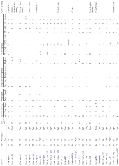
The 102 and 241 articles found, respectively, were manually screened for the absence of increased systemic endogenous estrogen production or estrogen intake in women with progressively increasing symptoms after menopause likely caused by endometriosis. The 29 re-ports found were searched for the age when the diagno-sis was made and for the number of years since surgical or spontaneous menopause. When the exact age of menopause was not available, we used 50 years (10 case reports, Table 1, indicated as *50*) to calculate years since menopause. A variation of a few years or omitting these reports did not change the conclusions. Years since menopause was considered important since inter-mittently increased ovarian estrogen secretion can occur during the first years after menopause. In addition, the following items were recorded: previous surgery espe-cially endometriosis surgery, drug intake, the presenting symptoms, the indication to start the investigation, pre-operative biochemistry and imaging, findings during surgery, and the final diagnosis by pathology. The sys-tematic review was submitted to PROSPERO but not ac-cepted since only a limited number of case reports were found. Our case reports were scrutinized in addition for the clinical exam with special attention to the presence or absence of vaginal atrophy, defined as an atrophic va-ginal appearance with para-basal cells on a wet smear.
The quality of evidence described in these case reports [17] is high since the descriptions have comprehensive details written by clinicians noting the uncommon find-ing durfind-ing surgery in postmenopausal women of severe endometriosis in the absence of increased circulating es-trogen concentrations. However, the reports obviously carry a publication bias, since only women with symp-toms sufficiently severe to perform surgery and in whom severe endometriosis was found were published. This re-view does not draw conclusions on the prevalence of symptomatic endometriosis after menopause, which is likely much higher. We know that unexpected endomet-riosis is frequent in women undergoing surgery after menopause [1].
We asked our colleagues (see the “Acknowledgments” section) with a known interest and a large experience of severe endometriosis surgery to retrieve all women in whom they had performed surgery for postmenopausal endometriosis without increased estrogen production or estrogen intake. This resulted in five additional cases and in a few cases of whom the records could not be retrieved.
Types of endometriosis: definitions used
limited to cystic ovarian endometriosis and larger nod-ules of deep endometriosis, the definitions used in this manuscript are as follows. Typical lesions are superficial (less than 5 mm) “powder burn” black dots in a fibrotic area. Subtle lesions are non-colored superficial lesions [18]. A discussion of the exact depth of subtle lesions, of their clinical significance [19], of the histological con-firmation of biopsies [20], of Müllerianosis [21], and of the significance of larger areas with subtle lesions is be-yond the scope of this article. Cystic endometrioses are the (larger) chocolate cysts of the ovary but not the smaller (3–6 mm) cysts under the peritoneum or close to the vagina in deep endometriosis. Deep endometriosis is defined as endometriosis deeper than 5 mm under the peritoneal surface [22]. A discussion of the limitations of depth to define deep endometriosis [12] is beyond the scope of this manuscript.
Results
Case reports of postmenopausal endometriosis in women without estrogen intake or signs of endogenous estrogen production
Only seven case reports could be collected, and ten sur-geons did not remember having seen such cases.
Case 1 (FA)
A 72-year-old woman with spontaneous menopause at age 54 was referred for increasing pelvic pain. She had her menarche at 11 years and did not have antecedents of dysmenorrhea, dyspareunia, dysuria, dyschezia, endo-metriosis, nor infertility. Her medical, surgical, and gynecological history was uneventful except a diverticu-litis and a volvulus 6 years before. She had never taken hormone replacement therapy (HRT), and the vaginal epithelium was atrophic. The gynecological exam and a transvaginal ultrasound confirmed a normal uterus and ovaries. However, it also showed a 1-cm arciform thicken-ing at the insertion of the right uterosacral ligament and a 24-mm nodule at 11 cm from the anus, on the recto-sigmoid, infiltrating the muscularis and affecting 15% of the circumference of the bowel. In the absence of other signs of malignancy, a laparoscopy was performed with a total hysterectomy, bilateral salpingo-oophorectomy, a seg-mental resection of the bowel, and a resection of the retro-cervical and right pararectal nodule. The pathology revealed active endometriotic glands and stroma in the retro-cervical and right pararectal nodules. The bowel le-sion had fibrosis secondary to diverticulitis. Recovery was uneventful, and she was discharged on day 5. After 4 weeks, she returned to her normal activities and is pain free.
Case 2 (FA)
A 64-year-old woman with spontaneous menopause at age 50 was referred for increasing pelvic pain,
dyspareunia, dyschezia, and repetitive constipation which had started some 5 years ago. She was a G3 P1 A2 with menarche at 11 years of age and a BMI of 37. At age 51, she had been operated for abdominal pain, nau-sea, and a large endometriotic mass close to the left kid-ney with signs of renal failure. An abdominal total hysterectomy with bilateral salpingo-oophorectomy and left nephrectomy had confirmed the diagnosis of endo-metriosis. She never had taken HRT, and the vaginal epi-thelium was atrophic. Clinical exam revealed a large painful nodule. MRI showed two nodules: one large nod-ule in the pouch of Douglas, invading the vaginal cuff and left parametrium and extending up to the hypogas-tric plexus and the left sacral nerves (S3 and S4); an-other nodule of 37 × 32 × 14 mm was seen in the rectum at 7.9 cm from the anal border, infiltrating the muscu-laris and submucosa. Transvaginal ultrasound examin-ation confirmed a 35 × 28 × 23 mm nodule in the vaginal cuff and a 41 × 10 × 19 mm nodule in the recto-sigmoid, compromising 50% of the circumference, in-filtrating the muscularis and submucosa at 9 cm from the anal border. A laparoscopic segmental bowel resection and resection of the deep endometriosis nodule invading the pelvic floor, the sacral nerves, and the left sciatic nerve con-firmed the diagnosis of endometriosis. Postoperative recov-ery was uneventful, and she was discharged on day 5 and is pain free for 5 months.
Case 3 (FA)
A 62-year-old woman with spontaneous menopause at age 45 was seen for increasing severe pelvic pain (10/10) and rectal pain with tenesmus. She was a P3G3 with me-narche at age 13, a BMI of 31, and hypertension. She had not taken HRT since menopause, and the vagina was atrophic. Four years before, she had undergone a recto-sigmoidectomy with resection of an endometriosis nodule from the right uterosacral ligament and rectovagi-nal septum. Not responding to treatment with GnRha after surgery, she returned in 2017. At MRI, a retro-cervical nodule infiltrating the apical portion of the vagina and a nodule (17 × 06 mm) in the sigmoid were found. Ultrason-ography confirmed the rectovaginal (15 × 4 mm) and the recto-sigmoid nodule (17 × 7 × 3 mm), compromising 25% of the circumference and infiltrating the muscularis. She is planned for surgery.
Case 4 (AU)
atrophic. The gynecological examination, MRI, and ultrasound were negative. A diagnostic laparoscopy re-vealed a large pelvic endometriosis plaque (5 × 4 cm) with small cystic spots (3–5 mm) in the pouch of Douglas and a deep endometriosis (1 × 1 cm) nodule around the right ureter, all of which were removed dur-ing the procedure. Pathology confirmed the diagnosis of endometriosis. The postoperative recovery was unevent-ful, and she is pain free for 1 year.
Case 5 (AU)
A 37-year-old woman with spontaneous menopause at age 32 visited for increasing pain symptoms for 1 year. She was a P0G0 with a menarche at age 9 and a BMI of 27. At 23 years of age, she had undergone an adnexect-omy for pain, and at age 27, we removed a rectovaginal deep endometriosis nodule extending around the left ur-eter (3 × 2 × 2 cm) and a cystic ovarian endometriosis from the remaining ovary. At age 32, a hysterectomy without ovariectomy was performed. Postoperatively, she became menopausal with increased FSH concentrations and a 17b-estradiol concentration of 80 pgr/ml, which is insufficient for endometrial proliferation. Following hys-terectomy, she was pain free for 4 years, after which she had progressively increasing pelvic pain on no hormonal therapy. Her clinical examination, MRI, and ultrasound demonstrated a 4 × 4 cm deep endometriosis nodule of the cecum. In order to avoid a fourth surgical interven-tion, she was treated with GnRha without success. We plan to try an aromatase inhibitor.
Case 6 (LA)
A 52-year-old woman with spontaneous menopause at age 48 visited for increasing pain symptoms for 1 year. She was a P3G8. At age 50, she had undergone a total laparoscopic hysterectomy because of pain and an ade-nomyotic uterus. She had not taken HRT, and clinical examination demonstrated vaginal atrophy. A large cys-tic ovarian endometriosis was diagnosed. The pathology of the excised cyst confirmed the endometriosis. The postoperative recovery was uneventful, and she now is pain free for 4 months.
Case 7 (MM)
A 67-year-old woman with spontaneous menopause at age 52 was referred because of chronic pelvic pain, dys-chezia, dyspareunia intermittent diarrhea since several years, and a suspicion of vaginal fornix endometriosis. She was a P0G0 with menarche at 12 years, a BMI of 32, and a history of sigmoid diverticulosis. She had not been taking HRT since menopause. The clinical examination revealed a vaginally visible retro-cervical nodule extend-ing to the left parametrium. By ultrasonography, this nodule (18 × 11 × 29 mm) was confirmed together with
another nodule (16 × 5 × 12 mm) at 8-cm distance from the anal verge. In addition to adenomyosis, a thickened endometrial layer suggestive for a polypoid hyperplasia, normal ovaries, and severe adnexal adhesions were found. MRI confirmed the retro-para-cervical nodule. After confirmation of endometrial hyperplasia without atypia on endometrial biopsy, a laparoscopic adhesioly-sis, total hysterectomy, bilateral salpingo-oophorectomy, ureterolysis with resection of the retro-para-cervical and vaginal bowel nodule, and shaving of a small rectal nod-ule were performed. Adenomyosis and deep infiltrating endometriosis were confirmed at histopathologic evalu-ation. The postoperative recovery was uneventful, and she was discharged on day 2. She is pain free for 2 years without signs of recurrence both clinically and at US.
A systematic review of clinically progressive
postmenopausal endometriosis without estrogen intake or increased production
Our review found 29 women (Table 1) with progres-sively increasing symptoms after menopause in whom endometriosis was diagnosed in the absence of HRT in-take or an increased endogenous estrogen production. The symptoms are variable and comprise increasing pain with urinary symptoms [26], an asymptomatic cystic ovarian endometrioma or a cystic ovarian endometrioma with pain [27–31], a small bowel obstruction [32, 33], a rectovaginal deep endometriosis [29,34], a sigmoid deep endometriosis [35], even a sigmoid obstruction more than 10 years after menopause [36], a deep endometriosis with progressive hydronefrosis [37], with renal failure [38] and with severe hypertension [39], urinary bleeding with an hydronephrosis and a polypoid intra-ureteral lesion [40], a vaginal endometriotic cyst [41], a lesion mimicking a bowel tumor [42], or an abdominal hemorrhage [43]. Some women were preoperatively suspected to have a can-cer either an adenocarcinoma [44] or a disseminated ovar-ian cancer although during surgery only superficial endometriosis lesions together with bilaterally large endo-metriomas were found [45]. Even endometriosis suspicious of a gastric cancer [46] or of a pancreatic tumor in a 69-year-old woman [47], endometriosis with inferior vena cava involvement [48], and endometriosis in the abdominal wall were described. A nodular wall endometriosis devel-oped in a woman without ovarian function treated with an aromatase inhibitor for breast cancer [49]. A new cystic endometrioma in the wall appeared spontaneously after the excision of two previous cysts. Plasma concentrations of 17b-estradiol were low (20–40 pg/ml); cyst fluid concen-trations were 80 pg/ml returning to normal after GnRha treatment [50]. A series of reports describe progressive endometriosis in women taking tamoxifen [51–54].
Figure1illustrates that a majority of women were over 60 years old and more than 10 years after menopause.
The treatment of symptomatic postmenopausal endo-metriosis seems to be surgical excision. If this cannot be performed, a treatment with aromatase inhibitors was suggested [55].
Discussion
The article of Kempers [1] and the three reviews on endometriosis found in women undergoing surgery after menopause [23–25] describe postmenopausal women who did not take HRT. It is not surprising that the prevalence decreases with age (Fig. 1) since the age of menopause is variable and since ovarian activity can be intermittently increased in the first years after pause. Unfortunately, the number of years since meno-pause is not always available. In addition, it is difficult to know whether the indication for surgery had been endo-metriosis, whether pain was caused by endoendo-metriosis, or whether endometriosis was a plausible explanation after surgery. They remain, however, remarkable documents with a detailed description of the clinical importance of endometriosis in women undergoing surgery after menopause [1]. The importance of postmenopausal endometriosis is also emphasized in this review of con-temporary articles. Endometriosis was considered to be the cause of postmenopausal surgery in 13 women out of 516 surgeries by Scott and Te Linde [56] and in 37 out of 1000 by Henriksen [57]. To interpret these data collected more than 60 years ago, we should remember that this was before the introduction of hormone re-placement therapy, estrogen assays, or ultrasound and with little knowledge of endocrinology such as
Fig. 1Women with progressive and symptomatic endometriosis after menopause (this series) and women operated after menopause in whom
peripheral conversion to estrogens and the intermittent and variable ovarian activity during the first years after menopause. This explains Kempers’ conclusion that these women must have had estrogen production some-where. He also clearly demonstrates that most women were young and (probably) early after menopause. Dif-ferences in life expectancy cannot explain the difDif-ferences between the data in the Kempers’article and in this art-icle since life expectancy after menopause was estimated to be 27 years in 1955 and 33 years today [58].
Our series describe women with progressive symptoms after menopause in whom endometriosis was diagnosed in the absence of estrogen intake or increased endogenous production. This is a rare condition, limited to case reports. Notwithstanding all limitations imposed by a recall bias, this also seems to be a rare condition when asking sur-geons with a large experience in endometriosis surgery. The biphasic frequency distribution of age and years after menopause moreover suggests two different endometriosis populations. The high incidence of endometriosis in women younger than 55 years old or early after menopause and decreasing thereafter is compatible with an estrogen-responsive tissue. The second group after 60 years or more than 10 years after menopause can only be explained by circulating estrogen-independent progression of endomet-riosis or a change in estrogen sensitivity or local produc-tion of estrogens. In more than half of the women, symptoms started more than 10 years after menopause. That women clearly remember when symptoms started moreover suggests that the progression of these endometri-osis lesions started abruptly at a specific point in time after menopause. This observation in postmenopausal women can be explained by the occurrence of a new genetic or epi-genetic incident as postulated by the epi-genetic–epigenetic theory to be the pathophysiology of endometriosis [12]. That such an incident is more likely to occur in women with a predisposition and with previous genetic–epigenetic incidents is supported by the observation that most of these women had been diagnosed with endometriosis before menopause. This genetic–epigenetic instability also is fully compatible with the rare occurrences of malignant trans-formation of endometriosis after menopause [59].
It is surprising that all reports of clinically progressive postmenopausal endometriosis lesions were either cystic ovarian endometriosis or deep endometriosis, without a single case report of clinically progressive superficial endo-metriosis. This contrasts with the high prevalence of typical lesions in premenopausal women who undergo a diagnostic laparoscopy for chronic pain. It is unclear, whether these women have less severe pain, and thus is not reported. The absence of reports thus can be a publication bias. An alter-native explanation could be that after menopause typical le-sions no longer cause pain. Only deep and cystic ovarian endometriosis seem to carry the risk that additional genetic
or epigenetic incidents cause them to start proliferation in the absence of increased circulating estrogens.
Our series permits the conclusion that (some) cystic or deep endometriosis lesions can progress notwithstanding the low postmenopausal estrogen concentrations which are insufficient to stimulate the endometrium or to lubri-cate the vagina. Conversion of precursor steroids to estro-gens in peripheral tissue, especially fat, might have played a role in some cases, as in the seventh case report with endometrial hyperplasia and obesity. In most cases, how-ever, peripheral conversion as a cause of endometriosis progression is unlikely considering the absence of endo-metrial stimulation and the onset of symptoms many years after menopause in non-obese women. An alterna-tive explanation could be a local estrogen production within the endometriosis lesions. Aromatase activity with local estrogen production and/or progesterone resistance in endometriosis lesions of young women are well known, although not yet described in postmenopausal endometri-osis. The mechanism and pathways for this aromatase activity and local estrogen production and lack of apop-tosis are moreover increasingly viewed as the result of genetic and/or epigenetic incidents or genetic polymorph-ism [2,60–62]. The case report of the wall endometrioma in a postmenopausal woman [50] confirms aromatase activity because of the increased, although still below 100 pg/ml, estrogen concentration in the cyst fluid.
These endometriosis lesions with progressive symp-toms and starting often more than 10 years after meno-pause strongly suggest heterogeneity of cystic and deep endometriosis. Indeed, most cystic and deep endometri-osis lesions are stimulated by estrogens and growth will be inhibited by progestins. This is moreover indirectly confirmed by the many reports of endometriosis in post-menopausal women taking hormone replacement therapy. However, some lesions grow in the absence of increased estrogen concentrations in plasma. The frequency of these circulating estrogen-independent lesions before meno-pause is unknown since they will only be diagnosed after menopause. This heterogeneity in cystic and deep endo-metriosis moreover is consistent with clinical observations during surgery. Heterogeneity of endometriosis lesions is also consistent with the variable response to medical therapy. Whereas the response of the endometrium to es-trogens and progestins is predictable, pain relief of endo-metriosis by medical therapy is much more variable. The occasional progression of deep endometriosis [63] or the severe bleedings [64] during pregnancy also supports het-erogeneity of lesions.
lesions can be explained with the genetic–epigenetic theory [6,12], postulating a cumulative series of genetic or epigen-etic incidents in the pathophysiology of endometriosis. Pre-disposition or susceptibility of endometriosis is considered to be caused by genetic and epigenetic incidents transmit-ted at birth. The transmittransmit-ted incidents are insufficient to express the disease, but a woman with more severe trans-mitted incidents is at higher risk to develop the disease when additional incidents occur later. The many differences in the endometrium of women with and without endomet-riosis can be considered as the expression of these incidents transmitted at birth. The variable genetic and/or epigenetic incidents can explain that endometriosis is heterogeneous. It also can explain that the growth of endometriosis can occur notwithstanding low circulating estrogen concentra-tions as evidenced by this series and by endometriosis in a man [65] not taking estrogens (the other six reported cases of endometriosis were in men who had been taking estro-gens for prostate cancer [66]). The onset of these progres-sive and symptomatic lesions more than 10 years after menopause suggests that new or additional incidents did occur. This poly-genetic and poly-epigenetic concept of pathophysiology of endometriotic disease also is fully com-patible with recent histochemical profiling of endometriosis [67] and the induction of epigenetic changes in endometri-osis [68].
Already in 1960 [1], Kempers described a series of women with active adenomyosis after menopause. Although the relationship between adenomyosis and endometriosis remains unclear, a similar pathogenesis of a combination of a series of genetic and/or epigenetic in-cidents can be postulated [69].
In conclusion, postmenopausal endometriosis progres-sing in the absence of estrogen intake or of a systemic increased production is an argument for the genetic– epigenetic theory to explain the pathophysiology of endometriosis. These lesions thus should be considered a benign tumor and no longer as normal endometrium or as “normal” metaplastic cells with an abnormal be-havior because of the environment or of the immun-ology. The direct clinical consequences of endometriosis progressing after menopause in the absence of increased estrogen concentrations are the heterogeneity of endomet-riosis lesions with a variable reaction during pregnancy and a variable response to medical therapy. Since this cir-culating estrogen-independent growth probably also oc-curs before menopause, women with medical therapy need an individual follow-up and therapy should be recon-sidered when lesions grow. After menopause, it is import-ant to recognize these lesions as different from a cancer.
Conclusions
Postmenopausal endometriosis can start to progress more than 10 years after menopause in the absence of
estrogen intake or of a systemic increased production. This suggests that an acute genetic or epigenetic inci-dent happened. The exact mechanism is not known, and it is unclear whether the growth of these lesions is gen independent or the consequence or an altered estro-gen sensitivity or of an altered estroestro-gen production within the lesion. It is unclear whether these are new le-sions or a change in existing lele-sions. This observation can be explained by the genetic–epigenetic theory which considers the pathophysiology of endometriosis as a cu-mulative series of genetic or epigenetic incidents. That the specific set of incidents is specific for each lesion can explain the heterogeneity of endometriosis lesions. After menopause, only incidents which stimulate growth in the absence of increased circulating estrogens will be-come clinically symptomatic. These lesions thus should be considered a benign tumor and not as “normal” metaplastic cells with an abnormal behavior because of the environment or of the immunology.
Acknowledgements
We thank Mauricio Abrao (Sao Paulo), Jacques Donnez (Belgium), Victor Gomel (Canada), Jörg Keckstein (Austria), Ceana and Camran Nezhat (USA), Antonio Setubal (Lisbon), Charles Koh (USA), Harry Reich and Assia Stepanian (USA) Arnaud Wattiez (Dubai) and Errico Zupi (Italy). Unfortunately they could not retrieve additional case reports.
Funding
No funding
Availability of data and materials
Records of case reports are available with the authors as indicated
Authors’contributions
FAA, PK, and AU contributed to the conception and design of the study. FAA, PA, and PK contributed to the acquisition of the data. All authors contributed to the drafting and revision and final approval of the manuscript.
Ethics approval and consent to participate
Not applicable
Consent for publication
All authors agreed for publication
Competing interests
The authors declare that they have no competing interests.
Publisher’s Note
Springer Nature remains neutral with regard to jurisdictional claims in published maps and institutional affiliations.
Author details
1Laparoscopy and Endometriosis Group Department, Santa Casa School of
Medical Sciences of São Paulo, São Paulo, Brazil.2Endoscopica Malzoni, Center for Advanced Endoscopic Gynecologic Surgery, Via C. Errico 2, 83100 Avellino, Italy.3Department of Operative Gynecology, Federal State Budget
Institution V. I. Kulakov Research Center for Obstetrics, Gynecology, and Perinatology, Ministry of Health of the Russian Federation, Moscow, Russia; and e Department of Reproductive Medicine and Surgery, Moscow State University of Medicine and Dentistry, Moscow, Russia.4Gruppo Italo Belga,
Villa del Rosario, Rome, Italy.5Università Cattolica, Rome, Italy.6Department
of Obstetrics and Gynecology, University of British Columbia Women’s Hospital, Vancouver, BC, Canada.7School of Medicine, University of
Tennessee Health Science Center, Memphis, TN, USA.8Institutional Review
Vuilenbos 2, 3360 Bierbeek, Belgium.10University of Oxford, Oxford, UK. 11Moscow State University, Moscow, Russia.
Received: 23 September 2018 Accepted: 10 January 2019
References
1. Kempers RD, Dockerty MB, Hunt AB, Symmonds RE (1960) Significant
postmenopausal endometriosis. Surg Gynecol Obstet 111:348–356
2. Bulun SE (2009) Endometriosis. N Engl J Med 360:268–279
3. Cumiskey J, Whyte P, Kelehan P, Gibbons D (2008) A detailed morphologic
and immunohistochemical comparison of pre- and postmenopausal
endometriosis. J Clin Pathol 61:455–459
4. Streuli I, Gaitzsch H, Wenger JM, Petignat P (2017) Endometriosis after
menopause: physiopathology and management of an uncommon
condition. Climacteric 20:138–143
5. Sampson JA (1925) Heterotopic or misplaced endometrial tissue.
Am J Obstet Gynecol 10:649–664
6. Gordts S, Koninckx P, Brosens I (2017) Pathogenesis of deep endometriosis.
Fertil Steril 108:872–885
7. Leyendecker G, Herbertz M, Kunz G, Mall G (2002) Endometriosis results
from the dislocation of basal endometrium. Hum Reprod 17:2725–2736
8. Gruenwald P (1942) Origin of endometriosis from the mesenchyme of the
celomic walls. Am J Obstet Gynecol 44:470–474
9. Cousins FL, DF O, Gargett CE (2018) Endometrial stem/progenitor cells and
their role in the pathogenesis of endometriosis. Best Pract Res Clin Obstet Gynaecol 50:12
10. Nisolle M, Donnez J (1997) Peritoneal endometriosis, ovarian endometriosis,
and adenomyotic nodules of the rectovaginal septum are three different
entities. Fertil Steril 68:585–596
11. Koninckx PR, Barlow D, Kennedy S (1999) Implantation versus infiltration: the
Sampson versus the endometriotic disease theory. Gynecol Obstet Investig
47(Suppl 1):3–9
12. Koninckx PR, Ussia A, Adamyan L, Wattiez A, Gomel V, Martin D (2018)
Pathogenesis of endometriosis: the genetic-epigenetic theory. Fertil Steril in press
13. Puttemans P, Benagiano G, Gargett C, Romero R, Guo SW, Brosens I (2017)
Neonatal uterine bleeding as a biomarker for reproductive disorders during adolescence: a worldwide call for systematic registration by nurse midwife.
J Matern Fetal Neonatal Med 30:1434–1436
14. Sandoval P, Jimenez-Heffernan JA, Guerra-Azcona G, Perez-Lozano ML,
Rynne-Vidal A, Albar-Vizcaino P, Gil-Vera F, Martin P, Coronado MJ, Barcena C, Dotor J, Majano PL, Peralta AA, Lopez-Cabrera M (2016) Mesothelial-to-mesenchymal transition in the pathogenesis of post-surgical
peritoneal adhesions. J Pathol 239:48–59
15. Cheng Y, Li L, Wang D, Guo Q, He Y, Liang T, Sun L, Wang X, Cheng Y,
Zhang G (2017) Characteristics of human endometrium-derived mesenchymal stem cells and their tropism to endometriosis. Stem Cells Int 2017:4794827
16. Lucas PA (2007) Stem cells for mesothelial repair: an understudied modality.
Int J Artif Organs 30:550–556
17. Guyatt GH, Oxman AD, Kunz R, Woodcock J, Brozek J, Helfand M,
Alonso-Coello P, Glasziou P, Jaeschke R, Akl EA, Norris S, Vist G, Dahm P, Shukla VK, Higgins J, Falck-Ytter Y, Schunemann HJ (2011) GRADE guidelines: 7. Rating the quality of evidence--inconsistency. J Clin Epidemiol
64:1294–1302
18. Martin DC, Hubert GD, Levy BS (1989) Depth of infiltration of endometriosis.
J Gynecol Surg 5:55–60
19. Koninckx PR, Donnez J, Brosens I (2016) Microscopic endometriosis: impact on
our understanding of the disease and its surgery. Fertil Steril 105:305–306
20. Martin DC (2003) Endometriosis: correlation between histologic and visual
findings at laparoscopy. Am J Obstet Gynecol 188:1663–1664
21. Batt RE, Smith RA, Buck Louis GM, Martin DC, Chapron C, Koninckx PR,
Yeh J (2007) Mullerianosis. Histol Histopathol 22:1161–1166
22. Koninckx PR, Martin DC (1992) Deep endometriosis: a consequence of
infiltration or retraction or possibly adenomyosis externa? Fertil Steril
58:924–928
23. Oxholm D, Knudsen UB, Kryger-Baggesen N, Ravn P (2007) Postmenopausal
endometriosis. Acta Obstet Gynecol Scand:1–7. 87:1158-64
24. Punnonen R, Klemi PJ, Nikkanen V (1980) Postmenopausal endometriosis.
Eur J Obstet Gynecol Reprod Biol 11:195–200
25. Morotti M, Remorgida V, Venturini PL, Ferrero S (2012) Endometriosis in
menopause: a single institution experience. Arch Gynecol Obstet
286:1571–1575
26. Maeda T, Uchida Y, Nakajima F (2009) Vesical endometriosis following the
menopause. Int Urogynecol J Pelvic Floor Dysfunct
27. Manero MG, Royo P, Olartecoechea B, Alcazar JL (2009) Endometriosis in a
postmenopausal woman without previous hormonal therapy: a case report. J Med Case Rep 3:135
28. Matsushima T, Asakura H (2016) Huge ovarian endometrioma that grew
after menopause: case report. J Obstet Gynaecol Res 42:350–352
29. Rosa e Silva JC, Carvalho BR, Barbosa HF, Poli-Neto OB, Rosa e Silva AC,
Candido dos Reis FJ, Nogueira AA (2008) Endometriosis in postmenopausal women without previous hormonal therapy: report of three cases.
Climacteric 11:525–528
30. Medina N, Martín A, Guillén V, Andújar M, García JA (2005) Ovarian
endometrioma in a postmenopausal woman unrelated to neoplasia or exogenous hormone therapy. Progresos de Obstetricia y Ginecologia
48:150–153
31. Bellina JH, Schenck D (2000) Large postmenopausal ovarian endometrioma.
Obstet Gynecol 96:846
32. Izuishi K, Sano T, Shiota A, Mori H, Ebara K (2015) Small bowel obstruction
caused by endometriosis in a postmenopausal woman. Asian J Endosc Surg
8:205–208
33. Popoutchi P, Lemos CR, Silva JC, Nogueira AA, Feres O, Rocha JJ (2008)
Postmenopausal intestinal obstructive endometriosis: case report and
review of the literature. Sao Paulo Med J 126:190–193
34. Torres-Rincón RA, Moreno-Rojas A, Salinas-Parra C (2017) Endometriosis of
the cecum in a postmenopausal women: case report and literature review.
Iatreia 30:333–339
35. Deval B, Rafii A, Felce DM, Kermanash R, Levardon M (2002) Sigmoid
endometriosis in a postmenopausal woman. Am J Obstet Gynecol
187:1723–1725
36. Bidarmaghz B, Shekhar A, Hendahewa R (2016) Sigmoid endometriosis in a
post-menopausal woman leading to acute large bowel obstruction: a case
report. Int J Surg Case Rep 28:65–67
37. Gudla VR, Tangudu S (2012) Postmenopausal endometriosis with ureteric
involvement. Radiol Case Rep 7:607
38. Bailey AP, Schutt AK, Modesitt SC (2010) Florid endometriosis in a
postmenopausal woman. Fertil Steril 94(2769):e2761–e2764
39. Khong SY, Lam A, Coombes G, Ford S (2010) Surgical management of
recurrent ureteric endometriosis causing recurrent hypertension in a
postmenopausal woman. J Minim Invasive Gynecol 17:100–103
40. Zhuang L, Eisinger D, Jaworski R (2017) A case of ureteric polypoid
endometriosis presenting in a post-menopausal woman. Pathology
41. Rabinerson D, Avrech O, Kaplan B, Braslavsky D, Goldman GA, Neri A (1996)
Endometrioma of the vagina in menopause. Acta Obstet Gynecol Scand
75:506–507
42. Jakhmola CK, Kumar A, Sunita BS (2016) Expect the unexpected:
endometriosis mimicking a rectal carcinoma in a post-menopausal lady.
J Minim Access Surg 12:179–181
43. Bhat RA, Teo M, Bhat AK (2014) Endometriosis after surgical menopause
mimicking pelvic malignancy: surgeons’predicament. Oman Med J 29:226–231
44. Suchonska B, Gajewska M, Zygula A, Wielgos M (2018) Endometriosis
resembling endometrial cancer in a postmenopausal patient. Climacteric
21:88–91
45. Agarwal Sharma R, Lee EY, Vardhanabhuti V, Khong PL, Ngu SF (2016)
Unusual case of postmenopausal diffuse endometriosis mimicking
metastastic ovarian malignancy. Clin Nucl Med 41:e120–e122
46. Mohamed AAA, Selim YARM, Arif MA, Albroumi SA (2016) Gastric wall
endometriosis in a postmenopausal woman. Egypt J Radiol Nucl Med
47:1783–1786
47. Plodeck V, Sommer U, Baretton GB, Aust DE, Laniado M, Hoffmann RT,
Platzek I (2016) A rare case of pancreatic endometriosis in a
postmenopausal woman and review of the literature. Acta Radiol Open 5:2058460116669385
48. Flyckt R, Lyden S, Roma A, Falcone T (2011) Post-menopausal endometriosis
with inferior vena cava invasion requiring surgical management. Hum
Reprod 26:2709–2712
49. Cameron M, Westwell S, Subramanian A, Ramesar K, Howlett D (2017)
Postmenopausal cutaneous endometriosis: mimicking breast metastasis.
50. Sasson IE, Taylor HS (2009) Aromatase inhibitor for treatment of a recurrent abdominal wall endometrioma in a postmenopausal woman. Fertil Steril
92:1170–1174
51. Jaegle WT, Barnett JC, Stralka BR, Chappell NP (2017) Polypoid
endometriosis mimicking invasive cancer in an obese, postmenopausal
tamoxifen user. Gynecol Oncol Rep 22:105–107
52. Bese T, Simsek Y, Bese N, Ilvan S, Arvas M (2003) Extensive pelvic
endometriosis with malignant change in tamoxifen-treated postmenopausal
women. Int J Gynecol Cancer 13:376–380
53. Ismail SM, Maulik TG (1997) Tamoxifen-associated post-menopausal
endometriosis. Histopathology 30:187–191
54. Buckley CH (1997) Tamoxifen-associated postmenopausal endometriosis.
Histopathology 31:296
55. Polyzos NP, Fatemi HM, Zavos A, Grimbizis G, Kyrou D, Velasco JG, Devroey P,
Tarlatzis B, Papanikolaou EG (2011) Aromatase inhibitors in post-menopausal endometriosis. Reprod Biol Endocrinol 9:90
56. Scott RB, Te Linde RW (1950) External endometriosis-the scourge of the
private patient. Ann Surg 131:697–720
57. Henriksen E (1955) Endometriosis. Am J Surg 90:331–337
58. Glei DA, Mesle F, Vallin J (2010) Diverging trends in life expectancy at age
50: a look at causes of death. In: Crimmins EM, Preston SH, Cohen B (eds) International differences in mortality at older ages: dimensions and sources. National Academies Press (US), Washington (DC)
59. Taylor AA, Kenny N, Edmonds S, Hole L, Norbrook M, English J (2005)
Postmenopausal endometriosis and malignant transformation of
endometriosis: a case series. Gynecol Surg 2:135–137
60. Bulun SE, Monsivais D, Kakinuma T, Furukawa Y, Bernardi L, Pavone ME,
Dyson M (2015) Molecular biology of endometriosis: from aromatase to
genomic abnormalities. Semin Reprod Med 33:220–224
61. Dyson MT, Roqueiro D, Monsivais D, Ercan CM, Pavone ME, Brooks DC,
Kakinuma T, Ono M, Jafari N, Dai Y, Bulun SE (2014) Genome-wide DNA methylation analysis predicts an epigenetic switch for GATA factor expression in endometriosis. PLoS Genet 10:e1004158
62. Xue Q, Zhou YF, Zhu SN, Bulun SE (2011) Hypermethylation of the CpG
island spanning from exon II to intron III is associated with steroidogenic factor 1 expression in stromal cells of endometriosis. Reprod Sci
18:1080–1084
63. Setubal A, Sidiropoulou Z, Torgal M, Casal E, Lourenco C, Koninckx P (2014)
Bowel complications of deep endometriosis during pregnancy or in vitro
fertilization. Fertil Steril 101:442–446
64. Brosens IA, Lier MC, Mijatovic V, Habiba M, Benagiano G (2016) Severe
spontaneous hemoperitoneum in pregnancy may be linked to in vitro fertilization in patients with endometriosis: a systematic review. Fertil Steril
106:692–703
65. Giannarini G, Scott CA, Moro U, Grossetti B, Pomara G, Selli C (2006) Cystic
endometriosis of the epididymis. Urology 68:203
66. Fukunaga M (2012) Paratesticular endometriosis in a man with a prolonged
hormonal therapy for prostatic carcinoma. Pathol Res Pract 208:59–61
67. Liu X, Zhang Q, Guo SW (2018) Histological and immunohistochemical
characterization of the similarity and difference between ovarian
endometriomas and deep infiltrating endometriosis. Reprod Sci 25:329–340
68. Zhang Q, Dong P, Liu X, Sakuragi N, Guo SW (2017) Enhancer of Zeste
homolog 2 (EZH2) induces epithelial-mesenchymal transition in endometriosis. Sci Rep 7:6804
69. Koninckx PR, Ussia A, Zupi E, Gomel V (2018) The relationship of endometriosis