Arzum G. Dogru
1, Selcuk Tunik
2, Veysi Akpolat
3, Mehmet Dogru
4,
Ebru E. Saribas
1, Filiz A. Kaya
1, Yusuf Nergiz
2The Effects of Pulsed and Sinusoidal Electromagnetic
Fields on E-cadherin and Type IV Collagen in Gingiva:
A Histopathological and Immunohistochemical Study
Wpływ impulsowego i sinusoidalnego pola elektromagnetycznego
na E-kadherynę i kolagen typu IV w dziąśle – badanie histopatologiczne
i immunohistochemiczne
1 Department of Periodontology, Faculty of Dentistry, University of Dicle, Turkey
2 Department of Histology and Embryology, Faculty of Medicine, University of Dicle, Turkey 3 Department of Biophysics, Faculty of Medicine, University of Dicle, Turkey
4 Department of Orthodontics, Faculty of Dentistry, University of Dicle, Turkey
A – research concept and design; B – collection and/or assembly of data; C – data analysis and interpretation;
D – writing the article; E – critical revision of the article; F – final approval of article; G – other
Abstract
Background. The potential beneficial effects of extremely low frequency pulsed and sinusoidal electromagnetic fields have been shown on many tissues. Gingival epithelium plays an important role in immunosurveillance of the periodontal tissues. The epithelium acts as a mechanical barrier through cell junctions such as E-cadherin.
Objectives. Investigation of the effects of extremely low frequency magnetic fields on gingiva.
Material and Methods. Twenty-seven male Wistar albino rats were used. The rats were divided into three groups: control group (n = 9), SEMF group (n = 9), PEMF group (n = 9). The SEMF and PEMF (pulse time: 25 µsn, pulse frequency: 50 Hz) groups were subjected to 1.5 mT, 50 Hz, exposure 6 h a day, 5 days a week for 28 days in meth-acrylate boxes. The gingival tissue pieces processed for routine histological and immunohistochemical examination and tissue sections were stained with H–E and Masson trichrome. In addition, E-cadherin and type IV collagen expressions were examined immunohistochemically.
Results. Intraepithelial lymphocytes and proliferation of epithelial cells increased in both electromagnetic field groups. The over-expressions of E-cadherin on gingival epithelium was detected in the PEMF and SEMF groups. The expression level of type IV collagen was not significant between the control and electromagnetic field treated groups, except for a significant increase in the basal cell layer of the PEMF group, as compared to the control and SEMF groups.
Conclusions. PEMF and SEMF have a local pro-inflammatory effect on gingiva, leading to an increase in E-cadherin level but not type IV collagen. Both PEMF and SEMF can be used as a supportive device in the treatment of gingival diseases, especially those which lead to defects in the epithelial barrier (Adv Clin Exp Med 2013, 22, 2, 245–252).
Key words: type IV collagen, E-cadherin, histopathology, pulsed and sinusoidal electromagnetic fields.
Streszczenie
Wprowadzenie. Potencjalne korzystne działanie impulsowych sinusoidalnych pól elektromagnetycznych bardzo niskiej częstotliwości wykazano w wielu tkankach. Nabłonek dziąseł odgrywa ważną rolę w dozorze immunolo-gicznym tkanek przyzębia. Nabłonek działa jako bariera mechaniczna w połączeniach komórkowych, takich jak e-kadheryna.
Cel pracy. Zbadanie wpływu pól magnetycznych bardzo niskiej częstotliwości na dziąsła.
Materiał i metody. Do badań włączono 27 szczurów albinosów płci męskiej szczepu Wistar. Szczury podzielono na trzy grupy: grupę kontrolną (n = 9), SEMF (n = 9), PEMF (n = 9). Osobniki w grupach SEMF i PEMF (czas
Adv Clin Exp Med 2013, 22, 2, 245–252 ISSN 1899–5276
OrIGINAl PAPErS
There has been an increasing attempt to re-search the possible roles and effects of extremely low frequency electromagnetic fields (ElF-EMF) on the tissues and organs of the body over the last three decades [1]. The potential effects of ElF-EMF were shown in several earlier studies. These effects are enhancement of DNA synthesis [2], decrease in bone resorption and maintenance of bone mass [3], protein synthesis [4–6] and gap junctional intercellular communication complex [7], and stimulation of nerve regeneration [8]. Most of the clinical studies and applications were performed with a pulsed electromagnetic field (PEMF), hence only little work has been carried out to evaluate the impacts of sinusoidal electro-magnetic field (SEMF). The effect of the mecha-nism of this type of electromagnetic field on tis-sues was determined to be different from those of PEMF [9]. As Steffensens et al. wrote, “clinical studies in humans have reported enhanced healing and bone reorganization of nonunion fractures and pseudoarthroses by electrical stimulation following long term unsuccessful conventional treatments” [10]. The epithelium acts as a me-chanical barrier through cell junctions such as E-cadherin [11, 12]. As Udey declared, “E-E-cadherin is a calcium-dependant homophilic cell adhesion molecule that helps in cell-cell interaction.” In addition, E-cadherin interaction is thought to be important for retention of the langerhans cells in the epithelial cells [13]. Gingival epithelium plays an important role in immunosurveillance of the periodontal tissues through the presence of immune cells and production of antimicrobial peptides [14]. The expression of E-cadherin has been shown to be indirectly affected by periodon-tal disease, presumably as a result of proteolytic destruction by putative periodontal pathogens [15, 16]. ElF-EMF application is described as a treatment method with desirable results where the traditional methods fail. Nevertheless, the possible role of EMF on healthy tissues has still
not been elucidated. The aim of this study was to investigate the effects of extremely low frequency pulsed and sinusoidal electromagnetic fields on E-cadherin, which mediate intercellular adhesion between the epithelial cells and type IV collagen expressions in gingiva.
Material and Methods
Animal Care and Preparations
for Experimental Animals
The experiments were performed on 27 male Wistar albino rats with initial weights of 150–230 g and aged 4 months approximately, obtained from the Medical Science Application and research Center of the University of Dicle. All of the rats were permitted free access to water and standard pellet food diet (TAVAS Inc., Adana, Turkey) dur-ing the experimental period. All of the rats used in this study were divided into three equal groups: control (Cnt) group (n:9), SEMF group (n:9), PEMF group (n:9). The SEMF and PEMF (pulse time: 25 µsn, pulse frequency: 50 Hz) groups were subjected to 1.5 mT, 50 Hz, exposure 6 h a day, 5 days a week for 28 days in methacrylate boxes (43 × 42 × 15 cm). The animals were kept in a 14/10h light/dark environment at a constant temperature of 22 ± 3°C, and 45 ± 10% humidity.
Generation and Application
of Magnetic Fields
The EMF was generated in a device designed and used as noted in an earlier publication, which had two pairs of Helmholtz coils 70 cm in diameter in a Faraday cage (130×65×80 cm) that was earthed to shield it against the electric component (Fig. 1). This magnet was constructed by winding 125 turns of insulated soft copper wire with a diameter of 1.5 mm. Coils were placed vertically as facing one
impulsu: 25 μsn, częstotliwość impulsu: 50 Hz) poddano działaniu pola magnetycznego 1,5 mT, 50 Hz, ekspozycja po 6 godz./dobę, 5 dni w tygodniu przez 28 dni w klatkach z metakrylanu. Tkankę dziąsła przygotowano do ruty-nowego badania histologicznego i immunohistochemicznego. Skrawki tkanek barwiono HE i wg Massona. Ponadto zbadano immunohistochemicznie ekspresję e-kadheryny i kolagenu typu IV.
Wyniki. liczba limfocytów śródnabłonkowych i proliferacja komórek nabłonkowych zwiększyły się w obu grupach poddanych wpływowi pola elektromagnetycznego. Nadmierną ekspresję e-kadheryny w nabłonku dziąsła wykryto w grupach PEMF i SEMF. Ekspresja kolagenu typu IV nie była znacząca między grupą kontrolną i grupami pod-danymi wpływowi pola elektromagnetycznego, z wyjątkiem znacznego zwiększenia się warstwy komórek podstaw-nych w grupie PEMF, w porównaniu do grupy kontrolnej i SEMF.
Wnioski. PEMF i SEMF wywierają miejscowe działanie prozapalne na dziąsła, co prowadzi do wzrostu stężenia e-kadheryny, ale nie kolagenu typu IV. Zarówno PEMF, jak i SEMF mogą być stosowane do wspomagania leczenia chorób dziąseł, zwłaszcza takich, które prowadzą do uszkodzenia bariery nabłonka (Adv Clin Exp Med 2013, 22, 2, 245–252).
another. The distance between the coils was 47 cm. An AC current produced by an AC power supply (DAYM, Turkey) was passed through the device. The current in the wires of the energized exposure solenoid was 40 A for 1.5 mT, which resulted in a 50 Hz MF. The MF intensities were measured once per week as 1.5 mT in 15 different points of the methacrylate cage with a Bell 7030 Gauss/ Teslameter (F.W. BEll, Inc., SYPrIS, Orlando, Florida, USA), to ensure homogeneity of the field during the course of the experiment by a person who was not involved in the animal experiment. The magnetic field measurements showed that, under the conditions of the experiment, the mag-netic field exposure system produced a stable flux density of 1.5 mT and stable frequency of 50 Hz with negligible harmonics and no transients. No temperature differences were observed between exposure and cages during the exposure [17]. For the control, nothing was applied to the rats in this group, and they completed their life cycle in the cage during the study period. The rats were free in a methacrylate cage inside the coils.
Tissue Preparation for
Histopathological Examination
A gingival tissue specimen was obtained from the same region of the gingiva after high dose Ket-amine HCl (Ketalar, Pfizer) sacrification. On exci-sion, tissues were fixed in 10% buffered formalin for 16 hours and then washed overnight. They were dehydrated with graded alcohol series pri-marily at 30% absolute ethanol, and next embed-ded into paraffin. All the sections (histological and immunohistochemical) were evaluated and photo-graphed by using a light microscope (Eclipse i80, Nikon, Japan).
Histological Examination
The paraffin blocks were cut into 5 μm sec-tions and stained with Hematoxylin–Eosin (H-E) and Masson Trichrome. All layers of the epithe-lium and underlying connective tissue were evalu-ated histopathologically under light microscope.
Immunohistochemistry
The tissues were put into a formalin solution for fixation and then embedded in paraffin wax. Then they were cut into 4–6 μm sections on positively charged glass slides. Sections were deparaffinized with xylene, followed by immersion in graded al-cohol for dehydration and incubation with EDTA (pH:8.0, Merck, Germany) for 5+4+3 minutes in a microwave oven (750 Watt) for antigen retrieval.
Next, sections were incubated for 20 minutes in 3% H2O2/Methanol to block endogenous peroxidase
activity, then rinsed in phosphate-buffered saline (PBS) for 5 min three times. The sections were later incubated with a blocking solution (normal goat serum, Invitrogen, Carlsbad, CA). Slides were then incubated overnight with primary antibodies, E-cadherin (Santa Cruz, 1/100, mouse monoclo-nal) and type IV collagen (Abcam, 1/500, rabbit monoclonal). After washing in PBS, the sections were treated with labeled-streptavidin kits (Invit-rogen, Carlsbad, CA). The reaction was visualized by incubating the sections for 7 min in a 0.1% 3,3’-diaminobenzidine and 0.02% hydrogen peroxide solution (DAB substrate kit, Invitrogen, Carlsbad, CA). Finally, the sections were counterstained with Hematoxylin (Sigma) and covered. Negative con-trol was obtained by the omission of primary an-tibodies that were replaced with PBS. E-cadherin and Type IV collagen staining status was identified as either negative or positive. Immunohistochem-istry positive staining was defined as the presence of a brown color detection chromogen (DAB) on the edge of the hematoxylin-stained cell nucleus, distributed within the cytoplasm or plasma mem-brane of the cells and assessed by light microscope. The stain intensity and proportion of immunopo-sitive cells were also assessed by light microscope. Intensity of staining was graded on a scale of 0–4, according to the following assessment: 0, no de-tectable staining; 1, weak staining; 2, moderate staining; 3, strong staining; 4, very strong staining. Immunostained slides were blindly evaluated un-der light microscope.
Statistical Analyses
Statistical analyses were performed with the Statistical Package for the Social Sciences for Win-dows (version 15.0, SPSS Inc., Chicago, Il, USA). The Mann-Whitney U test was used for the statis-tics as indicated, and the results were expressed as mean ±SD. A p value ≤ 0.05 was considered sig-nificant.
Results
Histopathological
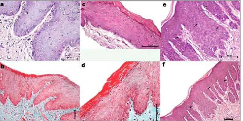
and rete pegs had normal appearance in the con-trol group (Fig. 1a–b). The number of rete pegs was determined to have increased in the gingival samples of both the PEMF and SEMF groups. In-traepithelial lymphocytes increased in both EMF groups as compared to the control. However, the increase in lymphocytes in the PEMF group was more than that seen in the SEMF group. Prolif-eration of epithelial cells was observed in the basal cell layer in the PEMF and SEMF groups but not in the control group. The difference of prolifera-tion was not significant as compared to each other (Table 1). The beneath of the epithelium, connec-tive tissue, exhibited a variable number of fibro-blasts, typical collagen bundles and blood vessels.
Inflammatory infiltrates were not existent in all groups (Fig. 1c–f).
Immunohistochemical
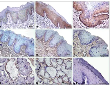
To determine the protein expression changes of E-cadherin and type IV collagen induced by an extremely low frequency pulsed electromag-netic field and a sinusoidal electromagelectromag-netic field, the authors detected these protein expressions by immunohistochemical assay. A low level of E-cadherin expression was observed in the epithe-lium of the gingiva of the control group sections especially in the basal cell layer (Fig. 2a). Over-expressions of E-cadherin on the gingival
epithe-Fig. 1. The representative sections for Masson trichrome and Hematoxylin-Eosin staining in all groups. Normal his-tological features of gingiva were observed in sections of control groups (a, b). The proliferation of epithelial cells was observed in the PEMF and SEMF groups, not in the control group. The number of rete pegs was similar in both EMF groups (c, d). Intraepithelial lymphocytes were increased in the PEMF group as compared to both the control and SEMF groups (e, f). Arrowheads: intraepithelial lymphocytes
Ryc. 1. reprezentatywne wycinki barwione wg Massona i hematoksyliną i eozyną we wszystkich grupach. Prawidłowe cechy histologiczne dziąsła obserwowano w wycinkach grup kontrolnych (A, B). Proliferację komórek nabłonkowych zaobserwowano w grupach PEMF i SEMF, ale nie w grupie kontrolnej. liczba wpukleń naskórka była podobna w obu grupach EMF (C, D). liczba śródnabłonkowych limfocytów była zwiększona w grupie PEMF w porównaniu zarówno z kontrolną, jak i grupą SEMF (E, F). Groty strzałek wskazują śródnabłonkowe limfocyty
Table 1. Histopathological findings in the different study groups
Tabela 1. Wyniki badań histopatologicznych w poszczególnych grupach
Control
(Grupa kontrolna) SEMF PEMF p
Proliferation of epithelial cells (Proliferacja komórek nabłonka) – ++ ++ <0.05
Intraepithelial lymphocytes (limfocyty śródnabłonkowe) + ++ +++ <0.05
Elongation of rete pegs (Wydłużenie wpukleń naskórka) + ++ ++ <0.05
Fibroblast (Fibroblast) + + + >0.05
Collagen bundles (Wiązki kolagenu) + + + >0.05
lium was detected in the PEMF and SEMF groups as compared to the control group (Fig. 2b–c). The difference of expression levels in the epithelium of gingiva between the PEMF and SEMF groups was not significant (Table 2). In addition, expres-sion of E-cadherin was detected in some cells of
the ducts of mucous glands (Fig. 2g). Type IV col-lagen expression was observed beneath the epithe-lium in the connective tissue layer, especially in collagen bundles and the basal membrane of the basal cell layer of the epithelium as well as the basal membrane beneath the cells composed of mucous
Fig. 2. The representative sections of immunohistochemistry for E-cad and Type IV collagen in all groups. Tissue expressing E-cad and type IV collagen appeared brown in color. The minimal staining for E-cadherin is found in the epithelium of the control group sections (d). Intense staining of E-cadherin was observed in the epithelium of both SEMF and PEMF group sections. E-cadherin expression was observed in the epithelium of ducts of the mucous glands in MF treated groups also to a moderate degree. Expression of type IV collagen was mainly observed in connective tis-sue, basement membranes of both ducts, epithelia and basal cell layer gingival epithelium (d–j). Arrows: expression of E cadherin and Type IV collagen
Ryc. 2. reprezentatywne wycinki immunohistochemiczne e-kadheryny i kolagenu typu IV we wszystkich grupach. Ekspresję tkankową e-kadheryny i kolagenu typu IV uwidoczniono w kolorze brązowym. Minimalne barwienie E-kadheryny zanotowano w nabłonku wycinka grupy kontrolnej (d). Intensywne barwienie E-kadheryny obserwowa-no w nabłonku zarówobserwowa-no w wycinku SEMF, jak i PEMF. Ekspresję E-kadheryny obserwowaobserwowa-no w nabłonku gruczołów śluzowych w grupach leczonych MF również w umiarkowanym stopniu. Ekspresję kolagenu typu IV obserwowano głównie w tkance łącznej, błonach podstawnych obu przewodów i warstwie podstawnej nabłonka dziąsłowego (d–j). Strzałki wskazują ekspresję E-kadheryny i kolagenu typu IV
Table 2. Expression patterns of E-cadherin
Tabela 2. Wzory ekspresji e-kadheryny
E-cadherin (E-kadheryna) Control
(Grupa kontrolna) SEMF PEMF p
Epithelium (Nabłonek) + ++ ++ <0.05
Basal cell layer (Warstwa podstawna) + ++ +++ <0.05
Endothelial cells (Komórki śródbłonka) + + + >0.05
acini (Fig. 3d–f, h). The expression level of type IV collagen was not significant between the control and EMF treated groups except for a significant increase in basement membranes of the basal cell layer of the PEMF group (Table 3), as compared to the other groups (control and SEMF).
Discussion
The authors investigated the effects of an ex-tremely low frequency pulsed electromagnetic field and sinusoidal electromagnetic field on the gingiva of healthy rats. Two of the main features of epithelial tissues were determined in the organ-isms. Firstly, cell-cell was closely opposed and at-tached via specialized junctions. The other was, epithelial cells produced the basement membrane which lies on and separates it from underlying connective tissue. Both features contribute to the barrier function of epithelial tissue [18]. Gingival tissue is constantly subject to mechanical and bac-terial aggression. In response to specific antigens or stimuli, inflammatory cells migrate and come into contact in localized areas where they phago-cyte the bacteria or damaged tissues. Cellular com-ponents of the innate system present in the gingi-val epithelium include cells such as dendritic cells and neutrophils [19].
Exposure to EMF has not affected their sys-temic hematologic parameters [20, 21]. However, decreases in the number of white cells were ob-served in rats exposed to high intensity electrical fields [22]. In addition, Ongaro et al. reported that EMFs have anti-inflammatory effects on osteoar-thritis synovial fibroblasts cells by modulating inflammatory and anti-inflammatory parameters [23]. The authors found that no inflammatory in-filtration in the connective tissue of gingiva was induced with EMF in the present study. Only slightly increased intraepithelial lymphocytes were found in EFM-treated groups as compared to the control group.
Collagen is a protein and synthesized by fibro-blasts, chondrofibro-blasts, osteoblasts and other cells. The molecular configuration of collagen fibers confers to them a tensile strength greater than that of steel. Consequently, collagen imparts a unique combination of flexibility and strength to the tis-sue where it lies. Type IV collagen bundles branch between type I collagen bundles and are continu-ous with those of the basement membrane and blood vessel walls [18].
The effects of electrical stimulation on peri-odontal tissue obtained from dogs with bony de-fects have been investigated. Histopathological examination of this study showed that connective tissue was organized as prominent collagen fiber bundles. In the present study, the authors ob-served that in the components of the connective tissue, including collagen bundles and fibroblast cells, the underlying epithelium was not impaired from both EMF-treated groups as compared to the control group. In addition, expression of collagen type IV was not significantly different between the groups. Moderate expression levels were observed in connective tissue control and EMFs [24].
The effects of PEMF on increased cell prolif-eration have previously been shown. In this study, the authors also observed a proliferation of cells in the PEMF and SEMF groups, especially in the basal cell layer of gingival epithelium [25].
E-cadherin is a calcium-dependant homo-philic cell adhesion molecule that contributes to cell-cell interaction. The role of E-cadherin is not only attributed to cell–cell adhesion, but also regulates proliferation, differentiation and polar-ization of epithelial cells [26]. The epithelium acts as a mechanical barrier through cell junctions, such as E-cadherin [11, 12]. E-cadherin interac-tion is also thought to be important for reteninterac-tion of the langerhans cells on the epithelial cells [13]. The high expression level of E-cadherin can be an appreciable indicator of the strong barrier func-tion of gingival epithelium [27]. The breakdown of the junctional complex between epithelial cells
Table 3. Expression patterns of type IV collagen
Tabela 3. Wzory ekspresji kolagenu typu IV
Type IV collagen (Kolagen typu IV) Control
(Grupa kontrolna) SEMF PEMF p
Collagen bundles (Wiązki kolagenu) + + + >0.05
Basement membranes of basal cell layer
(Błony podstawnej warstwy komórek) + + ++ <0.05
Basement membrane of ducts epithelium
References
[1] Manni V, Lisi A, Rieti S Serafino A, Ledda M, Giuliani L, Sacco D, D’Emillia E, Grimaldi S: low electromag-netic field (50 Hz) induced differentiation on primary human oral keratinocytes (HOK). Bioelectromagelectromag-netics 2004, 25, 118–126.
[2] Liboff AR, Williams T, Strong DM, Wistar R Jr.: Time-varying magnetic fields: Effect on DNA synthesis. Science 1984, 223, 818–820.
[3] Tabrah FL, Ross P, Hoffmeier M, Gilbert F Jr.: Clinical report on long-term density after short-term EMF appli-cation. Bioelectromagnetics 1998, 19, 75–78.
[4] Cossarizza A, Monti D, Bersani F, Paganelli R, Montagnani G, Cadossi R, Cantini M, Franceschi C: Extremely low frequency pulsed electromagnetic fields increase interleukin (Il-2) utilization and Il-2 receptor expression in mitogen-stimulated human lymphocytes from old subjects. FEBS lett 1989, 248, 141–144.
[5] Goodman R, Bassett CAL, Henderson S: Pulsing electromagnetic fields induce cellular transcription. Science 1983, 220, 1283–1285.
[6] Goodman R, Abbott J, Hendeson AS: Transcriptional patterns in the X chromosome of Sciara coprophila follow-ing exposure to magnetic field. Bioelectromagnetics 1987, 8, 1–7.
[7] Ubeda A, Angeles Trillo M, House DE, Blackman CF: A 50 Hz magnetic field blocks melatonin-induced enhance-ment of junctional transfer in normal C3H/10T1/2 cells. Carcinogenesis 1995, 16, 2945–2949.
[8] Phillips JL, Haggren W, Thomas WJ, Ishida-Jones T, Adey WR: Magnetic field-induced changes in specific gene transcription. Biochim Biophys Acta 1992, 1132, 140–144.
[9] Funk RH, Monsees T, Ozkucur N: Electromagnetic effects – from cell biology to medicine. Prog Histochem Cytochem 2009, 43, 77–264.
[10] Steffensen B, Caffesse RG, Hanks CT, Avery JK, Wright N: Clinical effects of electromagnetic stimulation as an adjunct to periodontal therapy. J Periodontol 1998, 59, 46–52.
[11] Kandikonda S, Oda D, Niederman R, Sorkin BC: Cadherin-mediated adhesion is required for normal growth regulation of human gingival epithelial cells. Cell Adhes Commun 1996, 4, 13–24.
[12] Hatakeyama S, Yaegashi T, Oikawa Y, Fujiwara H, Mikami T, Takeda Y, Satoh M: Expression pattern of adhe-sion molecules in junctional epithelium differs from that in other gingival epithelia. J Periodont res 2006, 41, 322–328.
[13] Udey MC: Cadherins and langerhans cell immunobiology. Clin Exp Immun 1997, 107, 6–8.
[14] Arun R, Hemalatha R, Arun KV, Kumar T: E-cadherin and CD1 expression in gingival epithelium in periodontal health, disease and post-treatment. Indian J Dent res 2010, 21, 396–401.
[15] Yilmaz O, Verbeke P, Lamont RJ, Ojcius DM: Intercellular spreading of porphyromonas gingivalis infection in primary gingival epithelial cells. Infect Immun 2006, 74, 703–710.
[16] Nakagawa I, Inaba H, Yamamura T, Kato T, Kawai S, Ooshima T, Amano A: Invasion of epithelial cells and proteolysis of cellular focal adhesion components by distinct types of Porphyromonas gingivalis fimbriae. Infect Immun 2006, 74, 3773–3782.
[17] Akpolat V, Celik MS, Celik MY, Ozerdem MS, Akdag MZ, Dasdag S, Yavas MC: Examination of long Term Magnetic Fields on rat Calvarial and Mandibular Bone Mass. Biotechnol Biotechnol 2010, 24(2), 1882–1885.
[18] Carranza FA, Newman MG: Clinical Periodontology 8th Edition. W.B. Saunders Company 12–35, Philadelphia, PA1996.
[19] Dale BA: Periodontal epithelium: a newly recognized role in health and disease. Periodontology 2000 2002, 30, 70–76.
[20] Ragan HA, Buschbom RL, Pipes MJ, Phillips RD, Kaune WT: Hematologic and serum chemistry studies in rats exposed to 60-Hz electric fields. Bioelectromagnetics 1983, 4, 79–90.
[21] Margonato V, Veicsteinas A, Conti R, Nicolini P, Cerretelli P: Biological effects of prolonged exposure to ElF electromagnetic fields in rats. I. 50 Hz electric fields. Bioelectromagnetics 1993, 14, 479–493.
[22] Seto YJ, Majean-Chargeois D, Lymangrover JR, Dunlap WP, Fox FT, Hsieh ST: Chronic 60 Hz electric field exposure-induced subtle bioeffects on hematology. Environ res 1986, 39, 143–152.
[23] Ongaro A, Varani K, Masieri FF, Pellati A, Massari L, Cadossi R, Vincenzi F, Borea PA, Fini M, Caruso A, De Mattei M: Electromagnetic fields (EMFs) and adenosine receptors modulate prostaglandin E(2) and cytokine release in human osteoarthritic synovial fibroblasts. J Cell Physiol 2012, 27, 2461–2469.
[24] Kaynak D, Meffert R, Gunhan M, Gunhan O: A histopathologic investigation on the effect of electrical stimula-tion on periodontal tissue regenerastimula-tion in experimental bony defects in dogs. J Periodontol 2005, 76, 2194–2204.
was responsible for the destruction of the barrier function of epithelial cells [28, 29]. The authors observed that E-cadherin expression was signifi-cantly increased in both PEMF and SEMF groups in the present study.
The authors concluded that extremely low frequency pulsed and sinusoidal electromagnetic
[25] Fitzsimmons RJ, Baylink DJ, Ryaby JT, Magee FP: EMF-stimulated bone cell proliferation, in Electricity and Magnetism in Biology and Medicine (M.J. Blank, ed.) 899–902, San Francisco Press, San Francisco 1993.
[26] Rodriguez-Boulan, Nelson EWJ: Morphogenesis of the polarized epithelial cell phenotype. Science 1989, 245, 718–725.
[27] Shapiro L, Fannon AM, Kwong PD, Thompson A, Lehmann MS, Grubel G, Legrend JF, Als-Nielsen J, Colman DR, Hendricksen WA: Structural basis of cell-cell adhesion by cadherins. Nature 1995, 374, 327–337.
[28] Obiso RJ Jr., Azghani AO, Wilkins TD: The Bacteroides fragilis toxin fragilysin disrupts the paracellular barrier of epithelial cells. Infect Immun. 1997, 65, 1431–1439.
[29] Wu S, Lim KC, Huang J, Saidi RF, Sears CL: Bacteroides fragilis enterotoxin cleaves the zonula adherens protein, E-cadherin. Proc Nat Acad Sci USA 1998, 95, 14979–14984.
Address for correspondence:
Selcuk Tunik
Department of Histology and Embryology Faculty of Medicine, University of Dicle 21280 Diyarbakir
Turkey
E-mail: [email protected] Tel.:+90 4122488001-4751 Conflict of interest: None declared received: 9.07.2012