CODEN [USA]: IAJPBB ISSN: 2349-7750
INDO AMERICAN JOURNAL OF
PHARMACEUTICAL SCIENCES
http://doi.org/10.5281/zenodo.1134390
Available online at:
http://www.iajps.com Research Article
DEVELOPMENT AND VALIDATION OF STABILITY
INDICATING RP-HPLC METHOD FOR SIMULTANEOUS
ESTIMATION OF SOFOSBUVIR AND LEDIPASVIR IN TABLET
DOSAGE FORM
M.Prasanthi Evangelin*
1, S.Manohar Babu
2, Konda Ravi Kumar
3 *1, 2SIMS College of Pharmacy, Mangaldas Nagar, Guntur, India.
2Hindu College of Pharmacy, Amaravathi Road, Guntur, A.P, India.
Abstract:
Simple, specific, accurate and precise reversed phase high pressure liquid chromatographic method has been developed for the simultaneous determination of Sofosbuvir and Ledipasvir in tablet dosage form by reversed phase C18 column (Kromasil C18 5μ, 250 mm x 4.6 mm). The sample was analyzed using 0.1% OPA: Acetonitrile in the ratio of 55:45 as a mobile phase at a flow rate of 1.0 ml/min and detection at 230 nm. Calibration curves were linear with correlation coefficient (r2) 0.999 over a concentration range of 100-600 µg/mL for Sofosbuvir and 0.999 over a concentration range of 22.5-135 µg/mL for Ledipasvir. The retention time was found to be 6 min. The mean recoveries were found to be 99.10% and 99.30% for Sofosbuvir and Ledipasvir respectively. The relative standard deviation (RSD) was found to be < 2.0% for both drugs. The proposed method was validated and successfully applied to the estimation of Sofosbuvir and Ledipasvir in tablet dosage form.
Keywords: Sofosbuvir, Ledipasvir, RP-HPLC, Validation, Stability.
*Corresponding Author:
M.Prasanthi,
SIMS College of Pharmacy,
Mangaldas Nagar, Guntur, India
E-mail: prasanthievangelin89 @gmail.com
Mobile: +91- 8688545553.
Please cite this article in press as M.Prasanthiet al., Development and Validation of Stability Indicating RP-HPLC Method for Simultaneous Estimation of Sofosbuvir and Ledipasvir in Tablet Dosage Form, Indo Am. J. P. Sci, 2017;
4(12).
INTRODUCTION:
Reverse phase is the choice for the majority of samples, but if acidic or basic analytes [1-3] are present then reverse phase ion suppression (for weak acids or bases) or reverse phase ion pairing should be used. The stationary phase should be C18 bonded[4,5]. Sofosbuvir is a prodrug nucleotide analog used as part of combination therapy to treat hepatitis C virus (HCV) infection or to treat co-infection of HIV and HCV. Sofosbuvir and other nucleotide inhibitors of the HCV RNA [6] polymerase exhibit a very high barrier to resistance development. Sofosbuvir has become available as a fixed dose drug combination product with ledipasvir (tradename Harvoni) used for the treatment of chronic Hepatitis C, an infectious liver disease caused by infection with Hepatitis C Virus (HCV). Ledipasvir is previously known as GS-5885, is an inhibitor of the Hepatitis C Virus (HCV) NS5A protein required for viral RNA replication and assembly of HCV virions.
Ledipasvir is available as a fixed dose drug combination product [7,8] with sofosbuvir (tradename Harvoni) used for the treatment of chronic Hepatitis C, an infectious liver disease caused by infection with Hepatitis C Virus (HCV). Approved in October 2014 by the FDA, ledipasvir and sofosbuvir are direct-acting antiviral agents indicated for the treatment of HCV genotype 1 with or without cirrhosis. Combination of these two drugs (Sofosbuvir 400mg and Ledipasvir 90mg is available in pharmacy under the brand name of Myhep Lvir, Ledifos tablet, Heterosofir plus and Hepcinat lp tablet. The chemical structures of both drugs were shown in Fig.1.
A
Literature survey revealed that there are only three HPLC methods [9-11] have been published regarding this research work. The present study aimed to develop a simple, sensitive, less retention time and accurate RP-HPLC method for the simultaneous estimation of sofosbuvir and ledipasvir in bulk and tablet dosage forms with high sensitivity, selectivity and stability that can be used for the routine analysis.
MATERIALS AND METHODS: Chemicals and Reagents
Pure standard samples of sofosbuvir and ledipasvir were obtained as gifted samples from Rhodes Pharmaceuticals Ltd. and its marketed formulations in the brand name of Harvoni [Label claim containing sofosbuvir 75mg and ledipasvir 325 mg] were procured from local pharmacy. Water (HPLC-Grade), Acetonitrile (HPLC-Grade; Rankem) and Methanol (HPLC-Grade Rankem), ortho phosphate buffer, Ortho-phosphoric acid (Rankem). All dilutions were performed in standard class-A, volumetric glassware. Instrumentation
The present assay was carried out on a Waters HPLC system [Model: 2695] equipped with 2487 photodiode array detector, automated sample injector and a column Kromasil C18 (250mmx4.6mm I.D; particle size 5μm)
respectively. Electronic Balance [Denver] and Ultra-Sonicator [SE60US; BVK enterprises] were also used in the present assay. The output of signal was monitored and integrated using waters Empower 2 software.
Buffer preparation
0.1%OPA Buffer:1ml of ortho phosphoric acid was diluted to 1000ml with HPLC grade water.
Mobile phase preparation
Prepare a filtered and degassed mixture of Buffer (pH -2.0), 0.1% OPA: Acetonitrile (55:45%).
Diluent preparation
Mobile phase is used as diluent.
Standard preparation
Accurately weighed 40mg of Sofosbuvir, 9mg of Ledipasvir and transferred to 10ml flasks and 3/4th of
diluents was added to these flask and sonicated for 10 minutes. Flask were made up with diluents and labeled as Standard stock solution. (40µg/ml of Sofosbuvir and 2500µg/ml Ledipasvir).
further the volume was made up with diluent and filtered by HPLC filters (40µg/ml of Sofosbuvir and 2500µg/ml of Ledipasvir).
RESULTS AND DISCUSSION: Method development
Initial trials were carried by the author in developing the proposed RP-HPLC method. The mobile phase was chosen after several trials with methanol, acetonitrile, water and buffer solutions in various proportions and at different pH values. A mobile phase consisting of 0.1%
OPA buffer (pH -2.0) and acetonitrile (55:45 v/v) was selected to achieve maximum separation and sensitivity. Flow rates between 0.5 and 1.5/min were studied. A flow rate of 1.0 ml/min at ambient temperature gave an optimal signal to noise ratio with a reasonable separation time. Using a Kromasil C18
column, the run time of analysis was 6 min. Detection wavelength of 230 nm was chosen for the analysis. A typical chromatogram for simultaneous estimation of sofosbuvir and ledipasvir obtained by using a mobile phase was shown in Fig.2.
Fig.2: Typical chromatogram of standard solution (sofosbuvir and ledipasvir)
Chromatographic conditions
The isocratic mobile phase consisted of 0.1% OPA buffer (pH -2.0) and Acetonitrile (55:45 v/v), flowing through the Kromosil C18 column (make: 250 mmx4.6
mm i.d; particle size 5μm) at a constant flowrate of 1.0 ml/min at ambient column temperature. The mobile phase was pumped through the column at a flowrate of 1.0ml/min with a sample injection volume of 10μl. Detection of the analytes (sofosbuvir and ledipasvir) was carried out at a wavelength of 230 nm.
Method validation
The proposed RP-HPLC method was validated, in accordance with USP guidelines for system.
System suitability
RP-HPLC method System performance parameters were determined by analyzing standard working solutions of sofosbuvir and ledipasvir. The chromatographic parameters, such as number of theoretical plates (n), resolution (Rs), USP plate count and USP tailing were determined. The results are shown in Table.1, indicating the good performance of the system.
Table1: System suitability data of sofosbuvir and ledipasvir
S.N o
Sample name
RT USP Plate count
USP tailing
1. Injection 1 (sofosbuvir)
2.442 7792 1.08 2. Injection 1
(Ledipasvir)
3.072 11272 0.94
Specificity
Blank and placebo interference
Linearity & Detector response
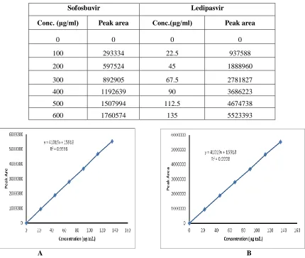
The linearity of the proposed method was accessed by calculating slope, intercept and correlation coefficient [r2] of standard curve. Sofosbuvir and ledipasvir
showed a linearity range in between 100-600 and 22.5-135μg/ml respectively and the slope and intercept of
the calibration plot of sofosbuvir and ledipasvir were 2966x+2193 and 41019x + 15917 with correlation coefficients obtained was greater than 0.999 respectively. The linearity results of both the drugs were shown in Tab.2 and Linearity curves of sofosbuvir and ledipasvir were depicted in Fig.3.
Table 2: Linearity results for Sofosbuvir and Ledipasvir
Sofosbuvir Ledipasvir
Conc. (μg/ml) Peak area Conc.(μg/ml) Peak area
0 0 0 0
100 293334 22.5 937588 200 597524 45 1888960 300 892905 67.5 2781827 400 1192639 90 3686223 500 1507994 112.5 4674738 600 1760574 135 5523393
A B
Fig.3: Linear calibration plot for Sofosbuvir (A) and Ledipasvir (B)
Sensitivity
The limit of detection (LOD) and limit of quantification (LOQ) were established at signal-to noise ratio of 3:1 and 10:1 respectively. The LOD of sofosbuvir and ledipasvir was found to be 0.02μg /ml & 0.28μg /ml respectively. The LOQ of sofosbuvir and ledipasvir was found to be 0.07 μg /ml & 0.85μg / ml respectively.
Table 3: System precision table of Sofosbuvir and Ledipasvir
S.No Area of Sofosbuvir Area of Ledipasvir
1. 1145677 3578272
2. 1152265 3587779 3.
1154492 3608922 4.
1148949 3624217 5. 1156499 3600959 6.
1160648 3601532 Mean 1153088 3600280 S.D 5357.08 16064.4 %RSD
0.5 0.4
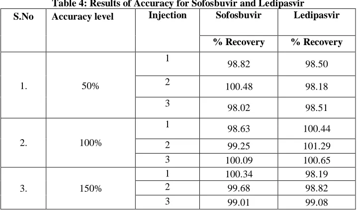
Accuracy
The accuracy of the proposed method for sofosbuvir and ledipasvir was assessed by recovery studies at three different levels i.e. 50%, 100%, 150%. The recovery studies were carried out in triplicate by adding Known amount of standard solution of sofosbuvir and ledipasvir to preanalysed tablet
solutions. The resulting solutions were then reanalysed by proposed method and the results are represented in Tab.4. The percentage recoveries were found in the range of 98.82 to 100.9% for sofosbuvir and 98.50 to 100.9% for ledipasvir respectively revealing that the developed RP-HPLC method was found to be accurate.
Table 4: Results of Accuracy for Sofosbuvir and Ledipasvir
S.No Accuracy level Injection Sofosbuvir Ledipasvir
% Recovery % Recovery
1. 50%
1
98.82 98.50 2
100.48 98.18 3 98.02 98.51
2. 100%
1 98.63 100.44 2 99.25 101.29 3 100.09 100.65
3. 150%
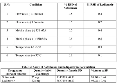
Table 5: Robustness data for Sofosbuvir and Ledipasvir
S.No Condition % RSD of Sofosbuvir
% RSD of Ledipasvir
1 Flow rate (-) 1.1ml/min 0.5 0.4
2 Flow rate (+) 1.3ml/min 0.5 0.7
3 Mobile phase (-) 35B:65A 0.5 0.4
4 Mobile phase (+) 45B:55A 0.5 0.5
5 Temperature (-) 25°C 0.3 0.3
6 Temperature (+) 35°C 0.1 0.1
Table 6: Assay of Sofosbuvir and ledipasvir in Formulation Drug name
(Harvoni tablets)
Quantity label claim(mg)
Quantity found± SD %Assay ± SD
Sofosbuvir 75 mg 1143799 ±0.50 99.10 ± 0.46 Ledipasvir 325 mg 3567195 ±0.30 98.98 ± 0.46
Robustness
The robustness of the developed method was evaluated by altering few experimental conditions and evaluating the resolution between two adjacent peaks of sofosbuvir and ledipasvir. The data results were shown in Tab.5.
Formulation assay
The validated method was applied on commercially available Harvoni tablets. The results of the assay undertaken yielded 99.10% and 99.30% of the label
claim for sofosbuvir and ledipasvir. Results of the
assay indicated that the method is quite selective for the analysis of Harvoni without interference from the excipients used to formulate and produce these tablets. The results were displayed in Tab.6 respectively.
CONCLUSION:
It was concluded that the proposed new RP-HPLC method developed for the quantitative determination of Sofosbuvir and ledipasvir in bulk as well as in its formulations was simple, selective, sensitive, accurate, precise and rapid. The method was proved to be superior to most of the reported methods. The mobile phase was simple to prepare and economical. The
as an alternative method to report routine estimation of Sofosbuvir and ledipasvir depending upon the availability of chemicals and nature of other ingredients present in the sample. The method was validated as per ICH guidelines, and validation acceptance criteria were met in all cases. Application of this method for estimation of Sofosbuvir and ledipasvir from tablet dosage form and stressed samples showed that neither the degradation products nor the excipients interfered in the estimation of drug. Hence, this method was specific, stability-indicating and can be successfully used for the estimation of drug in bulk and pharmaceutical dosage form.
REFERENCES:
4. Kumari R., Nguyen M.H., 2015. Fixed-dose combination of sofosbuvir and ledipasvir for the treatment of chronic hepatitis C genotype 1. Expert Opinion on Pharmacotherapy, 6(5), pp.739-748. 5.Younossi Z.M., Stepanova M., Marcellin P., Afdhal N., Kowdley K.V., Zeuzem S., Hunt S.L.,2015.Treatment with ledipasvir and sofosbuvir improves patient-reported outcomes: Results from the ION-1, -2, and -3 clinical trials. Hepatology. ,61(6),pp.1798-808.
6.Waheed Y., 2015. Ledipasvir and sofosbuvir: Interferon free therapy for hepatitis C virus genotype 1infection. World Journal of Virology. 2015, 12;4(1),pp.33-5.
7.Osinusi A., Townsend K.., Kohli A., Nelson A., Seamon C., Meissner E.G., Bon D., Silk R., Gross C.,Price A., Sajadi M., Sidharthan S., Sims Z., Herrmann E., Hogan J., Teferi G., Talwani R., Proschan, M., Jenkins V., Kleiner D.E., Wood B.J., Subramanian G.M., Pang PS, Mc Hutchison J.G., Polis M.A., Fauci A.S., Masur H, Kottilil S., 2015. Virologic response following combined ledipasvir and sofosbuvir administration in patients with HCV genotype 1 and HIV coinfection. Journal of the American Medical Association. 313(12),pp.1232-1239.
8.Bakht Zaman,Faisal Siddique.,Waseem Hassan..,2016.RP-HPLC method for simultaneous determination of sofosbuvir and ledipasvir in tablet dosage form and its application to in vitro dissolution studies.Chromatographia.79(23),pp.1605–1613. 9.Rezk M.R., Bendas E.R, Basalious E.B., Karim I.A.,2016.Quantification of sofosbuvir and ledipasvir in human plasma by UPLC-MS/MS method: Application to fasting and fed bioequivalence studies. J. Chromatogr B Analyt Technol Biomedical Life Sciences. 1028,pp.63-70.
10.Hassouna M. E. M., Abdelrahman M. M., Mohamed M. A., 2017. Assay and Dissolution Methods development and validation for simultaneous determination of Sofosbuvir and Ledipasvir by RP-HPLC method in tablet dosage forms. Journal of Forensic Science & Criminal Investigations. 1(3),pp.555-562.