35 INTERNATIONAL JOURNAL OF ADVANCES IN ENGINEERING RESEARCH
A NOVEL METHOD OF MRI IMAGE
SEGMENTATION USING K-MEANS ALGORITHM
1. J.PERSIS DASHNA PAULYN,2. S.PAVITHRA,3 S.ANUSUYA
[ASST.PROF]
DEPARTMENT OF COMPUTER SCIENCE AND ENGINEERING SAVEETHA SCHOOL OF ENGINEERING, THANDALAM, CHENNAI
Email id: [email protected],[email protected],[email protected]
ABSTRACT
The paper we have chosen portray regarding the competent methodology for automatic brain tumour segmentation for the extraction of tumor tissues from MRI pictures. Segmentation of pictures holds a very important position in image process. Segmentation is meted out victimization K-means algorithmic program. It becomes additional imperative whereas generally handling medical pictures wherever pre-surgery and post-surgery choices area unit needed for the aim of initiating and rushing up the recovery method. The algorithmic program is chosen to reinforce the growth boundaries fast as a flash in comparison to alternative agglomeration algorithms
Keywords: resonance Imaging (MRI), Image segmentation, k-means, Morphological operations,tumour.
INTRODUCTION
The sway and impact of digital pictures on fashionable society is unbelievable and image process is currently a important element in science and technology. The pc techniques have processed and manipulated that facilitate to check hidden diagnostic options that area unit tough or not possible to spot victimization plate like imaging ways. Image segmentation is outlined as a method that partitions a given image into finite range of non-overlapping regions with relevance grey worth, texture distribution etc. ROI (Region of interest) outline boundary of brain tumour and intra cerebral brain bleeding.
The present approaches area unit supported symbolic logic means that and Neural Networks
(NN) distribution,etc.
The image segmentation ways area unit classified into 3 categories: edge-based, region-based, pixel-based methods. In our planned methodology we have a tendency to area unit implementing K-means agglomeration in pixel-based ways. K-means agglomeration area unit easy and have procedure quality comparatively low compared to alternative region-based or edge-based ways. Growth detection victimization morphological operations open planned in [1][2]10]. Segmentation is a picture process operation that aims to partition a picture into unvaried regions composed of pixels with a similar characteristics consistent with predefined criteria. Most strategies of image segmentation need the adjustment of many management parameters to get sensible results but the multi device scales needs no fitting parameters. The multi scale analysis is to decompose the image on a wide range of scales, associate operation. At every level, the image is replaced by the approximation most applicable one are often drawn. Going the coarsest scale to the finer performances we have a tendency to access a lot of and a lot of correct the image. The analysis is
finished by conniving what differs from
36 INTERNATIONAL JOURNAL OF ADVANCES IN ENGINEERING RESEARCH
EXISTING SYSTEM
The existing methodology used numerous algorithmic programs like watershed algorithm, Fuzzy technique, EM algorithmic program, hierarchic agglomeration, median cut algorithmic program additionally as K-means algorithmic program [7][6][5][4][3][11][12][8][9]. To find the tumours, the authors used morphological operations like dilation, erosion, opening, closing. Erosion and Dilation were used as 2 elementary
Figure 1.a Block diagram of proposed approach.
In any medical application and military application and image sweetening plays a significant role. The present methodology refers to the method of clustering pixels of a picture such pixels that area unit within the same cluster area unit similar among them and area unit dissimilar to the pixels that belong to the opposite group of clusters. 2 steps were to calculate the centroids assignment step and update step. The generalized algorithmic program initiates k cluster centroids by at random choosing k feature vectors from X. Later, the feature Vectors Square {measure} sorted into k clusters employing a selected distance measure like geometer distance. The agglomeration procedure stops only if all cluster centroids tend to converge. Similarity is measured by distance associate degreed outlined by an N dimensional feature area. Feature distance calculation differs from spatial distance calculation. Feature distance calculation is predicated on options like colour or intensity and texture whereas spatial distance calculation is predicated on x; y (width, height) coordinates. Making associate degree apt distance calculation methodology is a very important task since it greatly affects final agglomeration result. The planned approach utilizes mathematical morphology operations for the pre-processing. Operations area unit applied on the grey scale pictures to phase the abnormal region.
The main argument of the present modification is on the reduction of intensive distance computation that takes place at every run (iteration) of K-means algorithmic program between every information and every one cluster centres. To scale back the intensive distance computation, a straightforward mechanism by that, at every iteration, the space between every information and therefore the cluster nearest there to computed and recorded during a arrangement recommended. Thus, on the subsequent iterations the space between every information and its previous nearest cluster is recomputed.
LITERATURE SURVEY
There are unit totally different algorithms to beat the issues caused in detection of tumor. The traditional watershed formula was accustomed check the medical image analysis is waterspread and so made a whole division of pictures. It conjointly makes use of automatic thresholding on the gradient magnitude map and post within which segmentation merging on the initial partitions to cut back the amount of false edges and over segmentation. the most draw back was once it had been applied to medical image victimisation k-means bunch it made a primary segmentation of the image. Subsequent formula was expectation maximization (EM) that was used for the segmentation of CT brain pictures victimisation k-means and EM bunch. The system has been tested with variety of real time pictures and has
achieved some promising results.
Pre-processing
Segmentatio n Technique
Post Processing
Tumour Detection
37 INTERNATIONAL JOURNAL OF ADVANCES IN ENGINEERING RESEARCH Followed by EM formula is that the median –cut formula that was used for the popularity of colour blood corpuscle pictures. During this method they used blood corpuscle pictures infected with protozoal infection parasites as cell pictures it conjointly made higher results. Within the more papers referred they conjointly mentioned a technique for segmentation of substantial Alba and grey matter from real mister pictures employing a LM-K suggests that technique-means is usually taken to the supervised system by victimisatio
lexenberg-marquardt improvement technique.
The existing approach utilizes mathematical morphology operations for the segmentation. The morphological operations area unit applied on the grey scale pictures to phase the abnormal regions. Erosion and dilation area unit the 2 elementary operations in Mathematical Morphology
PROPOSED SYSTEM
K-means is associate instance of exclusive bunch algorithms and is that the backbone of our paper's methodology. K suggests that bunch is that the most generally used and studied methodology among the bunch formulations that area unit supported minimizing a proper objective perform given in Equation. K-means bunch could be a key technique in pixel-based ways. In pixel-ways area unit easy and therefore the process quality is comparatively low compared with different region-based or edge-based ways, the applying is a lot of practicable. The annoyance within the existing methodology is it’s difficult to predict K price mechanically within the existing methodology. And conjointly troublesome in initial Partition that may lead to problem of ultimate clusters. It doesn't work well with clusters of various size
and density.
Thus in our projected methodology, we tend to area unit getting to style a technique within which k-value are often mechanically performed once the image is given as input. Input image is given that mechanically predict the amount of input clusters. K initial central points, either haphazardly or victimisation some heuristic information. It then teams every image element underneath the central purpose its nearest to. It calculates new central purposes by averaging the pixels sorted underneath every central point. the 2 former recursive steps area unit perennial alternately till convergence (central point’s no long modification by averaging. the employment of K-Means bunch methodology is fairly easy compared with oftentimes used fuzzy bunch ways. The execution time for K-means bunch is a smaller amount compared to the opposite bunch ways. The anticipated work conjointly reduces the process quality.
PROPERTIES OF K-MEANS:
It’s the partitioned clustering algorithm. • Simple to implement.
• It follows Euclidean distance measure(mean value-intensity of pixels)
• The existing work reduces the
38 INTERNATIONAL JOURNAL OF ADVANCES IN ENGINEERING RESEARCH
Algorithm
1. Apply morphological operation to smooth the image
2. Set initial number of clusters k=2
3. Obtain centres for the input image for the given CN
4. Form the clusters based on Euclidean distance
5. if result=’satisfied’ then halt else k=k+1 go to step 3
The limitations of K-means clustering require many iterative rounds. This work strives to reduce that limitation. Here the value of id is estimated if it satisfies the minimum value for Validity Measure=intra/inter.
The intra cluster distance is the average of distance between a point and its cluster centre. Here N is the number of data items, K is the number of clusters, and Ciis the cluster centre of cluster K.We obviously want to minimize this measure. We can also measure the intercluster distance, or the distance between clusters, which we want to be as big as possible. We calculate this as the distance between cluster centres, and take the minimum of this value, defined as
The proposed work also reduces the computational complexity and also provides an accurate method of extracting the Region of Interest (ROI). More importantly, the supervised segmentation method requires considerable amount of training and testing data which comparatively complicates the process. This revise can be applied to the minimal amount of data with steadfast results. Regarding the number of tumor pixels, K-means clustering gives a better result than the other methods
START
Perform mathematical operations K=2
Set k=K+1
Perform k-means clustering using Euclidean distance
Evaluate intra distance among clusters(id)
IF id==’YES’
END
No
2
i j
39 INTERNATIONAL JOURNAL OF ADVANCES IN ENGINEERING RESEARCH
EXPERIMENT AND RESULT ANALYSIS
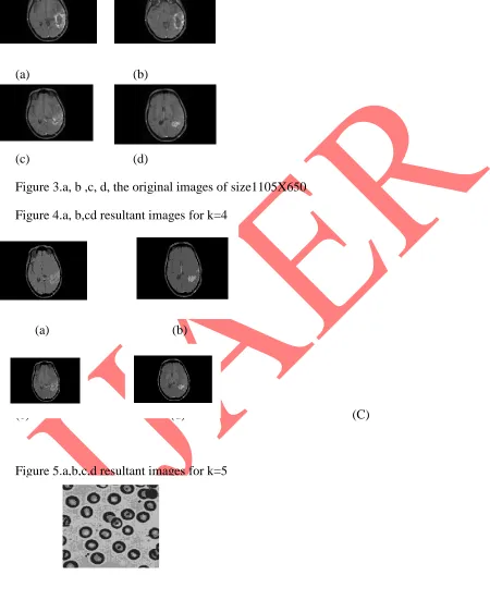
The performance of the proposed method is tested on the MRI brain image of 31 years old female.
The images shown in the figures 2. a,b,c,d are the different slices of MRI head image of size 1105x650.
(a) (b)
(c) (d)
Figure 3.a, b ,c, d, the original images of size1105X650
Figure 4.a, b,cd resultant images for k=4
(a) (b)
(c) (d) (C)
Figure 5.a,b,c,d resultant images for k=5
To test the robustness of the method the 2% of Gaussian noise applied on the image
40 INTERNATIONAL JOURNAL OF ADVANCES IN ENGINEERING RESEARCH Figure 7.a. Segmentation before pre-processing
7.bSegmentation after preprocessing for k=4
c
The output we have produced detects automatically the number of clusters affected by the tumor in the brain. After the detection of number of clusters, the tumor bounded region is isolated.
Here, the pre-processing is done using mathematical morphology. The square structuring element is used to smooth the image. The erosion is performed first to reduce the size of noise particles. Then image opening followed by closing is performed. Thus in our proposed method we produce the automatic detection of number input clusters affected by brain tumor tumor. We use segmentation by K-means clustering. It can be deduced from the results that unsupervised segmentation methods are better than the supervised segmentation methods. K-Means clustering method is undemanding, proficient and less error receptive when compared with frequently used clustering techniques. The tumor affected images are found out by applying the proposed Clustering algorithm using Matlab Simulator. The output we have produced detects automatically the number of clusters affected by the tumor in the brain .After the detection of number of cluster the tumor bounded region is isolated
Table 1. Performance Analysis
IMAGE METHOD EXECUTION
TIME Figure 2.a Proposed
SOM FCM
1.15 1.98 2.10 Figure 2.b Proposed
SOM FCM
1.18 1.92 2.00 Figure 2.c Proposed
SOM FCM
1.11 1.23 1.90 Figure 2.c Proposed
SOM FCM
1.14 1.33 1.97
41 INTERNATIONAL JOURNAL OF ADVANCES IN ENGINEERING RESEARCH
CONCLUSION
As a result we evaluate the proposed technique with other clustering methods and the results are very appreciable. Less execution time is achieved when compared with other clustering methods. Thus from the evaluated results it is clearly shown that the Proposed system is competent and is less fault perceptive.
REFERENCES
[1] Beham, M.P, Gurulakshmi, A.B., Morphological image processing approach on the detection of tumor and cancer cells, International Conference on Devices, Circuits and Systems (ICDCS), 2012.
[2] Vijay, J., Subhashini, J.An efficient brain tumor detection methodology using K-means clustering algorithm, 2013 International Conference on Communications and Signal Processing (ICCSP).
[3] M. Masroor Ahmed and Dzulkifli Bin Mohammad "Segmentation of Brain MRImages for Tumor Extraction by Combining K-means Clustering and Perona-Malik Anisotropic Diffusion Model" International Journal of Image Processing, Volume (2) Issue (l), 2010. [4] Arash Azim Zadeh Irani and Bahari Belaton "A K-means Based GenericSegmentation System" 2009 Sixth International Conference on Computer Graphics, Imaging and Visualization.
[5] Ming-Ni Wu, Chia-Chen Lin and Chin-Chen Chang "Brain Tumor Detection Using Colour-Based K-Means Clustering Segmentation" International Conference on Computer Graphics, 2009.
[6] S.Sathish Kumar, M.Moorthi, M.Madhu3and Dr.R.Amutha "An Improved method of Segmentation using Fuzzy-Neuro Logic" Second International Conference on Computer Research and development, 2009.
[7] S. Murugavalli and V. Raja Mani" An improved Implementation of Brain tumor Detection Using Segmentation Based on Neuro Fuzzy Technique", Journal of Computer Science 3, 2007 October.
[8] M. Schmidt, I. Levner, R. Greiner, A. Murtha, and A. Bistritz. “Segmenting Brain Tumors using Alignment-Based Features".International Conference on Machine Learning and Applications, Los Angeles, December 2005.
[9] S.R. Kannan, Jan. 2005 "Segmentation of MRl using new Unsupervised FuzzyC means algorithm" ICGST-GVIP Journal, Volume 5, Issue2.
[10] S.Saheb Baha and Dr. K.Satya Prasad, "Morphological image processing in Bio- Medical Application", Proceedings of PCEAIFTOMM-International conference - PICA- 2006, held on 11th to 14th of July at Nagpur and NCBME-2006 - National conference on Bio-Medical Engineering held on 28th to 29th of March at Mumbai.
[11] Juraj Horvath, 2006 "Image Segmentation using Fuzzy C-means" SAMI2006. [12] Lei jiang, Wenhui Yang , "A modified Fuzzy C-means algorithm for Magnetic Resonance Images" Proc. VlIth Digital Image Computing: Techniques and Applications, 2003.
[13] Xiang-Yang Wang, Hong-Ying Yang, Yu Zhang, Zhong-Kai Fu, image denoising using SVM classification in nonsubsampled contourlet transform