R E S E A R C H
Open Access
Developmental evolution of flowering plant
pollen tube cell walls: callose synthase (CalS)
gene expression patterns
Jason M Abercrombie, Brian C O
’
Meara, Andrew R Moffatt and Joseph H Williams
*Abstract
Background:A number of innovations underlie the origin of rapid reproductive cycles in angiosperms. A critical early step involved the modification of an ancestrally short and slow-growing pollen tube for faster and longer distance transport of sperm to egg. Associated with this shift are the predominantly callose (1,3-b-glucan) walls and septae (callose plugs) of angiosperm pollen tubes. Callose synthesis is mediated by callose synthase (CalS). Of 12CalSgene family members inArabidopsis, only one (CalS5) has been directly linked to pollen tube callose. CalS5 orthologues are present in several monocot and eudicot genomes, but little is known about the evolutionary origin ofCalS5or what its ancestral function may have been.
Results:We investigated expression ofCalSin pollen and pollen tubes of selected non-flowering seed plants (gymnosperms) and angiosperms within lineages that diverged below the monocot/eudicot node. First, we determined the nearly full length coding sequence of aCalS5orthologue from Cabomba caroliniana(CcCalS5) (Nymphaeales). Semi-quantitative RT-PCR demonstrated lowCcCalS5expression within several vegetative tissues, but strong expression in mature pollen.CalStranscripts were detected in pollen tubes of several species within Nymphaeales and Austrobaileyales, and comparative analyses with a phylogenetically diverse group of sequenced genomes indicated homology toCalS5. We also report in silicoevidence of a putativeCalS5orthologue from Amborella. Among gymnosperms, CalS5transcripts were recovered from germinating pollen ofGnetum andGinkgo, but a novelCalSparalog was instead amplified from germinating pollen ofPinus taeda.
Conclusion:The finding that CalS5 is the predominant callose synthase in pollen tubes of both early-diverging and model system angiosperms is an indicator of the homology of their novel callosic pollen tube walls and callose plugs. The data suggest thatCalS5had transient expression and pollen-specific functions in early seed plants and was then recruited to novel expression patterns and functions within pollen tube walls in an ancestor of extant angiosperms.
Background
The pollen tube is a unique feature of male gametophytes of seed plants. In cycads andGinkgo, pollen tubes are long-lived and function solely as haustorial, highly branched structures that grow invasively into female tis-sues [1-3]. In conifers and Gnetales pollen tubes function in a new way to deliver non-motile sperm to the egg (siphonogamy), while generally retaining a haustorial growth pattern [2,3]. Flowering plant (angiosperm) pollen tubes have lost most features of haustorial growth - their
pollen tubes are typically short-lived and seem to function exclusively to deliver sperm to the egg [4,5]. The origin of siphonogamy has been held up as a classic example of exaptation [6], because the plesiomorphic function of the pollen tube - nutritional support for the male gametophyte - was subsequently co-opted for a novel role in sperm delivery [3]. Yet siphonogamy is clearly a complex process, and it is not at all obvious which aspects have common origins, which represent modifications of an ancestral pat-tern, and which have arisen independently in separate lineages [1,3,4,7]. Understanding the homologies of pollen tube structure and growth pattern may provide deeper insights into the origin(s) of this remarkable innovation. * Correspondence: [email protected]
Department of Ecology and Evolutionary Biology, University of Tennessee, Knoxville, TN, USA
Angiosperm pollen tubes have a unique wall structure. Their thin growing tip is comprised almost entirely of pectins. Just behind the pectic tip, cellulose synthases operate to form a very thin, pecto-cellulosic primary wall. Then, still in the subapical region, (1,3)-b-glucan (callose) is synthesized beneath the thin primary wall to form a thick layer [8]. The mature pollen tube wall of most angiosperms is primarily made of callose (81% by weight inNicotiana; ref. 9). As an amorphous polysac-charide, callose can be synthesized more rapidly than an equivalent weight of fibrous cellulosic cell wall [9] and it provides resistance to tensile and compression stress [10]. Callose also severely reduces wall permeability and since angiosperm pollen tube walls are also prone to forming septae ("callose plugs”) [11], the plesiomorphic haustorial function of tubes is largely precluded. These patterns are general features of all angiosperms, from
Amborella and water lilies to Arabidopsis and maize [4,12]. Yet, despite their ubiquity, the ancestral function of callose walls and plugs is not obvious. Tubes that lack callose in their walls retain their function in some derived eudicot lineages, such as Lamiales [13] and in an Arabidopsis mutant line [14,15], though they have reduced competitive ability in the latter [14].
Pollen tubes in ovules of gymnosperms rarely contain callose in lateral walls, and callose plugs have never been reported [16]. Callose is found in the tip wall of growing pollen tubes of some conifers [17], a pattern never seen in angiosperms. Studies of in vitro-grown gymnosperm pollen tubes do sometimes find callose (or mixed-glucans) in lateral tube walls [17,18]. Importantly, the deposition of callose is generally transient in gym-nosperm male gametophytes and its extent and location varies even among closely related species [17-20]. Such transient and variable phenotypes contrast with the rela-tively invariant and persistent expression pattern seen in angiosperm pollen tubes.
Callose synthesis is mediated by the enzyme, callose synthase, encoded by the callose synthase gene, and origin-ally described as a glucan synthase-like gene (GSL) in
Nicotiana alata[21]. Honget al. (2001) named the callose synthase gene familyCalSafter identifying 12 gene family members inArabidopsis thaliana[22].AtCalS5and its characterized orthologues (NaGSL1fromN. alata) have been directly linked to pollen tube wall formation and cal-lose plug deposition, as well as to pollen exine develop-ment [14,15,23,24].
CalS5appears to have an ancient origin by duplication. A comparative phylogenetic analysis of allCalSparalogs from the genomes of the moss,Physcomitrella patens
[25] andArabidopsisfound thatAtCalS5was more clo-sely related to aPhyscomitrella CalSgene copy (PpCalS5) than to any otherArabidopsisparalog. Because callose was observed in the moss spore aperture region and
PpCalS5 was identified as a putative orthologue to
AtCalS5, PpCalS5 was hypothesized to play a role in moss spore germination [25]. If so, thenCalS5 involve-ment in pollen tube growth may ultimately derive from a more ancient function involving the germination process. As such, changes in gene regulation were likely prerequi-sites for the acquisition of novel callose deposition pat-terns in angiosperm pollen tube walls. Alternatively, the patterns arose via duplication and functional divergence of aCalSgene within seed plants, or perhaps within the stem lineage leading to angiosperms.
In this paper we present molecular evidence thatCalS5
orthologues are expressed in mature pollen and pollen tubes of several extant early-diverging angiosperms in Nymphaeales and Austrobaileyales and likely also in
Amborella trichopoda. CalS5 orthologues are also expressed in mature gymnosperm pollen, including one siphonagam (Gnetum) and one non-siphonogam (Ginkgo). In the siphonogamous conifer,Pinus, we report a poten-tially uniqueCalSgene expressed in germinated pollen. We discuss the implications of these findings for the evo-lution of the angiosperm pollen tube wall and suggest new avenues of research to clarify the functional roles ofCalS
in the seed plant male gametophyte.
Results
Putative orthologues ofCalS5are expressed in pollen and pollen tubes of early-diverging angiosperms
A nearly full length coding sequence was obtained from
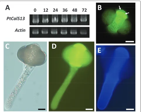
Cabomba caroliniana (Cabombaceae; Nymphaeales) (CcCalS5) comprising 5,562 bp which translated into a predicted 1854 amino acid polypeptide with 78% identity toArabidopsis thalianaCalS5 (AtCalS5) and 66% identity to the moss orthologue (PpCalS5) from thePhyscomitrella patensgenome [25]. The deduced polypeptide has a pre-dicted topology containing between 13 and 17 transmem-brane helices (as predicted by TMHMM v. 2.0; CBS, Lyngby, Denmark and SOSUI engine ver. 1.11; Nagoya University, Nagoya, Japan), a cytoplasmic N-terminal loop domain containing > 423 amino acids, and a large hydro-philic loop domain consisting of 758 amino acids, with loops between the helices ranging from 4 to 106 amino acids in length (Figure 1A).
RT-PCR was used to assess the presence ofCcCalS5
transcript in various tissues ofC. caroliniana, as well as in a pollen tube time-course experiment inNymphaea odor-ata(Figure 1B) using intron-spanning primers. A 1,497 bp nucleotide sequence that shared 100% nucleotide identity with theCcCalS5was amplified fromN. odoratapollen and was used to designate theNymphaea CalS5 ortholo-gue (NoCalS5) in our RT-PCR experiments (Figure 1B).
and leaf tissues during our RT-PCR optimization experi-ments (See Additional file 1). Expression ofCcCalS5 in bothCabombastem and leaf tissues was confirmed by sequencing clones of PCR products.NoCalS5was consis-tently expressed during a time-course ofin vitro-grown pollen tubes harvested at 0, 1, 3 and 6 hrs after inoculation in liquid medium (Figure 1B; right panel gel).
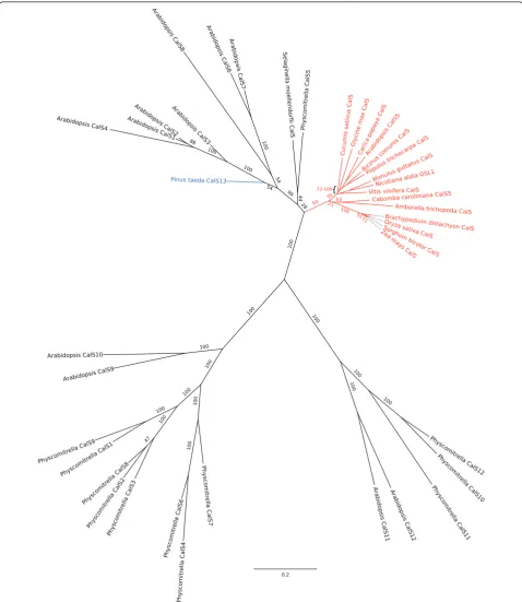
In the 18-taxon phylogenetic tree constructed to infer orthologous relationships, all putative angiosperm CalS5
orthologues plus CcCalS5, formed a clade with 99% bootstrap support (Figure 2). A partial sequence from
the putativeCalS5 orthologue in Amborella trichopoda
[26] also falls within the CalS5 clade (Figure 2). The putative CalS5 orthologues of Physcomitrella[25] and
Selaginella(this study) are strongly supported as falling in an angiosperm clade ofCalSparalogs, but not neces-sarily as sister to theCalS5clade.
RT-PCR also recovered CalS transcripts from the mature pollen of the early-diverging angiosperms, Austro-baileya scandens(Austrobaileyaceae; Austrobaileyales),
Nuphar advena (Nymphaeaceae; Nymphaeales), and
Trithuria austinensis(Hydatellaceae; Nymphaeales). These
6
28
35
14
40
758
25
14
40
106
34
4
41
423
A
B
CalS5
Actin
S L M A P
G 0 1 3 6
Fig 1.
!
"
#
$%
&
'()
(*
+
,
&
!
&
-
.
"
.
!.
&
.
.
!!
&!
&
"
""
&!
01
Figure 2Phylogenetic tree for full length CDSs ofArabidopsis,Physcomitrella CalSgene families andCalS5orthologues. Phylogenetic tree based upon alignment of predicted polypeptides for full length CDSs ofArabidopsis,Physcomitrella CalSgenes, and putativeCalS
partial sequences from the hydrophilic loop domain align with the partialNoCalS5sequence and are orthologous to
CalS5, based on phylogenetic analysis (Additional files 2 and 3).
Putative orthologues ofCalS5are expressed in pollen of Gnetum gnemonandGinkgo biloba
At 24 h after incubation,Ginkgopollen stained for callose in the aperture area, intine, and also in the walls that sepa-rated prothallial, generative, and tube cells (Figure 3A).
Gnetumpollen sheds its exine before tube growth, and prior to exine shedding, aniline blue staining was observed in the inner pollen wall (Figure 3B). After exine shedding, callose was not observed in the intine (Figure 3C, D).
Partial cDNA fragments from putativeCalS5 ortholo-gues were amplified from mature pollen ofGnetum gne-mon (GgCalS5) andGinkgo biloba (GbCalS5). Their predicted amino acid sequences aligned with the central loop domains of other CalS proteins, including those known to function during Arabidopsis pollen develop-ment (see Additional file 2). Phylogenetic analysis placed the Ginkgo andGnetum sequences within a strongly supported clade of angiosperm CalS5 sequences (Addi-tional file 3).
Interestingly, the aligned predicted protein sequences identified a short NASQ motif thatGinkgoshares with all
members of Nymphaeales but that is absent from all other CalS5 sequences (see Additional file 2). Prosite scans of the aligned sequences identified the shared motif as a putative N-glycosylation site (Prosite scan data not shown). Other putative functional motifs common within this alignment are CK2 and PKC phosphorylation sites that are highly conserved within all otherArabidopsis
CalS sequences, however one predicted cAMP-dependent phosphorylation site, K(R/K)ES, was unique to most taxa within the CalS5 clade (see Additional file 2).
A uniqueCalSorthologue is expressed inPinus taedapollen A 1,413 bp CalS transcript was strongly expressed in mature and germinated Pinus taedapollen (Figure 4A). Phylogenetic analysis of the 471 amino acid predicted polypeptide indicates 99% bootstrap support for its inclusion within a clade that does not include CalS5 (Figure 2). Because it is distantly related to any of the knownArabidopsisparalogs in that clade, we named the genePtCalS13.PtCalS13transcripts were abundant over a 72-hour time period of in vitro growth (Figure 4A), however repeated attempts to amplifyCalS5-like tran-scripts over the same developmental stages failed. All combinations of primers that amplifiedCalS5-like frag-ments fromGinkgo, Gnetum, and early-divergent angios-perm pollen cDNA were attempted.
A
B
D
C
Aniline blue staining was localized to the inner walls of the mature pollen grain, but not the tube wall (Figure 4B-D). In pollen, strong staining was observed within the third intine layer at the proximal face of the microgameto-phyte, whereas weaker staining was apparent in the aper-ture region, the leptoma (Figure 4B). Upon germination, the aperture area became strongly stained, but the thick lateral walls of the pollen tube did not (Figure 4C, D). Cal-cofluor staining for cellulose was strong throughout all time periods observed duringin vitropollen tube growth (Figure 4E).
Comparative analysis of predicted functional motifs among callose synthases
Protein motif searches [27,28] were performed on the sequences of CalS5 orthologues, as well as on all other
callose synthases inPhyscomitrellaand Arabidopsisto evaluate conserved patterns of short linear motifs that may have functional significance. Comparison of the cal-lose synthase protein sequences revealed conservation of seven different site patterns in all callose synthases: N-glycosylation, cAMP- and cGMP-dependent protein kinase phosphorylation, protein kinase C phosphoryla-tion (PKC), casein kinase II phosphorylaphosphoryla-tion (CK2), tyr-osine kinase phosphorylation, N-myristoylation, and amidation (data not shown). The majority of putative functional motifs were found within the N-terminal loop and the large central loop domain. The central loop domains of all callose synthases were consistently enriched with predicted PKC and CK2 phosphorylation sites. There were no clear distinguishing features of CalS5 orthologues with respect to selected patterns of
A
B
A
01224364872
B
PtCalS13
Actin
C
D
E
predicted linear motifs when compared to the other cal-lose synthases.Zea mays,Sorghum bicolor, and Selagi-nella moellendorfiiall appear to have truncated CalS5 proteins, with the entire N-terminal domains completely absent. This may reflect incorrect annotation, given the conserved nature of this large functional domain.
In silicoidentification and phylogenetic analysis of putative CalS5 orthologues
BLAST searches of NCBI [29] and Phytozome [30] data-bases revealed putativeCalS5orthologues from a phylo-genetically diverse group of plant species which ranged from the unicellular green algae,Chlamydomonas rein-hardtiito most completely sequenced angiosperms. Amino acid sequences from these putative orthologues were aligned with all known callose synthase gene family mem-bers fromPhyscomitrella,Arabidopsis, as well as other putativeCalS5orthologues for other angiosperms and the spikemoss,Selaginella moellendorfii. Also included in the alignment were two sequences of particular interest, the 471 amino acid PtCalS13 sequence obtained fromPinus taedapollen cDNA, and a putativeCalS5orthologue iden-tifiedin silicofrom the early-diverging angiosperm, Ambor-ella trichopoda. TheAmborellatranslated uniscript was obtained from the 454-EST build from the Ancestral Angiosperm Genome Project website [26] and comprised 196 amino acids. Several other translated uniscripts from
Amborelladisplayed homology to CalS5, but because con-tig assembly was not feasible, they were not included in this analysis (data not shown).
Discussion
CcCalS5 contains both general CalS functional motifs and CalS5-specific motifs
All CalS proteins studied to date share a common topol-ogy with a large N-terminal hydrophilic domain followed by two clusters of transmembrane (TM) domains that flank a large central hydrophilic domain [15,22,23,31,32]. The large hydrophilic loop is thought to accommodate interactions with other proteins, such as Rop1, UDP-glu-cose transferase (UGT1), sucrose synthase, and annexin, which enable the formation of a CalS enzyme complex [31]. Scans for functional motifs in CcCalS5 support the model of Verma and Hong [31], in which most known cal-lose synthase proteins exhibit similarities in putative glyco-sylation and phosphorylation sites, particularly within the N-terminal domain and the large central loop.
With respect to pollen tube growth, cAMP is a known signalling molecule for pollen tube guidance and growth [33]. In our alignment of the loop domains, we identified one putative cAMP and cGMP-dependent phosphoryla-tion site common to all seed plant members of the CalS5 clade, but absent from PpCalS5 of Physcomitrellaand from all other CalS paralogs (see Additional file 2). We
also identified a putative CalS5 N-glycosylation site unique to seed plants - present inGinkgoand all members of the Nymphaeales clade (see Additional file 2) but absent from
Austrobaileya and all monocots and eudicots. It is not known whether these variants cause functional differences inCalS5expression.
CalSgenes involved in male gametophyte development Five of 12 callose synthase genes (CalS5,CalS9,CalS10,
CalS11, andCalS12) have been shown to function during microsporogenesis and microgametogenesis in Arabidop-sis[15,34-37]. In contrast to otherCalSgenes,CalS11and
CalS12contain only two or three exons [22] and are Ca2
+
-dependent [38].CalS11 and CalS12 are genetically linked and perform partially redundant roles in the forma-tion of walls separating microspores of the tetrad and in late maturation of the male gametophyte [34].CalS12is also activated during wound response and on stigmatic papillae [39].
The other 10CalSgenes contain between 39 and 50 exons and are Ca2+-independent [40,41].CalS5is sporo-phytically expressed to form the callose wall of pollen mother cells and microspore tetrads and is also the predo-minant gametophytically-expressed transcript in germinat-ing pollen and growgerminat-ing pollen tubes [14, 15, this study].
CalS9andCalS10function early in microgametogenesis [35,36] since mutant lines independently exhibited func-tional aberrations during the entry of microspores into mitosis. Mutants ofCalS9caused failure of mitosis II as well as abnormal positioning of nuclei in mature pollen and precocious germination, inside the anther [37].
CalS10is known to be involved in cell plate formation [22]. Silencing ofCalS9andCalS10using gene-specific dsRNAi constructs also resulted in a dwarfed growth habit, suggesting that both also function in the sporophytic phase [35]. These studies show that male gametophyte development in Arabidopsis is mediated by transient expression of a number of CalS genes, whereas in all angiosperms studied to dateCalS5is abundant and predo-minant during pollen germination and in all stages of pol-len tube growth.
ThePinus taedamale gametophyte expresses a unique CalSgene in pollen
We found strong expression of a novelCalSgene in mature and in germinated pollen ofP. taeda.PtCalS13
was strongly supported as falling within a clade ofAtCalS
paralogs that does not include any of the known gameto-phytically-expressed paralogs (AtCalS5andAtCalS9-12).
PtCalS13cannot be a deeply divergent copy ofCalS5or
CalS9-12, but is most likely a novel copy ofCalS (alter-natively, it may be a deeply divergent orthologue of
PtCalS13functions in place ofCalS5. However, more work is needed to test this hypothesis.
Pinusspecies are quite variable in the location and extent of pollen callose deposition [42,43]. InP. taeda, cal-lose was present in the intine and male gametophyte walls before pollen germination. Upon germination, it became strongly expressed in the aperture region surrounding the exiting pollen tube, but did not extend into the tube wall, as it does in angiosperms [41,44,45]. Thus, the callose dis-tribution pattern associated withPtCalS13expression is quite different from theCalS5pattern in germinating angiosperm pollen.
GinkgoandGnetumexpress an orthologue ofCalS5in pollen
There is strong evidence that theCalSgenes expressed in mature pollen ofGnetumandGinkgoare orthologous to angiosperm CalS5since their protein sequences were nested within the angiosperm CalS5 clade. Callose deposi-tion is apparently restricted to pollen in these species. In
Gnetumit was present in the inner wall of pollen before exine shedding, but after shedding it was absent from the intine which is continuous with the emerging pollen tube wall. InGinkgo, callose was abundant in the intine and in the walls of the male gametophyte. A study ofin vitro pol-len tube growth inGinkgofound that their tube walls stained weakly for aniline blue but reacted strongly to cal-cofluor white andb-(1,3)(1,4) antibodies, suggesting the presence ofb-(1,3)(1,4) mixed glucans [18].
The evolutionary developmental origins of callose synthase expression in angiosperm pollen tubes
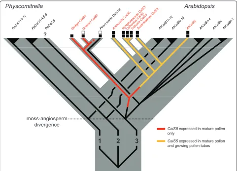
Fast growing pollen tubes are arbiters of the rapid repro-ductive cycles of angiosperms and their unique wall struc-ture may have been a trigger for extensive pollen tube growth rate evolution in the group [1,4,46]. Gene family expansions are thought to have been important for trait diversification early in angiosperm history or pre-history [47,48]. SixCalSparalogs from three ancient land plant lineages - five fromArabidopsis[22] and one fromPinus
(this study) - are now known to be expressed in male gametophytes of seed plants (Figure 5). To date, only
CalS5has been shown to be expressed in pollen tubes, and only in a few model system eudicots [15,23,24]. The finding that pollen tubes of a broad set of extant early-diverging angiosperms also utilizeCalS5in their pollen tubes supports the homology of callose walls and plugs in flowering plants [4,5].
CalS5transcripts were also found in mature and/or ger-minating pollen, of two distantly-related gymnosperms,
GinkgoandGnetum. The predominant anatomical loca-tion of callose inGinkgowas in the intine and internal gametophytic cell walls, whereas the pollen tube-forming intine of germinated Gnetum pollen did not contain
callose. In all early-divergent angiosperms used in this study, callose was present within the intine of germinating pollen and continuous with the callose inner wall of the pollen tube [4,43,49]. Gametophytic expression ofCalS5
has a similar pattern in Arabidopsis and tobacco [14,15,23,24,32,37,50]. Thus, it seems likely thatCalS5had an ancestral expression pattern within the inner pollen wall that later became modified via the evolution of gene regulation to function in growing pollen tubes of an ances-tor of extant angiosperms (Figure 5).
A number of hypotheses have been proposed as to what the ancestral function of callose in germinating pollen or spores might have been. In the moss,Physcomitrella, cal-lose was deposited in the inner exine layer (not the intine) near the aperture at the proximal pole of the spore just before germination [25]. Callose was inferred to function in spore germination and it was suggested that aCalS5
orthologue was involved [25]. Expression data are needed to determine if these results reflect an ancient aspect of land plant spore germination or an apomorphic feature of moss spores (Figure 5). We also found callose thickenings in the aperture area of germinatingPinuspollen, but these were associated withPtCalS13, not aCalS5orthologue (Figure 5). Callose is localized to the inner intine of mature pollen of some Pinusspecies [51-53], the outer layer inP. sylvestris[17,54], and is absent from the intine of
P. wilsonii[55]. Paciniet al. (1999) concluded that such variable and transient expression of callose inPinuspollen indicates that it functions as a reserve polysaccharide, rather than serving a structural or prophylactic function [45]. Górska-Brylass (1970) argued its presence in the proximal outer intine was due to non-retrieval of callose plates from the prior divisions of prothallial cells [56]. Alternatively, its co-localization with degenerate prothallial cells may indicate the prior involvement of callose as a wall sealant that initiates cell death, a pattern also seen during megasporogenesis in seed plants [57].
To date no study has convincingly shown callose to be the predominant and permanent constituent of any gym-nosperm pollen tube wall, nor is there any finding of a callose plug in a gymnosperm pollen tube. For example, callose is reported from young but not old tubes inPinus
andCycas[17,20]; it is a transient feature of long-lived
male gametophyte, which typically lives within a pollen grain attached to female tissues at the pollination site [3]. In most conifers, sperm are formed late in male gameto-phyte ontogeny and must travel from the pollen grain to the tip late in life. Thus, the biology of the fertilization process in most gymnosperms prevents their pollen tubes from utilizing callose as a semi-permanent struc-tural feature of their walls.
At some point(s) along the lineage leading to angios-perms, two shifts in CalS5 localization occurred - callose deposition became restricted to a short subapical region of the growing pollen tube tip and to a small distal region of the tube where a callose plug forms. Importantly, both of these changes in localization must also have involved changes in callose retrieval, giving rise to persistent cal-lose walls and plugs. One model for the origin of angios-perm pollen tube morphology is that an ancestral conifer-like pollen tube became transformed by the
evolution of faster growth rates [46], causing the callose synthesis and retrieval machinery to be displaced from the newly-forming tube tip to a subapical position [1,17]. Comparative analyses support the notion that early angiosperm pollen tubes grew faster than those of their gymnosperm-like ancestor (extant gymnosperm pollen tubes are characterized by exceptionally slow pollen tube growth rates) [5]. What is not clear is whether non-retrieval of callose is a cause or a consequence of the ori-gin of faster growth rates.
Given the increased tendency of molecular phyloge-netic analyses to place conifers/Gnetales in an isolated position relative to angiosperms [58], it is worth consid-ering that angiosperm pollen tube morphology may have evolved independently from an ancestralGinkgo/ Cycad-like haustorial tube [20], rather than from a transitional siphonogamous, conifer-like predecessor. Resolving the question of pollen tube origins will require careful
Physcomitrella
Arabidopsis
*
?moss-angiosperm
1
2
3
divergence
CalS5expressed in mature pollen only
CalS5expressed in mature pollen and growing pollen tubes and growing pollen tubes
developmental analyses of pollen tube growth and the many genes that mediate differences among extant seed plant groups. It would be especially interesting to look at the evolution of the callase (b-1,3-glucanase) gene family, which catalyzes the retrieval of callose [59].
Conclusion
This study supports the homology of callose pollen tube walls and plugs across flowering plants at the level of
CalS5gene expression. SinceCalS5was also found to be actively transcribed in mature pollen ofGinkgoand Gne-tum, we suggest that CalS5 localization to the inner intine of mature germinating pollen was present in a distant angiosperm ancestor, perhaps extending into the walls of young pollen tubes, and was later co-opted as a non-tran-sient feature of angiosperm pollen tubes (Figure 5).CalS5
is a structural gene that originated by duplication long before the origin of extant angiosperms. Thus, the novel callose deposition patterns of angiosperm pollen tubes must be a consequence of the evolution of novel regula-tion of an ancient gene. It remains to be seen to what extent this involved duplication and divergence of other genes involved in the callose synthesis or retrieval path-ways, and to what degree it was or was not a developmen-tal outcome of the evolution of faster growth rates.
Methods Plant material
Whole flowers fromAustrobaileya scandensWhite were collected near Millaa Millaa, Queensland, Australia (17° 31’15”S, 145° 33’53”E).A. scandenspollen tubes were grown in hanging drops of BK media [60] containing 2.5% sucrose inside closed petri plates for 5 to 12 hours. Pollen fromTrithuria austinensisSokoloff was collected in Bran-chinella Lake, shire of Manjimup, Western Australia (34° 16’S, 116° 42’E). Pollen or pollen tubes of these two spe-cies were centrifuged briefly and resuspended in RNLater
(Ambion, Austin, TX, USA) and RNA was isolated within several weeks. RNA was isolated from fresh pollen or pol-len tubes for the remaining species below. Polpol-len from
Ginkgo bilobaL. was collected from trees growing on the University of Tennessee campus in Knoxville, TN, USA.
Nuphar advenaAiton. flowers were collected near Sparta, TN, USA (35° 55’11”N, 85° 20’41”W) and pollen tubes were grown in BK media with 5% sucrose for two hours before RNA isolation.Nymphaea odorataAiton. flowers were collected from a pond in Knoxville, TN, USA (35° 53’ 51” N, 84° 10’23” W).Cabomba carolinianaA. Gray plants were grown in greenhouse water tanks and were originally collected from Racoon Creek, Jackson County, AL, USA (34° 46’N, 85° 50’W) or purchased from Caro-lina Biological Supply (Burlington, NC, USA).Gnetum gnemonL. flowers with dehiscent anthers were collected from greenhouse-grown plants in DEPC-treated water,
vortexed to separate pollen from all other flower parts, and briefly centrifuged prior to RNA isolation. Pollen from
Pinus taedaL. andP. strobusL. was collected from trees on the University of Tennessee campus.Pinuspollen was grown in a liquid medium containing 10% sucrose, 15 mM MES, 1 mM H3B, 1 mM CaCl2, pH 4.0 in petri
plates sealed with parafilm.
In vitropollen tube experiments withNymphaeaand Cabomba
Due to the poor germination observed for bothNymphaea
andCabombapollen in standard BK media, stigmatic fluid from first dayNymphaeaflowers was collected on site with a disposable pipette and used as the pollen tube growth medium for these species. Both species exhibited 70 to 90% germination success when grown in the fresh stigmatic fluid, and thus fresh stigmatic fluid was used for all pollen tube experiments described here. Stigmatic fluid was collected shortly after flower opening (9 to 10 am), and centrifuged for three minutes at 13,000 rpm to remove any contaminating debris and/or pollen grains. Anthers fromCabomba andNymphaeawere removed with forceps and placed into 1.7 ml tubes that contained 1 ml of stigmatic fluid. Anther number was used to stan-dardize samples for pollen density during tube growth. Tubes were vortexed briefly to separate pollen grains from anthers, anthers were then removed, and contents were transferred to a small petri plate for pollen tube growth at room temperature.Nymphaeapollen tubes were grown for various lengths of time (one, three, and six hours post-inoculation) for gene expression experiments. For RNA extractions, pooled pollen tube samples were harvested at each time point, centrifuged at a 2,000 rpm for 30 seconds to maintain pollen tube integrity, and immediately frozen in liquid nitrogen after growth medium was removed.
Bioinformatics and primer design
In order to search for orthologousCalS5gene sequence in the early-diverging angiosperms, the local BLAST tool on the ancestral angiosperm genome project website [26] was used to blast theArabidopsisCalS5 protein sequence (tBlastn) against all available 454-Sanger hybrid data-bases. After performing non-redundant nucleotide NCBI BLAST searches of individual uniscript hits, primers were designed to amplify a Nuphar advenauniscript sequence (c78546) with the lowest E-value corresponding to AtCalS5. The primers designed towards Nuphar advenasequence (Sec16F; Sec17R) amplified a 600 bp highly conserved region of sequence within the predicted hydrophilic domain of the putativeCalS5orthologue. These primers were also used for amplifying putative
Nymphaea odorata, andTrithuria austinensis. Another forward primer (Sec17F) nested within this sequence and the reverse primer (Sec17R) enabled PCR amplification of the 250 bp Gnetum sequence. Various multiple sequence alignments were carried out on all callose synthase DNA and protein sequences inPhyscomitrella patensand Arabidopsisto aid in primer selection. To amplify thePinus taeda CalScDNA, two EST sequences showing the highest homology toAtCalS5andPpCalS5
sequence (AI812992 and FJ114840) obtained from non-redundant NCBI BLAST of Pinus were aligned with
PpCalS5andAtCalS5. Forward FJ114840-F and reverse primer AI812992-R amplified a product that reflected the alignment. To prevent amplification of other pollen-expressedCalSgenes (CalS9,CalS10,CalS11,CalS12), particular attention was given to these sequences during primer design for PCR applications. All primer design and sequence alignments were performed using Vector NTI software (Invitrogen, Carlsbad, CA, USA). A com-plete set of primers used in this study is listed in Addi-tional file 4.
Molecular analyses
Total RNA from pollen and pollen tubes was isolated using Tri ReagentJ (Ambion, Austin, TX, USA) and a modified CTAB protocol was required forCabomba vege-tative tissues [61]. For cDNA synthesis, 1.5μg of total RNA was used, according to the manufacturer’s protocol (ArrayScript; Ambion, Austin, TX, USA). Prior to cDNA synthesis, all samples were subjected to DNase I (Turbo-Free DNase kit; Ambion). RNA was assessed for quality with agarose gel electrophoresis and quantified with a NanoDrop (Thermo Scientific, Waltham, MA, USA) spec-trophotometer. A 3’RACE procedure was used according to the First Choice RLM RACE kit (Ambion, Austin, TX, USA) to acquire the 3’end of theCabomba caroliniana CalS5gene. However, due to difficulties with the 5’RACE protocol 5’products were amplified using a forward pri-mer designed towards highly conserved sequence within a multiple sequence alignment of CalS5 orthologues (CalS515F). Inverse PCR was performed to obtain the 5’ end of the full length cDNA using a digest/re-ligation/ digest strategy with HindIII/PstI, respectively [62]. Although attempts to obtain the 5’end of the cDNA were incomplete, the inverse PCR procedure did enable the sequencing of introns that flanked the 5’-most exon. Iden-tification of these intron-exon boundaries enabled the design of intron-spanning primers for semi-quantitative RT-PCR. All PCR reactions that required cloning were performed with Herculase II DNA fusion polymerase (Agilent, Santa Clara, CA, USA). Products were gel puri-fied using a Qiaex II gel purification kit (Qiagen, Valencia, CA), cloned into pcr8-GW-TOPO (Invitrogen, Carlsbad, CA, USA), and sequenced on an ABI 3100 capillary
sequencer at the University of Tennessee Molecular Biol-ogy Resource Facility. cDNA tissue sources are listed in Additional file 5. All sequences were deposited in Gen-bank [http://www.ncbi.nlm.nih.gov/genGen-bank/index.html].
Semi-quantitative RT-PCR
To prevent amplification of possible orthologous pollen-expressedCalS genes (CalS9, CalS10, CalS11, CalS12), both protein and nucleotide alignments were used to design primers to amplify a 631 bp intron-flanking sequence that shared a predicted low homology to non-target CalScDNA. Primers were designed to amplify an
Actingene isolated from Nuphar advenapollen.Actin
was also used as a reference gene to confirm equal tem-plate loading in Pinus taedaRT-PCR. A master mix of PCR reagents (described above) was used to amplify the
CalS5fragment and Actincontrol gene in separate reac-tions at equal template concentrareac-tions and cycling para-meters. Threshold cycle optimization was determined by performing PCR amplifications over a range of cycles. The number of PCR cycles selected corresponded to where the trend line exhibited the highest correlation to exponential amplification. Bands representing each tis-sue type were purified, cloned, and the sequence was verified to confirm single product amplification.
Phylogenetic analysis
Alignments of predicted amino acid sequences were per-formed using MAFFT (Cambridge, Engand) [63]. Mes-quite (Vancouver, BC, Canada) [64] was used to truncate taxon names. Gblocks software (Barcelona, Spain) [65] with gap mode ALL was used to exclude poorly aligned regions. Model testing was performed using ProtTest 2.4 (Vigo, Spain) [66] using only those substitution models present in RAxML (San Diego, CA, USA) [67,68]. RAxML was used on the CIPRES web ser-ver [68,69] to infer phylogenetic trees and do fast boot-strapping (maximum likelihood method). Trees were visualized using FigTree (Edinburgh, Scotland, UK) [70].
Additional material
Additional fie 1: N-terminal alignment of CcCalS5 with AtCalS5 and PpCalS5, andCcCalS5expression in various tissues ofCabomba caroliniana.A) Amino acid alignment showing the expected missing sequence of the N-terminal end of the CcCalS5 cDNA.B) Agarose gel showing amplified PCR products that were cloned and sequenced to confirm the presence ofCcCalS5transcript in vegetative and reproductive tissues ofCabomba. S, stem tissue, L, leaf tissue, M, meristem tissue, A, pre-dehiscent anther, P, pollen from dehiscent anther.
Additional file 3: Phylogenetic tree from central loop domains of
ArabidopsisandPhyscomitrella CalSgenes and putativeCalS5
orthologues. Phylogenetic tree based on alignment of predicted polypeptides for central loop domains of knownArabidopsis,
Physcomitrella CalSgenes and putativeCalSorthologues identified in this study.
Additional file 4: Primers used in this study. Primers used to amplify
CalSorthologues and conduct semi-quantitative RT-PCR.
Additional file 5: Sources for putativeCalSorthologues amplified in this study. Tissue sources and cDNA fragment sizes for putative CalS orthologues amplified from taxa in this study.
Abbreviations
AsCalS5:Austrobaileya scandensCalS5; CalS: callose synthase; CK2: casein kinase II; PKC: protein kinase C; CcCalS5:Cabomba carolinianaCalS5; GbCalS5:Ginkgo bilobaCalS5; GgCalS5Gnetum gnemonCalS5; GSL: glucan synthase-like; NoCalS5:Nymphaea odorataCalS5; PtCalS13:Pinus taeda
CalS13; TaCalS5:Trithuria austinensisCalS5; UGT1: UDP-glucose transferase I.
Acknowledgements
The authors wish to thank Richard Moore, Barry Bruce and three anonymous reviewers for their insightful comments. For technical assistance we thank Mark Lazzaro (gymnosperm pollen tube growth), Ken McFarland (greenhouse), Joe May (sequencing of clones), Nick Buckley (lab assistance) and Karen Hughes (use of lab equipment). We also thank Mackenzie Taylor for
Trithuria austinensissamples and David McFarland for access to theNymphaea odoratapopulation. The work of all authors was supported by US National Science Foundation awards DEB 0640792 and IOS 1052291 to J. H. W.
Authors’contributions
JA conceived of the study, carried out the experiments, and drafted the manuscript. AM performed troubleshooting of RNA isolation protocols and assisted in experiments. BO performed phylogenetic analyses and tree construction. JW also conceived of the study, provided experimental guidance, and shared in writing the manuscript. All authors read and approved the final manuscript.
Competing interests
The authors declare that they have no competing interests.
Received: 1 February 2011 Accepted: 1 July 2011 Published: 1 July 2011
References
1. Knox RB:Pollen-pistil interactions.InCellular Interactions.Edited by: Linskens HF, Heslop-Harrison J. Berlin, Germany: Springer-Verlag; 1984:77-92. 2. Pettitt JM:Detection in primitive gymnosperms of proteins and
glycoproteins of possible significance in reproduction.Nature1977,266:530. 3. Friedman WE:The evolutionary history of the seed plant male
gametophyte.Trends Ecol Evol1993,8:15-21.
4. Williams JH:Novelties of the flowering plant pollen tube underlie diversification of a key life history stage.Proc Natl Acad Sci USA2008,
105:11259-11263.
5. Williams JH:Amborella trichopoda(Amborellaceae) and the evolutionary developmental origins of the angiosperm progamic phase.Am J Bot
2009,96:144-165.
6. Gould SJ, Vrba ES:Exaptation: a missing term in the science of form.
Paleobiology1982,8:4-15.
7. Friedman WE, Floyd SK:Perspective: the origin of flowering plants and their reproductive biology - a tale of two phylogenies.Evolution2001,
55:217-231.
8. Meikle PJ, Bonig I, Hoogenraad NJ, Clarke AE, Stone BA:The location of 1,3-β-glucans in the walls of pollen tubes ofNicotiana alatausing a
1,3-β-glucan-specific monoclonal antibody.Planta1991,185:1-8. 9. Schlupmann H, Bacic A, Read SM:Uridine diphosphate glucose
metabolism and callose synthesis in cultured pollen tubes ofNicotiana alataLink et Otto.Plant Phys1994,105:659-670.
10. Parre E, Geitmann A:More than a leak sealant. The mechanical properties of callose in pollen tubes.Plant Physiol2005,137:274-286.
11. Mogami N, Miyamoto M, Onozuka M, Nakamura N:Comparison of callose plug structure between dicotyledon and monocotyledon pollen germinatedin vitro.Grana2006,45:249-256.
12. Mascarenhas JP:Molecular mechanisms of pollen tube growth and differentiation.Plant Cell1993,5:1303-1314.
13. Prósperi CH, Coccuci AE:Importancia taxonomica de la calosa de los tubos polinicos enTubiflorae.Kurtziana1979,12-13:75-81.
14. Nishikawa S, Zinkl GM, Swanson RJ, Maruyama D, Preuss D:Callose (beta-1,3 glucan) is essential forArabidopsispollen wall patterning, but not tube growth.BMC Plant Biol2005,5:22.
15. Dong X, Hong Z, Sivaramakrishnan M, Mahfouz M, Verma DP:Callose synthase (CalS5) is required for exine formation during
microgametogenesis and for pollen viability inArabidopsis.Plant J2005,
42:315-328.
16. Fernando DD, Quinn CR, Brenner ED, Owens JN:Male gametophyte development and evolution in extant gymnosperms.Intl J Plant Dev Biol
2010,4:47-63.
17. Derksen J, Li YQ, Knuiman B, Guerts H:The wall ofPinus sylvestrispollen tubes.Protoplasma1999,208:26-36.
18. Yatomi R, Nakamura S, Nakamura N:Immunochemical and cytochemical detection of wall components of germinated pollen of gymnosperms.
Grana2002,41:21-28.
19. Martens P, Waterkeyn L:Structure du pollen“ailé”chez les Coniféres.
Cellule1962,62:173-222.
20. Pettitt JM:Ultrastructural and immunocytochemical demonstration of gametophytic proteins in the pollen tube wall of the primitive gymnospermCycas.J Cell Sci1982,57:189-213.
21. Saxena IM, Brown RMJ:Cellulose synthases and related enzymes.Curr Opin Plant Biol3:523-531.
22. Hong Z, Delauney AJ, Verma DP:A cell plate-specific callose synthase and its interaction with phragmoplastin.Plant Cell2001,13:755-768. 23. Brownfield L, Ford K, Doblin MS, Newbigin E, Read S, Bacic A:Proteomic
and biochemical evidence links the callose synthase inNicotiana alata
pollen tubes to the product of theNaGSL1gene.Plant J2007,
52:147-156.
24. Brownfield L, Wilson S, Newbigin E, Bacic A, Read S:Molecular control of the glucan synthase-like protein NaGSL1 and callose synthesis during growth ofNicotiana alatapollen tubes.Biochem J2008,414:43-52. 25. Schuette S, Wood AJ, Geisler M, Geisler-Lee J, Ligrone R, Renzaglia KS:
Novel localization of callose in the spores ofPhyscomitrella patensand phylogenomics of the callose synthase gene family.Ann Bot2009,
103:749-756.
26. Ancestral angiosperm genome project.[http://ancangio.uga.edu/]. 27. PBIL Network protein sequence analysis.[http://expasy.org/tools/
scanprosite/].
28. Falquet L, Pagni M, Bucher P, Hulo N, Sigrist CJ, Hofmann K, Bairoch A:The PROSITE database, its status in 2002.Nucleic Acids Res2002,30:235-238. 29. National Center for Biotechnological Information.[http://www.ncbi.nlm.
nih.gov/].
30. Phytozome: a tool for green plant comparative genomics.[http://www. phytozome.org].
31. Verma DP, Hong Z:Plant callose synthase complexes.Plant Mol Biol2001,
47:693-701.
32. Doblin MS, De Melis L, Newbigin E, Bacic A, Read SM:Pollen tubes of
Nicotiana alataexpress two genes from differentβ-glucan synthase families.Plant Phys125:2040-2052.
33. Mountinho A, Hussey PJ, Trewavas AJ, Malho R:cAMP acts as a second messenger in pollen tube growth and reorientation.Proc Natl Acad Sci USA2008,98:10481-10486.
34. Enns LC, Kanaoka MM, Torii KU, Comai L, Okada K, Cleland RE:Two callose synthases, GSL1 and GSL5, play an essential and redundant role in plant and pollen development and in fertility.Plant Mol Biol2005,58:333-349. 35. Toller A, Brownfield L, Neu C, Twell D, Schulze-Lefert P:Dual function of
Arabidopsisglucan synthase-like genes GSL8 and GSL10 in male gametophyte development and plant growth.Plant J2008,54:911-923. 36. Huang L, Chen XY, Rim Y, Han X, Cho WK, Kim SW, Kim JY:Arabidopsis
glucan synthase-like 10 functions in male gametogenesis.J Plant Physiol
37. Xie B, Wang X, Hong Z:Precocious pollen germination inArabidopsis
plants with altered callose deposition during microsporogenesis.Planta
2010,231:809-823.
38. Delmer DP:Cellulose biosynthesis.Annu Rev Plant Physiol1987,38:259-290. 39. Jacobs AK, Lipka V, Burton RA, Panstruga R, Strizhov N, Schulze-Lefert P,
Fincher GB:AnArabidopsisCallose Synthase, GSL5, Is Required for Wound and Papillary Callose Formation.Plant Cell2003,15:2503-2513. 40. Li YQ, Moscatelli A, Cai G, Cresti M:Functional interactions among
cytoskeleton, membranes, and cell wall in the pollen tube of flowering plants.Int Rev Cytol1997,176:133-199.
41. Schlupmann H, Bacic A, Read SM:A novel callose synthase from pollen tubes ofNicotiana.Planta1993,191:470-481.
42. Ferguson C, Teeri TT, Siika-aho M, Read SM, Bacic A:Location of cellulose and callose in pollen tubes and grains ofNicotiana tabacum.Planta
1998,206:452-460.
43. Williams JH, McNeilage RT, Lettre MT, Taylor ML:Pollen tube growth and the pollen-tube pathway ofNymphaea odorata(Nymphaeaceae).Bot J Lin Soc2010,162:581-593.
44. Heslop-Harrison J:Aspects of the structure, cytochemistry and
germination of the pollen of rye(Secale cerealeL.).Ann Bot1979,44:1-47. 45. Pacini E, Franchi GG, Ripaccioli M:Ripe pollen structure and
histochemistry of some gymnosperms.Plant Syst Evol1999,217:81-99. 46. Mulcahy DL:The rise of angiosperms: a genecological factor.Science
1979,206:20-23.
47. Soltis DE, Albert VA, Leebens-Mack J, Bell CD, Paterson AH, Zheng CF, Sankoff D, dePamphilis CW, Wall PK, Soltis PS:Polyploidy and angiosperm diversification.Am J Bot2009,96:336-348.
48. Van de Peer Y, Fawcett JA, Proost S, Sterck L, Vandepoele K:The flowering world: a tale of duplications.Trends Plant Sci2009,14:680-688. 49. Taylor ML, Williams JH:Consequences of pollination syndrome evolution
for post-pollination biology in an ancient angiosperm family.Int J Plant Sci2009,170:584-598.
50. Cai G, Faleri C, Del Casino C, Emons AM, Cresti M:Distribution of callose synthase, cellulose synthase and sucrose synthase in tobacco pollen tube is controlled in dissimilar ways by actin filaments and microtubules.Plant Physiol2010,155:1169-1190.
51. Pettitt JM:Pollen tube development and characteristics of protein emission in conifers.Ann Bot1985,56:379-397.
52. Waterkeyn L:Callose microsporocyteaire et callose pollinique.InPollen Physiology and Fertilization.Edited by: Linskens HF. London, Amsterdam: N Holland Publishing; 1964:52-58.
53. Martens P, Waterkeyn L, Huyskens M:Organization and symmetry of microspores and origin of intine inPinus sylvestris.Phytomorph1967,
17:114-118.
54. Rowley JR, Skvarla JJ, Walles B:Microsporogenesis inPinus sylvestrisL. VIII. Tapetal and late pollen grain development.Plant Syst Evol2000,
225:201-224.
55. Fang KF, Wang YN, Yu TQ, Zhang LY, Baluska F, Samaj J, Lin JX:Isolation of de-exined pollen and cytotogical studies of the pollen intines ofPinus bungeanaZucc. Ex Endl. andPicea wilsoniiMast.Flora2008,203:332-340. 56. Górska-Brylass A:The“callose stage”of the generative cells in pollen
grains.Grana1970,10:21-30.
57. Wu H, Cheung AY:Programmed cell death in plant reproduction.Plant Mol Bio2000,44:267-281.
58. Mathews S, Clements MD, Beilstein MA:A duplicate gene rooting of seed plants and the phylogenetic position of flowering plants.Phil Trans Roy Soc B2010,365:383-395.
59. Albersheim P, Darvill A, Roberts K, Sederoff R, Staehelin A:Plant Cell Walls: from Chemistry to BiologyNew York, NY: Garland Science; 2011. 60. Brewbaker JL, Kwak BH:The essential role of calcium ion in pollen
germination and pollen tube growth.Am J Bot1963,50:859-865. 61. Gasic K, Hernandez A, Korban S:RNA extraction from different apple
tissues rich in polyphenols and polysaccharides for cDNA library construction.Plant Mol Bio Rep2004,50:859-865.
62. Ochman H, Gerber AS, Hartl DL:Genetic applications of an inverse polymerase chain reaction.Genetics1988,120:621-623.
63. Katoh K, Kuma K, Toh H, Miyata T:MAFFT version 5: improvement in accuracy of multiple sequence alignment.Nucleic Acids Res2005,
33:511-518.
64. Maddison WP, Maddison DR:Mesquite: a modular system for evolutionary analysis.2009 [http://mesquiteproject.org], Version 2.6.
65. Talavera G, Castresana J:Improvement of phylogenies after removing divergent and ambiguously aligned blocks from protein sequence alignments.Syst Biol2007,56:564-577.
66. Abascal F, Zardoya R, Posada D:ProtTest: selection of best-fit models of protein evolution.Bioinformatics2005,21:2104-2105.
67. Stamatakis A:RAxML-VI-HPC: Maximum Likelihood-based Phylogenetic Analyses with Thousands of Taxa and Mixed Models.Bioinformatics2006,
22:2688-2690.
68. Stamatakis A, Hoover P, Rougemont J:A fast bootstrapping algorithm for the RAxML web-servers.Syst Biol2008,57:758-771.
69. The CIPRES Portals. CIPRES.[http://www.phylo.org/sub_sections/portal]. 70. FigTree version 1.3.1. Distributed by the authors.[http://tree.bio.ed.ac.uk]. 71. Stevens PF:Angiosperm phylogeny website.2010 [http://www.mobot.org/
MOBOT/research/APweb/], 2001 onwards. Version 11.
doi:10.1186/2041-9139-2-14
Cite this article as:Abercrombieet al.:Developmental evolution of flowering plant pollen tube cell walls: callose synthase (CalS) gene expression patterns.EvoDevo20112:14.
Submit your next manuscript to BioMed Central and take full advantage of:
• Convenient online submission
• Thorough peer review
• No space constraints or color figure charges
• Immediate publication on acceptance
• Inclusion in PubMed, CAS, Scopus and Google Scholar
• Research which is freely available for redistribution