Abbreviations: AML – acute myeloid leukemia, CBF – core binding factor, eGFP – enhanced green fluorescent protein, GTU – GFP transducing units, HDAC – histone deacetylase, IRES – internal ribosome entry site, NHR – nervy homology region, RHD – runt homology domain, VSV-G – vesicular stomatitis virus G glycoprotein.
Specific Silencing of leukemic oncogeneS uSing RnA-inteRfeRence AppRoAch
T. D. LebeDev1, P. v. SPIrIN1, N. N. OrLOva1, a. S. GOrNOSTaeva1, C. STOCkING2, v. S. PraSSOLOv1*
1engelghardt Institute of Molecular biology, russian academy of Sciences, Moscow, russia; 2Heinrich-Pette Institute of experimental virology and Immunology, Hamburg, Germany;
*e-mail: [email protected]
rNa-interference is an effective natural mechanism of post-transcriptional modulation of gene expres-sion. rNa-interference mechanism exists as in high eukaryotes both animals and plants as well in lower eu-karyotes and viruses. rNa-interference is now used as a powerful tool in the study of functional gene activity, and many essential for fundamental biology results were obtained with this approach. also it is widely believed that rNa-interference could be used in working out of new therapeutic medicine against malignant, infectious and hereditary diseases. One of the main problems of these developments is search of effective methods of sirNa transfer into the target cells. at the present time different sorts of transfections or viral transduction are used for these purposes. The results of comparison of inhibition of expression of oncogene aML-eTO by synthetic sirNa and by recombinant lentiviruses coding for corresponding shrNa are presented in the article.
k e y w o r d s: retroviral vectors, rNa-interference, embryonic mouse fibroblasts.
T
he controlled silencing of target genes is im-portant both for molecular biological studies and for related applied sciences: in particu-lar, modern biomedicine.Among the many gene silencing approaches (which include the use of anti-sense RNA, ri-bozymes, chemical blockers, and targeted mu-tagenesis), the most efficient approach is based on RNA-interference.
RNA- interference is a highly specific mecha-nism for the posttranscriptional silencing of target genes. It involves the degradation of the target gene mRNA. The degradation of mRNA occurs in a complex formed by short-interfering RNA oligo-nucleotides (siRNA) and cellular proteins such as endonucleases. The nucleotide sequence of siRNA is complementary to that of target gene mRNA.
In the past couple of years, the use of siRNA has become widespread in studies of gene func-tioning and gene interaction. The use of siRNA as next generation therapeutic agents in biomedicine is also being explored. It is possible that, in the near future, siRNA will be used for treating viral and oncological diseases.
Currently, short synthetic 21–22-bp double-stranded siRNA molecules are widely used to si-lence mammalian genes. A number of commercial firms synthesize siRNA oligonucleotides. These commercial firms have siRNA design tools
availab-le on their websites (e.g., www.qiagen.com). Syn-thetic siRNA oligonucleotides are transferred into cells in vitro by lipofection. Since siRNA induces the degradation of mRNA (and not the protein directly), the silencing effect does not occur imme-diately after cell transfection. The silencing effect is generally noticeable within 18 hours of transfec-tion: however, in the case of stable proteins, the silencing effect may be noticeable only 24-48 hours after transfection. The longevity of siRNA silenc-ing is comparatively short, and different sources claim that the silencing effect lasts for 3-5 cell di-visions. It should be noted that the longevity of siRNA silencing may depend on many factors, in particular the nature of the cells being transfected. Approaches have been developed to synthetically modify siRNA oligonucleotides, which enhance the longevity of siRNA silencing in cells. Such synthetically modified siRNA oligonucleotides are useful for the post-transcriptional silencing of genes that encode proteins with a long half-life.
molecules. shRNA molecules form a hairpin struc-ture consisting of two complimentary strands sepa-rated by a loop. The cellular endonuclease dicer is responsible for the cleavage of shRNA molecules. As a result, the loop gets removed from shRNA molecules and double-stranded siRNA molecules are formed. These siRNA molecules are capable of initiating the degradation of target gene mRNA.
Within the framework of our project, we were able to silence the expression of activated onco-genes AML1-ETo (t8;21) and RUNX1(K83N) with the help of RNA interference. These activated oncogenes are frequently found in acute myeloid leukemia patients. We were able to compare the efficiency of gene silencing: (a) after the lipofec-tion of oncogene-expressing model cell lines with synthetically modified double-stranded siRNA oligonucleotides and (b) after the transduction of oncogene-expressing model cell lines with shRNA-expressing recombinant lentiviral vector particles. oncogene-expressing model cell lines were obtained from murine SC1 embryonic fibroblast cell lines after their transduction with bicistronic retroviral vector particles. These retroviral vectors contained a bicistronic expression cassette com-prised of the gene of interest and an eGFP marker gene separated by an IRES sequence and driven by a common promoter. The following genes were selected as genes of interest:
(1) The AML1-ETo fusion gene, which is formed as a result of the t(8:21) chromosomal translocation
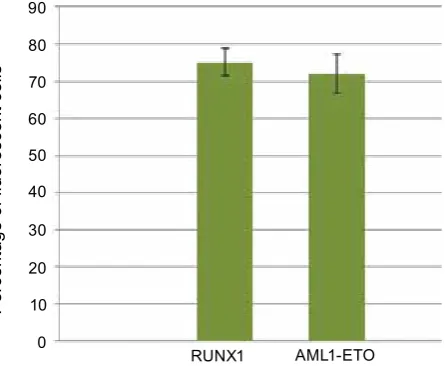
(2) The activated RUNX1(K83N) oncogene, which is formed as a result of a point mutation in the RUNX1 gene and leads to the substitution of lysine to asparagine in the 83 position of the RUNX1 protein. Since both the gene of inter-est and eGFP marker gene are driven by a com-mon promoter, the expression levels of the gene of interest in cells can be evaluated based on the intracellular expression of the eGFP marker gene.
The typical results of transduction efficien-cy and transgene expression are shown in Fig. 1. Throughout the course of our work, we used the murine SC1 embryonic fibroblast cell line, the HEK 293 cell line, and two transgenic SC1 cell lines expressing either activated oncogene AML1-ETo or RUNX1(K83N). All four cell lines were cultured in standard DMEM medium containing 10% FBS, 4 mM L-glutamine, 1mM sodium pyru-vate, 100 mkg/ml streptomycin, and 100 units/ml penicillin at 37 °С in a 5% СО2 atmosphere.
The design and synthesis of double-stranded siRNA molecules was carried out in collaboration with the Institute of Chemical Biology and Fun-damental Medicine, Siberian Branch of the Rus-sian Academy of Sciences (ICBFM SB RAS). The
oligonucleotides were synthesized at ICBFM SB RAS using the phosphamide method. During oli-gonucleotide synthesis, a unique method was used to methylate the oligonucleotides at certain posi-tions. These modifications significantly increase the lifespan of synthetic siRNA in cells. The nuc-leotide sequences of siRNA oligonucnuc-leotides:
AML1-ETo sense 5′
-CCUCGAAAUCGU-mACUmGAGUAG-3′;
AML1-ETo antisense 5′
-UCUCmAGUmAC-GAUUUCGAGGUU-3′;
ETo sense 5′
-GGCCmAGCGGUmACmA-GUCCmAGAU-3′;
ETo antisense 5′
-UUmGGACUmGUmAC-CGCUmGGCCUG-3′;
AML(5′) sense 5′
-GAACCmAGGUUmGC-mAAGAUUGAA-3′;
AML(5′) antisense 5′-
AAAUCUUmGC-mAACCUmGGUUCUU-3′;
AML(3′) sense 5′
-AGCCCGGGAGCUUm-GUCCUCUU-3′;
AML(3′) antisense 5′
-AAGGACmAAGCUC-CCGGGCUUmG-3′.
Anti-AML1-ETo and anti-RUNX1 siRNA duplexes were transferred into transgenic SC1 cell lines by lipofection using Lipofectamin2000 (Invit-rogen) as per the manufacturers protocol. The final concentration of siRNA duplexes in the transfec-tion culture medium was 200 nM. Four hours after transfection, the culture medium was changed to a fresh medium. The transfected cell lines were cultured at 37 °С in a 5% СО2 atmosphere for 72 hours.
Fig. 1. efficiency of transgene expression in SC-1 murine fibroblast cell lines transduced with aML1-eTO- and rUNX1-k83N-expressing recom-binant retroviral vector particles
AML1-ETO RUNX1
Pe
rc
en
ta
ge o
f fl
uo
re
sc
en
t c
el
ls
90
80
70
60
50
40
30
20
10
The level of oncogene expression in the cell lines was evaluated by reverse transcription PCR (RT-PCR) and flow cytometry. The total mRNA from the cell lines was extracted using TRIZoL (Invitrogen) as per the manufacturers protocol. The extracted mRNA was used to synthesize the first strands of cDNA using the ImProm-II™ re-verse transcriptase (Promega) kit.
PCR with sequence-specific primers was car-ried out to identify the nucleotide sequences of the genes of interest and eGFP marker gene in the total cDNA. The following primers were used for RUNX1 (K83N):
sense-AGTCCTACCAATACCTGGGA; antisense-TCTCAGCTGTGGTGGTGAAG, for AML1/ETo:
sense-CATTTCACCCGAGATAGGAG; antisense-AAGTCCTCGGCGTCACTGAT, for eGFP:
sense-ACCTACGGCCTGCAGTGCTT; antisense-TGCCGTTCTTCTGCTTGTCG. PCR products were then visualized after 1.5% aga-rose gel electrophoresis. The results were processed using the Gel-Pro Analyzer 4.0 software, which gives a maximum optical density (maxoD) reading . The results were normalized against β-actin.
Cell fluorescence was measured using an epics 4XL Beckman coulter flow cytometer (United States). The WinMDI2.8 software program was used for data collection and data analysis. The dia-grams of the percentage of fluorescent cells in the cell populations are shown in Fig. 2. Cell
popula-tions transfected with respective siRNA duplexes are shown along the X axis, while the percentage of fluorescent cells in the population is shown on the y axis.
Results obtained by flow cytometry correlate well with the results obtained by RT-PCR. The electrophoregram of RT-PCR products is shown in Fig. 3. The reduction of oncogene mRNA is clearly visible in the SC1-AML1/ETo (t8;21) transgenic cell lines transfected with siRNA oligonuc leotides
targeting the 5′-end of RUNX1 mRNA, ETo
mRNA and the site of junction of AML1-ETo mRNA. oncogene mRNA reduction is also seen in the SC1-RUNX1(K83N) transgenic cell lines transfected with siRNA oligonucleotides targeting the 5′- and 3′-ends of RUNX1 mRNA.
While there is a significant reduction of fluorescence in a portion of the cell population (two times or more) after the addition of synthetic siRNA , there is no such reduction in fluorescence in the rest of the cell population (Fig. 2). This coincides with the results obtained by RT-PCR (Fig. 3). The most likely reason for this is the low efficiency of siRNA delivery into cells. According to the manufacturers of Lipofectamin2000 (Invit-rogen), the lipofection efficiency with siRNA oli-gonucleotides is 50–60% and depends on the na-ture of the cells being transfected. Apart from this, there is the possibility of a reduction of synthetic siRNA activity due to their enzymatic degrada-tion, which is catalyzed by cellular endonucleases, despite the stabilizing modifications introduced to the siRNA oligonucleotides. According to our
Fig. 2. results obtained after flow cytometric analysis of cell suspensions. The percentage of fluorescent cells in the aML1-eTO and rUNX1 (k83N) expressing murine fibroblast cell lines 48 hours after transfection with various sirNa duplexes
control AML5′ AML3′ ETO
siRNA
Pe
rc
en
ta
ge o
f fl
uo
re
sc
en
t c
el
ls
80
60
40
20 100
0 120
AML1-ETO AML1-ETO
control AML5′ AML3′ ETO siRNA
Pe
rc
en
ta
ge o
f fl
uo
re
sc
en
t c
el
ls
80
60
40
20 100
0 120
data, the maximum interfering activity of syntheti-cally modified siRNA is observed 48–72 hours post transfection.
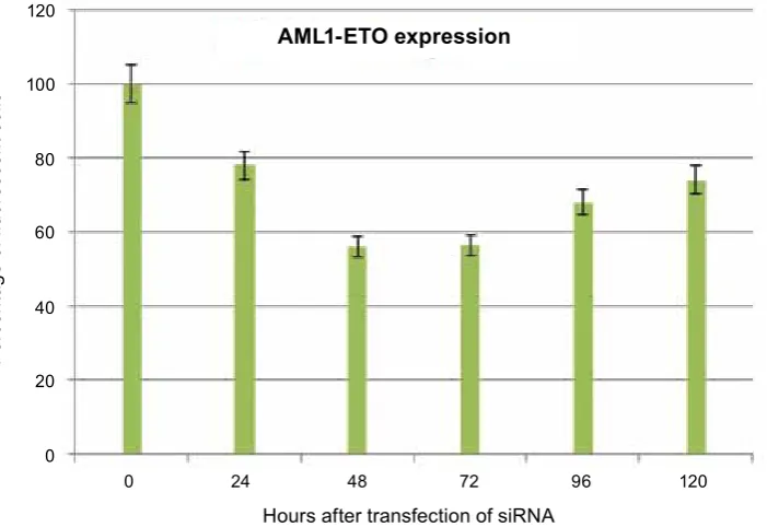
The gene silencing kinetics of synthetic siRNA oligonucleotides targeting the junction point of AML1-ETo mRNA is illustrated in Fig. 4. The moment of siRNA transfection has been taken as zero-time reference.
It has been shown that the maximum silencing activity of synthetic siRNA is 48–72 hours post transfection, when the percentage of fluorescent cells in the population is around 56%. The per-centage of fluorescent cells in the population in-creases to 67, 92 and 74.39% 96 and 120 hours post transfection, respectively. The results shown are averaged from three parallel measurements and are in reference to a control.
Fig. 3. The electrophoregram of rT-PCr products of total cDNa obtained from oncogene expressing model cell lines transfected with sirNa. 1 – sirNa(aML1-5′), 2 – sirNa(aML1-3′), 3 – sirNa(aML1-eTO), 4 – sirNa(eTO), 5 – Control (without sirNa)
750 500
SC1-AML1-ETO
RUNX1
500
SC1-RUNX1(K83N)
M 1 2 3 4 5 1 2 3 4 5
AML1-ETO
β-actin
Fig. 4. The gene silencing kinetics of synthetic sirNa oligonucleotides targeting the junction point of aML1-eTO mrNa
0 24 48 72 96 120 Hours after transfection of siRNA
Pe
rc
en
ta
ge o
f fl
uo
re
sc
en
t c
el
ls
80
60
40
20 100
0 120
AML1-ETO expression
The efficiency of RNA-interference can be increased by increasing the efficiency of siRNA delivery into the model cell lines. For this purpose, taking into account the efficiency of the synthetic siRNA duplexes, two shRNA-expressing lentivi-ral vectors were constructed targeting the junc-tion point of AML1-ETo mRNA and the 5′-end of RUNX1 mRNA, respectively. The lentivectors were constructed by cloning the shRNA-expressing DNA sequence into the pLSLP vector.
DNA sequence which encodes anti AML1-ETo shRNA:
- AML1-ETo-sense 5′
-p-GATCCGC-
CTCGAAATCGTACTGAGGCTTCCTGT-CATCTCAGTACGATTTCGAGGTTTTTG-3′
AATTCAAAAACCTCGAAATCGTACTGAGA- TGACAGGAAGCCTCAGTACGATTTCGAG-GCG-3′
DNA sequence which encodes anti-RUNX1 shRNA:
- AML-5-sense 5′
-p-GATCCGGAACCAG-
GTTGCAAGATTCCTTCCTGTCAAAATCTTG-CAACCTGGTTCTTTTTG-3′
- AML-5-antisense 5′
-p-AATTCAAAAA-
GAACCAGGTTGCAAGATTTTGACAGGAA-GGAATCTTGCAACCTGGTTCCG-3′
The design of the shRNA-coding DNA se-quences was done through an internet resource (http://gesteland.genetics.utah.edu/sirnA_scales/ index.html). Standard genetic engineering tech-niques were used for all cloning procedures. The shRNA-expressing lentiviral vectors are shown in Fig. 5.
The transient cotransfection of HEK293T cells with shRNA-expressing lentiviral vectors and packaging plasmids was done for the pro-duction of shRNA-expressing lentiviral vector particles. The transfection was carried out using Lipofectamin2000 (Invitrogen) as per the manu-facturers’ protocol. Viral stocks were harvested for three days and used for the infection/transduc-tion of the transgenic model cell lines expressing the activated oncogenes AML1-ETo and RUNX1 (K83N). Forty-eight hours after transduction/in-fection, the model cell lines were placed in cul-ture media containing puromycin (10 mkg/ml) for five days. After selection for puromycin resistance, the shRNA expressing transduced model cell lines were analyzed by flow cytometry.
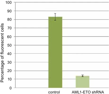
The percentage of fluorescent cells in the SC1-AML1/ETo(t8;21) transgenic cell line
de-Fig. 5. shrNa expressing lentiviral vectors: A – Lentiviral vector expressing shrNa; B – sirNa targeting dif-ferent oncogene zones
AML3′ AML1-ETO ETO AML5′ AML3′ А
B
creased 6 times after their transduction with shR-NA-expressing lentiviral vector particles targeting the junction point of AML1-ETo mRNA (Fig. 6). Similar results were seen when the SC1-AML1/ ETo(t8;21) and SC1-RUNX1(K83N) cell lines were transduced with shRNA-expressing lentivi-ral vector particles targeting the 5′-end of RUNX1 mRNA. The percentage of fluorescent cells in the cell lines decreased 8 and 8,5 times, respectively. It should be noted that the Sc1-RUNX1(K83N) cell line showed no reduction in fluorescence when transduced with shRNA- expressing
lentivi-Fig 6. Histogram illustrating the reduction in fluo-rescence in the aML1-eTO expressing SC-1 murine fibroblast cell line before and after transduction with shrNa-expressing lentiviral vector particles targeting the junction point of the aML1-eTO mrNa
AML1-ETO shRNA control
Pe
rc
en
ta
ge o
f fl
uo
re
sc
en
t c
el
ls
90 80
70
60
50 40
30 20
10
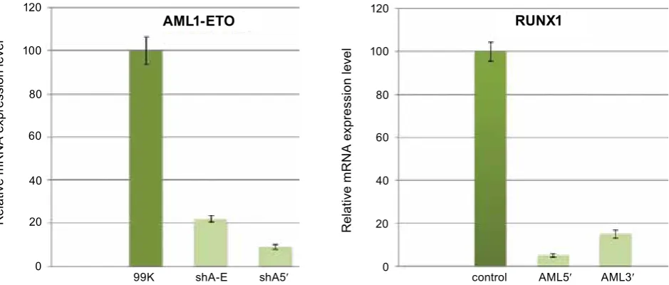
Fig. 7. The real Time rT-PCr products of total cDNa obtained from rUNX1(k83N) and aML1-eTO onco-gene expressing SC1 model cell lines transduced with shrNa expressing lentiviral vector particles: 99k – control (non-transduced rUNX1(k83N) expressing SC1 cell line); sha-e – rUNX1(k83N) expressing SC1 cell line transduced with shrNa expressing lentiviral vector particles targeting the junction point of aML1-eTO mrNa; sha5′ – rUNX1(k83N) expressing SC1 cell line transduced with shrNa expressing lentiviral vector particles targeting the 5′-end of rUNX1 mrNa; last three samples - analysis of aML1-eTO(t8;21) expressing SC1 cell lines (98k – non transduced control, sha-e, sh5′ – transduced with corresponding lentiviral vector particles)
99K shA-E shA5′
R
el
at
iv
e m
R
N
A e
xp
re
ss
io
n l
ev
el
80
60
40
20 100
0 120
AML1-ETO
control AML5′ AML3′ 80
60
40
20 100
0 120
RUNX1
R
el
at
iv
e m
R
N
A e
xp
re
ss
io
n l
ev
el
ral vector particles targeting the junction point of AML1-ETo mRNA. The results obtained by flow cytometry correlate well with the results obtained by Real Time RT-PCR (Fig. 7).
our results illustrate the high efficiency of stable synthetically modified siRNA duplexes for silencing the activated oncogenes frequently found in acute myeloid leukemia patients.
Validated siRNA sequences were used to de-sign and synthesize shRNA- coding DNA sequenc-es. The synthesized DNA sequences were cloned into a recombinant lentiviral vector. The shRNA-expressing lentiviral vector particles targeting the junction point of AML1-ETo mRNA and 5′-end of RUNX1 mRNA were used to transduce the on-cogene-expressing model cell lines. The transduced model cell lines were analyzed by RT-PCR and flow cytometry. The analysis revealed a significant reduction in the expression of activated oncogenes in the transduced cell lines. This is indicative of the high efficiency of the constructed lentiviral vector constructs, which can be used for silencing target genes by RNA interference.
This work was supported by the Programms of the Presidium of the russian academy of Scien ces «Molecular and Cell biology», «Funda-mental research basics in Nanotechnology and Nanomaterials », and the russian Foundation for basic research (grant N 11-04-01365-a and N 13-04-00599-a).
СПЕЦИФИЧЕСКОЕ ПОДАВЛЕНИЕ ЛЕЙКЕМИЧЕСКИХ ОНКОГЕНОВ С ИСПОЛЬЗОВАНИЕМ ПОДХОДА РНК-ИНТЕРФЕРЕНЦИИ
Т. Д. Лебедев1, П. В. Спирин1,
Н. Н. Орлова1, А. С. Горностаева1,
С. Стокинг2, В. С. Прассолов1*
1Институт молекулярной биологии им. В. А. Энгельгардта, РАН, Москва, Россия;
2Институт экспериментальной вирусологии и иммунологии Генриха-Петте, Гамбург, Германия;
*e-mail: [email protected]
ме-тодов переноса siРНК в клетки-мишени. Для этих целей используются разные виды транс-фекции или вирусная трансдукция. В статье представлены результаты сравнения угнетения экспрессии онкогена AML-ETo с помощью синтетической siРНК и кодирования с помо-щью рекомбинантных лентивирусов для соот-ветствующей shРНК.
К л ю ч е в ы е с л о ва: ретровирусные век-торы, РНК-интерференция, фибробласты эмб-риона мыши.
СПЕЦИФіЧНЕ ПРИГНіЧЕННя ЛЕЙКЕМіЧНИХ ОНКОГЕНіВ іЗ ВИКОРИСТАННяМ ПіДХОДу РНК-іНТЕРФЕРЕНЦії
Т. Д. Лебедєв1, П. В. Спірін1,
Н. Н. Орлова1, А. С. Горностаєва1,
С. Стокінг2, В. С. Прассолов1*
1Інститут молекулярної біології ім. В. О. Енгельгардта, РАН, Москва, Росія;
2Інститут експериментальної вірусології і імунології Генріха-Петте, Гамбург, Німеччина;
*e-mail: [email protected]
РНК-інтерференція є ефективним при-родним механізмом посттранскрипційної модуляції генної експресії. Механізм РНК-інтерференції є у вищих евкаріотів – тва-рин і рослин, а також у нижчих евкаріотів і вірусів. На сьогодні РНК-інтерференція використовується як потужний інструмент для вивчення функціональної активності генів. За допомогою цього підходу вдалося до-сягти результатів, що мають істотне значен-ня для фундаментальної біології. Відомо, що РНК-інтерференція може використовувати-ся у виробництві лікарських препаратів для лікування злоякісних, інфекційних та
спадко-вих хвороб. Однією з основних проблем цих розробок є пошук ефективних методів пере-носу siРНК в клітини-мішені. Для цих цілей використовуються різні види трансфекції або вірусна трансдукція. У статті представ-лено результати порівняння пригнічення експресії онкогену AML-ETo за допомогою синтетичної siРНК та кодування за допо-могою рекомбінантних лентивірусів для відповідної shРНК.
К л ю ч о в і с л о в а: ретровірусні векто-ри, РНК-інтерференція, фібробласти ембріона миші.
1. vilgelm a. e., Chumakov S. P., Prasolov v. S.
// Mol. Biology. – 2006. – 40, N 3. – P. 1–18. 2. volkov a. a., kruglova N. S., Meschaninova M. I.
et al. Selective protection of nuclease-sensitive sites in siRNA prolongs silencing effect // oligonucleotides. – 2009. – 19, N 2. – P. 191– 202.
3. Shuey D. J., McCallus D. e., Giordano T. // Drug Discov. Today. – 2002. – 7, N 20. – P. 1040–1046.
4. Stanisławska J., Olszewski W. L. // Arch. Immunol. Ther. Exp. (Warsz). – 2005. – 53, N 1. – P. 39–46.
5. elbashir S. M., Harborth J., Weber k., Tuschl T.
// Methods. – 2002. – 26, N 2. – P. 199–213. 6. brummelkamp T. r., bernards r., agami r. //
Science. – 2002. – 296, N 5567. – P. 550–553. 7. Satake N., Maseki N., kozu T. et al. // Br. J.
Haematol. – 1995. – 91, N 4. – P. 892–898. 8. Peterson L. F., Zhang D. e. // oncogene. –
2004. – 23, N 24. – P. 4255–4262.