www.fm.viamedica.pl
Address for correspondence: Dr Marek Opałka, Department of Animal Physiology, University of Warmia and Mazury in Olsztyn, ul. Oczapowskiego 5, 10–718 Olsztyn, Poland, tel: +89 523 49 23, fax: +89 523 39 37, e-mail: [email protected]
The morphometric parameters of adrenal
cortex in sows: in normal condition
and after prolactin infusion
Marek Opałka
1, Barbara Kamińska
1, Teresa Doboszyńska
2, Luiza Dusza
11Department of Animal Physiology, University of Warmia and Mazury in Olsztyn, Poland
2Institute of Animal Reproduction and Food Research, Polish Academy of Sciences, Olsztyn, Poland [Received 24 October 2001; Accepted 5 November 2001]
The aim of this study was to examine the effect of experimental hyperprolactine-mia on the sterological parameters of porcine adrenal cortex. In cyclic sows, after preovulatory luteinizing hormone (LH) peak, porcine prolactin (PRL, 0.3 mg) or saline were administered iv. for 48 h at 2 h intervals. Next sows were slaughtered and adrenal glands were dissected. Stereological analysis of the left adrenal gland did not reveal any significant differences between control and PRL-treated sows. Experimental hyperprolactinemia did not affect the volume of particular cortical zones, the number and the volume of adrenocortical cells or the average vol-ume of their cell nuclei. Moreover, we present for the first time a detailed stereo-logical description of adrenal cortex in sows.
key words: prolactin, stereology, adrenal cortex, sows
INTRODUCTION
Adrenal cortex steroidogenesis is under multihormo-nal regulation with the well-established stimulatory role of ACTH. Adrenocortical steroid secretion may also be controlled by prolactin (PRL) acting through its adre-nal receptor [8]. There is in vivo and in vitro evidence that PRL affects adrenal steroidogenesis in guinea pigs [17], rats [1], cows [5], baboons [16], and humans [4, 18]. In vitro studies performed on rats and guinea pigs have shown that PRL stimulated adrenal se-cretion of corticosterone [15], androstenedione, de-hydroepiandrosterone (DHEA) and cortisol [14]. The release of DHEA and/or dehydroepiandrosterone sulphate (DHEAS) by adrenals was also elevated in the presence of PRL in primates [4, 6, 16]. Moreover, chronic PRL treatment caused a notable hypertrophy of zona glomerulosa cells of the rat adrenal cortex [13]. PRL was also involved in the regulation of adrenal
steroidogenesis in pigs. In a previous paper PRL ad-ministration increased plasma level of cortisol in the same sows [7]. However, in available literature there are no data pertaining to the effect of PRL on mor-phometric parameters of adrenal cortex in the pig.
Therefore the aim of this study was to examine the effect of experimental hyperprolactinemia on the stereologic parameters of adrenal cortex in sows during the early luteal phase of the oestrous cycle.
MATERIAL AND METHODS
Investigations were conducted following institu-tional guidelines and the protocol was approved by the Animal Investigation Committee. The study was performed on 10 multiparous crossbred sows. On the 13th day of the oestrous cycle jugular veins of the animals were cannulated. Cannulas were used for administration (48 h in 2 h intervals) of PRL (0.3 mg) or saline (control sows). Injections of PRL (or soline) began 4–20 h after the preovulatory LH surge. Oc-currence of the preovulatory LH surge (the highest concentrations of LH) was estimated by monitoring of vaginal mucus electrical resistance as described earlier [3]. At the end of the administration period sows were slaughtered by electrical shock. Slaugh-ter procedure was performed in such a manner as to avoid the occurrence of stress-related changes. Adrenals were immediately dissected. One adrenal gland of each sow was designated for histological examination. Adrenals freed of adhering fat were fixed in Bouin’s solution and embedded in paraffin. Subsequently, serially cut sections (5–6 µm) were stained with haematoxylin and eosin for stereology as well as the Masson method for microscopic ex-amination. Additionally, the green filter (GIF) was used for histological observation.
Stereologic studies were performed in two sta-ges. In stage I, using a magnification of 100 ¥ and a square lattice test system of type A, the volume of the zona glomerulosa (ZG), zona fasciculata (ZF), zona reticularis (ZR), and adrenal medulla (M) were estimated by differential point counting, as described previously [12, 19]. The specific weight of the adre-nal gland was assumed to be 1.039 mg/mm3. In stage II, the size and number of adrenocortical cells were estimated on a screen at 3000 ¥, using the multi-purpose test system M42. Stereologic results were expressed per one adrenal gland.
Results were expressed as means ± SEM. Mor-phometric data (n = 5/group) were log transformed and then submitted to the T-test for independent variables (Statistica, StatSoft Inc., Tulsa, OK., USA).
RESULTS
Analysis of the morphometric parameters
Results of histological examination are presented in Table 1. Statistical analysis of the data did not reveal any significant differences between control and PRL--treated sows. Hyperprolactinemia induced by 2-day administration of exogenous PRL did not affect the volume of particular cortical zones, the number and the volume of adrenocortical cells as well as the ave-rage volume of their cell nuclei.
Microscopic observation
In all examined sows the adrenal gland had three cortical zones clearly identifiable by arrangement and stainability of cells: zona glomerulosa, zona fascicu-lata and zona reticularis (Fig. 1). The thickness of the cortex and all the zones remained fairly constant in adrenals from both normal sows and sows after PRL administration.
Subcapsularly lying the zona glomerulosa (Fig. 1A) was created by oval or cuboidal epithelial cells
6213.7 ± 222.2 567.5 ± 36.4 2614.8 ± 61.6 1219.1 ± 217.4 1255.4 ± 154.5
5769.0 ± 727.5 489.1 ± 61.5 2425.6 ± 421.4 1350.1 ± 433.0 992.8 ± 113.8
9.15 ± 0.54 42.25 ± 1.47 19.44 ± 3.03 20.07 ± 2.09
8.40 ± 0.35 42.86 ± 5.76 22.36 ± 5.72 17.46 ± 1.17
966 ± 81 1682 ± 110
908 ± 71
976 ± 68 1957 ± 304
923 ± 78
162 ± 16 149 ± 14 102 ± 13
167 ± 10 176 ± 25 114 ± 12
16.75 ± 0.96 46.75 ± 5.40 36.51 ± 5.20
16.64 ± 2.20 44.47 ± 8.01 38.89 ± 7.53
558.5 ± 71.5 1502.4 ± 90.5 1303.7 ± 341.8 3364.6 ± 359.1
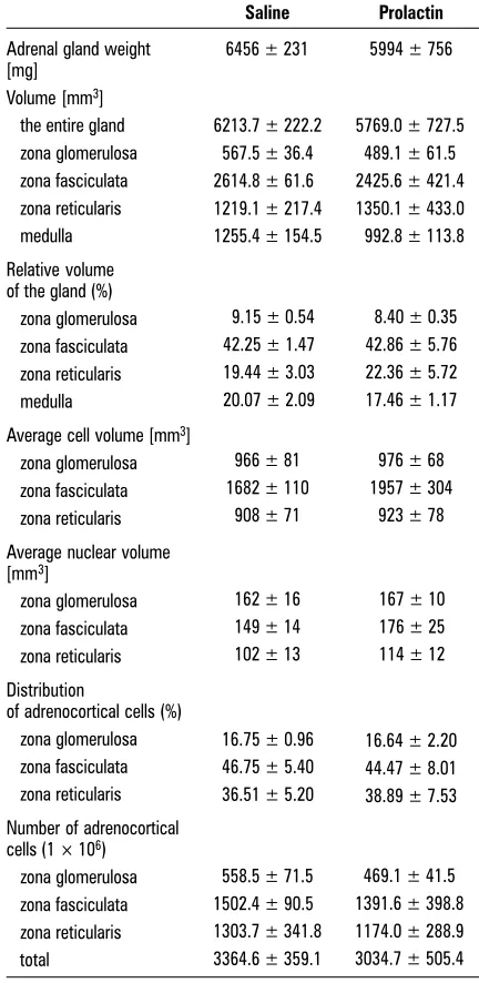
469.1 ± 41.5 1391.6 ± 398.8 1174.0 ± 288.9 3034.7 ± 505.4 Table 1. The morphometric parameters of the cortex of the left adrenal gland of saline and prolactin-treated sows during early luteal phase of the oestrous cycle. Results expressed as means ± SEM
Saline Prolactin
Adrenal gland weight 6456 ± 231 5994 ± 756 [mg]
Volume [mm3]
the entire gland zona glomerulosa zona fasciculata zona reticularis medulla
Relative volume of the gland (%) zona glomerulosa zona fasciculata zona reticularis medulla
Average cell volume [mm3]
zona glomerulosa zona fasciculata zona reticularis
Average nuclear volume [mm3]
zona glomerulosa zona fasciculata zona reticularis
Distribution
of adrenocortical cells (%) zona glomerulosa zona fasciculata zona reticularis
Number of adrenocortical cells (1 ¥ 106)
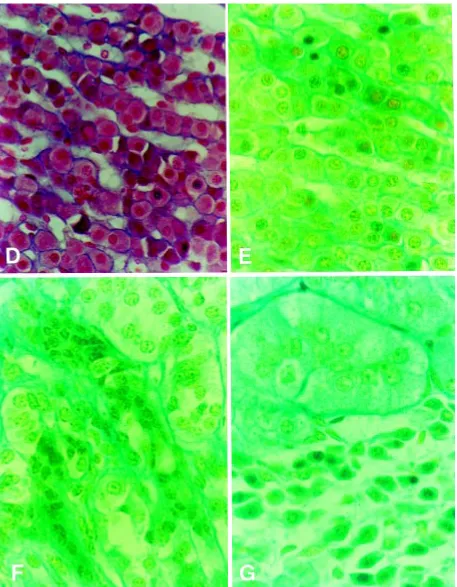
Figure 1. Adrenal cortex cross-section after staining acc. To Masson’s method (D) and Masson/GIF (green filter). D and E. Zona fasciculata;
F. Fragments of zona fasciculata and zona reticularis; G. The conglomeration of loose cells near the zona glomerulosa (D–G ¥ 750).
collected in cluster (glomerules) surrounded by de-licate connective tissue fibres. Between the zona glomerulosa and zona fasciculata of frequent
cords, mainly with cuboidal cells of light frontier cytoplasm and round nucleus (Fig. 1A). However, some cords were formed by narrow cylindrical cells (Fig. 1B) or cords consisted of numerous cells with pycnotic nucleus and shrink cytoplasm (Fig. 1D, E). In some areas of the zona fasciculata the compaction of the connective tissue was visible (Fig. 1F). The zona reticularis was formed by epi-thelial cells lying longways of thinlywall blood vessels. There, various types of cells were visible (Fig. 1C).
DISCUSSION
The present study for the first time gives a de-tailed stereologic description of the adrenal cor-tex in the pig. Only Baba [2] reported planimetric data on the zonation of the pig adrenal cortex — in his study the area of cortex occupied by the glomerulosa and fasciculata — reticularis zones was 11.9 and 88.0%, respectively. Like in other mammalian species, in the pig adrenal cortex the best developed is the fasciculata zone, the cells of which are the largest and the most numerous (c. 47%) among the parenchymal cells of the cor-tex. The zona reticularis cells comprise about 37% of all parenchymal cells of the gland while glom-erulosa cells only c. 17%. Thus, the zonal and cel-lular composition of the porcine adrenal cortex resembles that of some other mammalian spe-cies [for review see 11].
As demonstrated in the present study, hyperpro-lactinemia induced by two-day PRL administration changed neither the cellular composition nor the parenchymal cell size of the porcine adrenal cortex. In the rat, short-term PRL-treatment did not alter zona glomerulosa morphology or plasma aldoste-rone concentration but within 15 days PRL adminis-tration resulted in a hypertrophy of the zona glom-erulosa cells and elevation of blood aldosterone lev-el [13]. The lack of changes in the structure of the pig adrenal cortex observed in the present experi-ment is probably connected with a too short PRL stimulation of the adrenal glands. As demonstrated in the previous paper, two-day PRL administration increased both plasma cortisol concentration and adrenal content of cortisol in the some sows used for present study as well as PRL-stimulated cortisol release by suspension of porcine adrenocortical cells [7]. Thus, the stimulatory effect of PRL on secretion and plasma concentration of cortisol was not accom-panied by quantitative morphological alternations in the adrenal cortex.
ACKNOWLEDGEMENTS
We are sincerely indebted to Prof. LK Malendowicz (School of Medicine, Poznań, Poland) for enabling us to conduct morphometric analysis of adrenals in his laboratory. This study was supported by State Committee for Scientific Research, Poland (project No. 2030.804).
REFERENCES
1. Albertson BD, Sienkiewicz ML, Kimball D, Munabi AK, Cassorla F, Loriaux DL (1987) New evidence for a di-rect effect of prolactin on rat adrenal steroidogenesis. Endocrine Res, 13: 317–333.
2. Baba AI (1974) Histologische und histometrische Veränderungen der Nebennieren bei kümmernden Ferkeln. Zbl Vet Med, 21A: 331–335.
3. Dusza L, Opałka M, Kamińska B, Kamiński T, Ciereszko RE (1996) The relationship between electrical resistance of vaginal mucus and plasma hormonal parameters during periestrus in sows. Theriogenology, 45: 1491–1503. 4. Glasow A, Breidert M, Haidan A, Anderegg U, Kelly
PA, Bornstein SR (1996) Functional aspects of the ef-fect of prolactin (PRL) on adrenal steroidogenesis and distribution of the PRL receptor in the human adrenal gland. J Clin Endocrinol Metab, 81: 3103–3111. 5. Higuchi K, Nawata H, Kato K, Ibayashi H (1985) Ovine
prolactin potentiates the action of adrenocorticotropic hormone on the secretion of dehydroepiandrosterone sulfate and dehydroepiandrosterone from cultured bo-vine adrenocortical cells. Horm Metab Res, 17: 451–453. 6. Higuchi K, Nawata H, Maki T, Higashizima M, Kato KI, Iba-yashi H (1984) Prolactin has a direct effect on adrenal an-drogen secretion. J Clin Endocrinol Metab, 59: 714–718. 7. Kamińska B, Opałka M, Ciereszko RE, Dusza L (2000) The involvement of prolactin in the regulation of ad-renal cortex function in pigs. Dom Anim Endocrinol, 19: 147–157.
8. Klemcke HG, Pond WG, Nienaber JA (1989) Porcine prolactin receptors: characterisation, changes during neonatal development and effects of hypoprolactine-mia. Comp Biochem Physiol, 92A: 197–206.
9. Kochman H, Kochman K (1977) Purification of ovine and bovine prolactins on DEAE-cellulose chromato-graphy and preparative polyacrylamide gel electro-phoresis. Bull Polish Acad Sci ser Biol Sci, 25: 67–70. 10. Li Ch (1976) Studies on pituitary lactogenic hormone:
The primary structure of the porcine hormone. Int J Peptide Protein Res, 8: 205–224.
11. Malendowicz, LK (1994) Cytophysiology of the mam-malian adrenal cortex as related to sex, gonadectomy and gonadal hormones. PTPN, Poznań.
12. Malendowicz LK, Robba C, Nussdorfer GG (1986) Sex differences in adrenocortical structure and function. XXII. Light- and electron-microscopic morphometric studies on the effect of gonadectomy and gonadal hormone replacement on the rat adrenal cortex. Cell Tissue Res, 244: 141–145.
plasma hormone concentrations. Acta Endocrinol, 111: 101–105.
14. O’Connel Y, Mc Kenna TJ, Cunningham SK (1994) The effect of prolactin, human chorionic gonadotropin, insulin and insulin like growth factor-1 on adrenal ste-roidogenesis in isolated guinea pig adrenal cells. J Ste-roid Biochem Molec Biol, 48: 235–240.
15. Ogle TF, Kitay JI (1979) Interactions of prolactin and adrenocorticotropin in the regulation of adrenocortical secretions in female rats. Endocrinology, 104: 40–44. 16. Pepe GJ, Albrecht ED (1985) Prolactin stimulates
adre-nal androgen secretion in infant baboons. Endocrino-logy, 117: 1968–1973.
17. Sautin YuYu, Chelnakowa IS, Tron’ko ND, Mikosha AS (1992) Trophic effect and modulation of ACTH--dependent stimulation of steroidogenesis by pro-lactin in guinea pig adrenal cortex. Endocrine Reg, 26: 35–39.
18. Schiebinger RJ, Chrousos GP, Cutler GB Jr, Loriaux DL (1986) The effect of serum prolactin on plasma ad-renal androgens and the production and metabolic clearance rate of dehydroepiandrosterone sulfate in normal and hyperprolactinemic subjects. J Clin Endo-crinol Metab, 62: 202–209.