Analysis of the function of IL-10 in chickens using speci
fi
c neutralising
antibodies and a sensitive capture ELISA
Zhiguang Wu
a, Tuanjun Hu
a, Lisa Rothwell
a, Lonneke Vervelde
a, Pete Kaiser
a,
Kay Boulton
a, Matthew J. Nolan
b, Fiona M. Tomley
b, Damer P. Blake
b, David A. Hume
a,*aThe Roslin Institute and Royal (Dick) School of Veterinary Studies, University of Edinburgh, Easter Bush, Midlothian EH25 9RG, UK bDepartment of Pathology and Pathogen Biology, Royal Veterinary College, University of London, Hatfield AL9 7TA, UK
a r t i c l e i n f o
Article history:
Received 9 March 2016 Received in revised form 18 April 2016
Accepted 18 April 2016 Available online 22 April 2016
Keywords:
Chicken Interleukin-10 Monoclonal antibody Capture ELISA Neutralising antibody
a b s t r a c t
In mammals, the inducible cytokine interleukin 10 is a feedback negative regulator of inflammation. To determine the extent to which this function is conserved in birds, recombinant chicken IL-10 was expressed as a secreted human Ig Fc fusion protein (chIL-10-Fc) and used to immunise mice. Five monoclonal antibodies (mAb) which specifically recognise chicken IL-10 were generated and charac-terised. Two capture ELISA assays were developed which detected native chIL-10 secreted from chicken bone marrow-derived macrophages (chBMMs) stimulated with lipopolysaccharide (LPS). Three of the mAbs detected intracellular IL-10. This was detected in only a subset of the same LPS-stimulated chBMMs. The ELISA assay also detected massive increases in circulating IL-10 in chickens challenged with the coccidial parasite,Eimeria tenella. The same mAbs neutralised the bioactivity of recombinant chIL-10. The role of IL-10 in feedback control was testedin vitro. The neutralising antibodies prevented IL-10-induced inhibition of IFN-gsynthesis by mitogen-activated lymphocytes and increased nitric oxide production in LPS-stimulated chBMMs. The results confirm that IL-10 is an inducible feedback regulator of immune response in chickens, and could be the target for improved vaccine efficacy or breeding strategies.
©2016 The Authors. Published by Elsevier Ltd. This is an open access article under the CC BY license (http://creativecommons.org/licenses/by/4.0/).
1. Introduction
Interleukin-10 (IL-10) is an anti-inflammatory cytokine that controls the nature and extent of inflammatory responses during infection with viruses, bacteria, fungi, protozoa and helminths (Couper et al., 2008; Moore et al., 2001) and has a particular central role in intestinal immunity and homeostasis (Manzanillo et al., 2015). IL-10 wasfirst described as an inhibitor of cytokine synthesis in mice, a product of the TH2 subset of T cells that inhibited synthesis of proinflammatory cytokines by TH1 cells (Fiorentino et al., 1989). IL-10 is now recognised as a multifunc-tional cytokine produced by many immune cell types including macrophages, monocytes, dendritic cells (DCs), TH1, TH2, TH17 and regulatory T cell subsets and B cells and a feedback regulator of diverse immune responses to infections (Reviewed inCouper et al., 2008; Moore et al., 2001; Saraiva and O'Garra, 2010).
Accordingly, vaccine efficacy in mammals has been associated with genetic variation in IL-10 production, and anti-IL-10 treat-ment has been shown to increase the nature, magnitude and
ef-ficacy of vaccine responses against a diversity of pathogens (Chen et al., 2014; Darrah et al., 2010; Pitt et al., 2012; Roberts et al., 2005; Stober et al., 2005).
Infectious diseases are a major threat to intensive poultry pro-duction. There is less evidence of the function of IL-10 in birds. The cDNA of chicken IL-10 (chIL-10) was isolated from cecal tonsils of Eimeria tenella-infected chickens (Rothwell et al., 2004) and the expressed protein product inhibited IFN-gsynthesis by mitogen-activated lymphocytes (Rothwell et al., 2004). Inducible expres-sion of IL-10 mRNA has been described in viral, bacterial and parasitic infections of birds (Barjesteh et al., 2013; Humphrey et al., 2014; Parvizi et al., 2015). However, the lack of reagents has pre-vented analysis of the production and function of IL-10 protein. In the current study we describe the generation of monoclonal anti-bodies to chIL-10 and their applications in western blot, capture ELISA and neutralising assays. The results indicate that IL-10 has similar functions in birds as in mammals. As predicted from the *Corresponding author.
E-mail address:[email protected](D.A. Hume).
Contents lists available atScienceDirect
Developmental and Comparative Immunology
j o u r n a l h o m e p a g e : w w w . e l s e v i e r . c o m / l o c a t e / d c i
http://dx.doi.org/10.1016/j.dci.2016.04.016
original studies on infected birds (Rothwell et al., 2004), Eimeria infection of chickens produced a massive increase in circulating IL-10, providing a biomarker for disease status. Accordingly, we sug-gest that manipulation of IL-10, either through genetic or thera-peutic intervention, could have application in reducing disease burden in chickens.
2. Materials and methods
2.1. Expression and purification of recombinant chIL-10-Fc and recombinant chIL-10-V5H6 protein
ChIL-10 cDNA was sub-cloned into the vector, pKW06 (John Young, unpublished) to express the protein in mammalian cells with a C-terminal human IgG1 Fc tag. For screening purposes, to eliminate anti-human Ig antibodies, a V5His tagged chIL-10 was produced in the vector pKW08 (John Young and Tuanjun Hu, un-published). ChIL-10 cDNAs (Rothwell et al., 2004) were expressed with the native signal peptide allowing secretion of the fusion proteins. Both constructs were expressed in COS-7 cells following transfection using the DEAE-dextran method (Rothwell et al., 2004). Recombinant chIL-10-Fc protein was purified using a HiTrap Protein G affinity column and the chIL-10-V5H6 protein with a HisTrap excel column (GE Healthcare Life Sciences).
2.2. Monoclonal antibody production, isotyping, purification, and labelling
Immunisation with chIL-10-Fc and fusion to generate hybrid-omas was carried out by Dundee Cell Products (DCP, Dundee, UK). Following fusion, hybridoma cultures were tested with recombi-nant chIL-10-V5H6 by dot-blot. Supernatants from these cultures were screened by ELISA with chIL-10-V5H6 and chIL-10-Fc and Western-Blot. Positive cultures were selected for further cloning. The antibody isotype was determined using the IsoStrip Mouse Monoclonal Antibody Isotyping Kit (Roche). Monoclonal hybrid-omas were cultured in D-MEM with 10% Ig-depleted FBS. Mono-clonal antibodies were purified using HiTrap Protein G affinity columns and dialysed against PBS using 30 kDa molecular weight cut-off (MWCO) Slide-A-Lyser cassettes (Pierce, ThermoFisher Sci-entific). The concentrations of mAbs were determined by absor-bance at 280 nm with a Nanodrop and then aliquots of the purified antibodies were biotinylated using Sulfo-NHS-LC-LC-biotin (Thermo Scientific). All procedures were performed according to the manufacturer's instructions.
2.3. Western blot
Recombinant chIL-10-V5H6 was treated with SDS-PAGE reducing buffer, denatured for 5 min at 100C and loaded onto a 4e15% pre-cast Mini-PROTEAN TGX Gels (Bio-Rad) and transferred onto a nitrocellulose membrane (Immunobilon-P, Millipore) using a Trans-Blot Semi-Dry Electrophoretic Transfer Cell (Bio-Rad). After blocking with 0.5% skimmed milk power/PBS solution, the mem-brane was stained with 1.0
mg/ml of each mAb, followed by
incu-bation with goat anti-mouse IgG1-horseradish peroxidase (Southern Biotec) diluted in 0.5% skimmed milk powder/PBS. Detection was carried out using enhanced chemiluminescence (ECL) (GE Healthcare Life Sciences), according to the manufacturer's instructions.2.4. Detection of chicken IL-10 by indirect ELISA and development of capture ELISA assays
Indirect ELISA was performed as described previously (Rothwell
et al., 2001). Briefly, assay plates (Nunc Immuno MaxiSorp, Thermo Electron LED) were coated with recombinant chIL-10 or control protein in Carbonate/Bicarbonate buffer and incubated overnight at 4C. Plates were washed in PBS containing 0.05% Tween-20 (PBS-T) and blocked with 0.5% casein/PBS at room temperature (RT) for 1 h. Purified mAb was added to the plate at a 1.0
mg/ml and incubated at
RT for 1 h. After three washes with PBS-T, plates were incubated with goat anti-mouse IgG-HRP at RT for 1 h. After a further three washes, plates were visualised by TMB substrate (ThermoScienti-fic) and reaction was stopped by 2 N H2SO4. Plates were read at
450 nm in a SpectraMax 250 microplate spectrophotometer system (Molecular Devices, Sunnyvale, CA, USA). Two capture ELISA assays was developed using ROS-AV162 or 164 as capture antibody and biotinylated ROS-AV163 as detecting antibody. Briefly, assay plates were coated with capture antibody ROS-AV162 or ROS-AV164 at 4
mg/ml overnight at 4
C. Plates were washed and blocked as in the indirect ELISA. Plates were incubated with recombinant IL-10 standards, or test samples at RT for 1 h, then washed and incu-bated with biotinylated detecting antibody ROS-AV163 at 1mg/ml
at RT for 1 h. After three washes, plates were incubated with streptavidineHRP (1:10,000, Pierce) for a further hour at RT before adding substrate 1-step TMB (Thermo Scientific) and then sulfuric acid stop solution. Absorbance was read at 450 nm.2.5. Detection of IL-10 production by bone marrow derived macrophages (BMMs)
Chicken BMMs were cultured from bone marrow cells from ED20 Novogen embryos in the presence of recombinant chicken CSF-1 as previously described (Garceau et al., 2010). On day 7 of culture, cells were stimulated with 0.5
mg/ml of LPS (
Eimeria coli serotype 055:B5, Sigma) to induce IL-10 expression. Cell culture supernatant from BMMs cells was collected at various times and 10 was detected using the capture ELISA. To detect intracellular IL-10 protein, the BMM cells were stimulated with 0.5mg/ml of LPS for
2 h before Brefeldin A (10mg/ml, Sigma-Aldrich) was added to the
culture to block secretion. Cells were resuspended in Fixation/ Permeabilization solution (BD Bioscience) for 20 min at 4 C, washed twice with 1BD Perm/Wash™buffer, and incubated with ROS-AV160-164 at 1.0mg/ml in 1
BD Perm/Wash™for 30 min at 4C. Cells were washed as before and resuspended in 0.4mg/ml
Alexa Fluor 647 goat anti-mouse IgG1 (Invitrogen/A-21240). Cells were washed then three times before analysing on FACScalibur (BD Bioscience).2.6. Infection of chicken with E. tenella and measurement of circulating IL-10
2.7. IL-10 bioassay and mAb neutralisation assay
ChIL-10 bioassay was performed as described previously (Rothwell et al., 2004). Briefly, splenocytes were isolated from 8 week old birds over Histopaque and resuspended at 5106cell/ml in DMEM containing 2 mg/ml BSA, 1%L-glutamine, 1 U/ml
peni-cillin, and 1
mg/ml streptomycin. Cells were added to round-bottom
96-well plates (100ml/well) containing serial 5-fold dilutions of
rchIL-10 or negative controls (including control protein tagged with HuIgG1 Fc, control protein tagged withV5H6, pCIneo supernatant), in afinal volume of 200ml in the presence of 0.5
mg/ml of ConA
(Sigma-Aldrich) or no ConA. Plates were incubated at 41C for 72 h for analysis of IFN-gcontent in the supernatant by capture ELISA (IFN-gamma chicken antibody pair, Novex, Life technologies, UK) as per manufacturer's instructions. For neutralisation assay, rchIL-10 was pre-incubated with 2-fold diluted mAbs or a control mAb with a starting concentration of 4mg/ml at 37
C for 2 h before adding lymphocytes and ConA.2.8. Nitric oxide assay
Chicken BMMs were cultured on 96 well plates as above. On day 7 of culture, cells were stimulated with 0.5
mg/ml of LPS (
E. coli serotype 055:B5, Sigma) with or without mAbs ROS-AV160 to 164. Supernatant was collected at 24 h after treatment to measure ni-trite, a stable metabolite of nitric oxide, by Griess assay. Briefly, 100ml of culture supernatant from each well was transferred to a
new flat-bottom 96-well plate. Griess reagent was prepared by mixing equal volumes of components A (0.2%a-napthyl ethylene
diamine dihydrochloride in dH2O) and B (2% sulfanilimide in 5%phosphoric acid in dH2O). Equal amount of Griess reagent was
added to each wells. After 10 min incubation at room temperature, the plates were read at 570 nm in a SpectraMax 250 microplate spectrophotometer system (Molecular Devices, Sunnyvale, CA, USA). Sodium nitrite (Promega) was used as a standard to deter-mine nitrite concentrations.
3. Results and discussion
3.1. Production of mouse anti-chicken IL-10 monoclonal antibodies
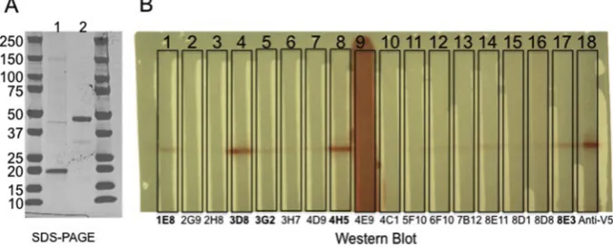
The predicted molecular weight of the expressed chIL-10-Fc and chIL-10-V5H6 proteins are 46.53 kDa and 20.97 kDa. Following expression and purification as described in the Methods, the purity and molecular weight were confirmed by SDS-PAGE (Fig. 1A). The purified chIL-10-Fc fusion protein was used to immunise mice for generation of mAbs. Seventeen initial hybridoma supernatants reactive with chIL-10-Fc were tested for specific binding to chIL-10
by WB and ELISA (Figs. 1B and2). Eight supernatants recognised chIL-10 but not control-Ig by ELISA andfive of these also bound chIL-10-V5H6 by WB. The corresponding lines were cloned andfive IgG1 monoclonal antibodies (ROS-AV160 (4H5), ROS-AV161 (3D8), ROS-AV162 (8E11), ROS-AV163 (8E3), ROS-AV164 (1E8)) were produced.
3.2. Characterisation of mAbs by indirect ELISA and western blot (WB)
All five monoclonal antibodies reacted specifically with re-combinant chIL-10, by ELISA (Fig. 3) and bound both the 47 kDa of chIL-10-Fc and 21 kDa of chIL-10-V5H6 (Supplemental material Fig. S1). Based upon epitope blocking experiments (data not shown), ROS-AV160 and ROS-AV161 recognise the same or closely-related epitopes on IL-10, whereas ROS-AV162, ROS-AV163 and ROS-AV164 recognised different epitopes. Accordingly, these anti-bodies in combination were considered appropriate to establish a capture ELISA.
3.3. Detection of native chIL-10 by capture ELISA and intracellular staining
Two capture ELISA assays for chIL-10 were developed using different combinations of mAbs. Both assays give the same sensi-tivity so only data from one assay (capturing with ROS-AV164 and detecting with biotinylated ROS-AV163) was carried forward. The sensitivity range of this assay was between 8 pg/ml to 1 ng/ml (Fig. 4A). Many cells of the innate immune system, including macrophages, dendritic cells, mast cells, NK cells, eosinophils and neutrophils express IL-10 (Saraiva and O'Garra, 2010). In mammals at least, macrophages can express IL-10in vitrofollowing activation of specific PRRs with LPS (Chang et al., 2007) or other TLR agonists (Boonstra et al., 2006). In the chicken, activation of TLR3 and TLR21 through poly I:C (double-stranded RNA) or CpG-ODN (a CpG-motif containing oligodeoxydinucleotide) respectively induced IL-10 mRNA in monocytes (He et al., 2012). In splenocytes, IL-10 mRNA was induced transiently in response to various bacterial lip-opolysacharides (Barjesteh et al., 2013).
The ELISA was used to detect secretion of chIL-10 by LPS-stimulated BMMs (Fig. 4B). ChIL-10 in the supernatant was rapidly induced from 2 h post stimulation with LPS, reached a maximum after 4 h and went down gradually. In stimulated human (Xue et al., 2015) and mouse macrophages (Ravasi et al., 2002), only a subset of cells express each individual inducible cytokine. Intra-cellular cytokine detection by flow cytometry allows studying cytokine production at single cell level together with other cyto-kines or cell surface markers. The anti-chIL-10 antibodies were
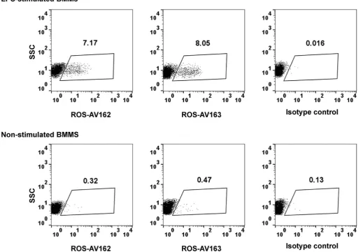
used to detect intracellular chIL-10. ChBMMs were stimulated with LPS for 2 h and BFA was used to block the secretion of chIL-10 (Fig. 5). Both ROS-AV162 and 163 detected intracellular chIL-10, but surprisingly only around 10% of the LPS-stimulated chBMMS were positive. This result suggest that the secreted IL-10 may be produced by only a subset of the stimulated macrophages, and might therefore act in a paracrine manner on neighbouring cells.
3.4. Detection of circulating IL-10 after E. tenella infection
IL-10 is a key immunoregulator during infection with pathogens including intracellular protozoa (Gazzinelli et al., 1996; Wilson et al., 2005). As in mammalian species, chicken IL-10 has emerged to be a crucial player in the Th bias during infection with Eimeriaspp., preventing the development of strong, IFN-g-driven responses, which have been shown to be crucial for control of
Fig. 2.Screening of hybridoma supernatant by ELISA. Values represent the mean of duplicate wells. Clone 1E8, 3D8, 3G2, 4D9, 4H5, 8E11, 8D8 and 8E3 recognised chIL-10 but not control-Fc.
Fig. 3.Specificity of mouse anti-chIL-10 mAb as demonstrated by indirect ELISA against rchIL-10-Fc, control-Fc, chIL-10-V5H6 and control-V5H6. Allfive monoclonal antibodies specifically recognised recombinant chIL-10 not Fc or V5H6 tagged control protein. Values represent the mean of duplicate wells.
Eimeriainfections (Rothwell et al., 2004). IL-10 was undetectable in the serum of healthy uninfected birds. As expected from previous detection of the IL-10 mRNA in infected birds (Rothwell et al., 2004), the level of circulating IL-10 was substantially increased around 5 days following either low or high dose challenge (Fig. 6).
These observations indicate that circulating IL-10 could provide a marker for infection, and that Eimeriainfections could produce systemic immunosuppression.
Fig. 5.Detection of intracellular chIL-10 in LPS-stimulated chBMMs. After 7-days culture, ChBMMs were stimulated with LPS for 2 h or non-stimulated before adding Brefeldin A for further 4 h. Cells werefixed, permeabilised and stained as described in materials and methods. Cells debris were gated out. SSC: side scatter.
Fig. 6.Circulating IL-10 was detected in the serum from chicken infected with
E. tenellaat 5 dpi. Six birds were used for each group. Student's t-test was used to determine significance (P< 0.05, indicated by an asterisk) of IL-10 production in infected birds, when compared to the relevant control.
3.5. The impact of IL-10 neutralisation of T cell and macrophage activation
Mammalian IL-10 strongly inhibited proliferation of CD4þT cells and production of cytokines including IFN-g, IL-2, IL-4 and TNF-a (Couper et al., 2008; Joss et al., 2000; Moore et al., 2001). As in mammals, chIL-10 inhibits the expression of IFN-g by mitogen-activated splenocytes (Rothwell et al., 2004). Each of the forms of IL-10 produced for this study inhibited IFN-gexpression by sple-nocytes after mitogen stimulation (ConA). A 1/80 dilution of neoIL-10 or 32 ng/ml of chIL-neoIL-10-V5H6 or 8 ng/ml of chILneoIL-10-Fc completely inhibited IFN-gexpression (Fig. 7). Preincubation with ROS-AV160 and 161 did not neutralise rchIL-10 even at a concentration of 4
mg/ml. However ROS-AV162, 163 and 164 completely neutralised
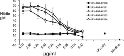
the inhibition effect by rchIL10 at high concentrations, and this effect titrated out with increasing dilution of mAbs (Fig. 8). These neutralising antibodies provide useful tools to study the regulatory role of IL-10 in chicken immune system as well as the molecular interaction with its receptor.To test the potential paracrine role of IL-10, implied by the expression in subsets of macrophages above, we tested the impact of adding the neutralising antibodies to stimulated macrophages. Nitric oxide production in mice contributes to macrophage cyto-toxicity towards pathogens including parasites, fungi and bacteria. Mouse macrophages produce NO after activation with LPS. This activity is not shared by the macrophages of large animals, including humans and pigs (Kapetanovic et al., 2012; Schroder et al., 2012). IL-10 inhibits NO production of activated mouse macrophages and compromises cytotoxic functions (Gazzinelli et al., 1992; Oswald et al., 1992; Romani et al., 1994). Neutralisa-tion of endogenous IL-10 by anti-IL-10 antibodies up-regulated nitric oxide production in murine macrophagesin vitroand pro-tected susceptible mice from challenge with Candida albicans (Romani et al., 1994). Anti-IL-10 treatment in vivo results in a
substantial and significant increase in LPS-induced serum NO levels in a mouse model (ter Steege et al., 1998). Like mouse macrophages, chicken macrophages produced abundant NO following activation with LPS or cytokines (Lillehoj and Li, 2004; Sung et al., 1991; Weining et al., 1996). As shown in Fig. 9, this activity is conve-niently assayed in chBMMs. Incubation with the neutralising mAbs ROS-AV162, 163 and 164 significantly up-regulated the NO con-centrations by LPS-stimulated BMMs in a dose-dependent manner, where the non-neutralising antibodies had no effect.
4. Conclusion
We have producedfive mouse mAbs to chicken IL-10 using chIL-10-Fc as immunogen. All five monoclonal antibodies recognised specifically chIL-10 via ELISA and WB. Two capture ELISA assays were developed to quantify native IL-10. Three mAbs (ROS-AV162, 163 and 164) neutralised the bioactivities of chIL-10 on ConA stimulated splenocytes and upregulated the nitric oxide production by LPS-stimulated chBMMs. These antibodies are new tools to investigate the important role of IL-10 in the chicken immunity and diseases. The data indicate that IL-10 is immunosuppressive in chickens as it is in mammals, and suggest that intervention in IL-10 production could be used to manipulate chicken immune responses.
Acknowledgements
This project was funded by the Wellcome Trust with grant number 099164/Z/12/Z with support from the BBSRC Animal Research Club under grants BB/L004003/1 and BB/L004046/1. We thank Dundee Cell Product (DCP, Dundee, UK) for production of mAbs (immunisation, first screening and cloning of monoclonal hybridomas). We thank Dr. Tina Sørensen Dalgaard (Department of Animal Science, Aarhus University, Denmark) for useful discussions on intracellular cytokine staining.
Appendix A. Supplementary data
Supplementary data related to this article can be found athttp:// dx.doi.org/10.1016/j.dci.2016.04.016.
References
Barjesteh, N., Hodgins, D.C., St Paul, M., Quinteiro-Filho, W.M., DePass, C., Monteiro, M.A., Sharif, S., 2013. Induction of chicken cytokine responses in vivo and in vitro by lipooligosaccharide ofCampylobacter jejuniHS:10. Vet. Micro-biol. 164, 122e130.
Boonstra, A., Rajsbaum, R., Holman, M., Marques, R., Asselin-Paturel, C., Pereira, J.P., Bates, E.E., Akira, S., Vieira, P., Liu, Y.J., Trinchieri, G., O'Garra, A., 2006. Macro-phages and myeloid dendritic cells, but not plasmacytoid dendritic cells, pro-duce IL-10 in response to MyD88- and TRIF-dependent TLR signals, and TLR-independent signals. J. Immunol. 177, 7551e7558.
Chang, E.Y., Guo, B., Doyle, S.E., Cheng, G., 2007. Cutting edge: involvement of the
Fig. 8.Neutralising effect of mAbs to chIL-10. ChIL-10 was incubated with mAbs for 2 h before IL-10 bioassay as inFig. 7. Assays were carried out in triplicate, and representative data from three independent experiments is shown.
type I IFN production and signaling pathway in lipopolysaccharide-induced IL-10 production. J. Immunol. 178, 6705e6709.
Chen, S., Wang, X., Wu, X., Wei, M.Q., Zhang, B., Liu, X., Wang, Y., 2014. IL-10 sig-nalling blockade at the time of immunization inhibits Human papillomavirus 16 E7 transformed TC-1 tumour cells growth in mice. Cell. Immunol. 290, 145e151. Couper, K.N., Blount, D.G., Riley, E.M., 2008. IL-10: the master regulator of immunity
to infection. J. Immunol. 180, 5771e5777.
Darrah, P.A., Hegde, S.T., Patel, D.T., Lindsay, R.W., Chen, L., Roederer, M., Seder, R.A., 2010. IL-10 production differentially influences the magnitude, quality, and protective capacity of Th1 responses depending on the vaccine platform. J. Exp. Med. 207, 1421e1433.
Fiorentino, D.F., Bond, M.W., Mosmann, T.R., 1989. Two types of mouse T helper cell. IV. Th2 clones secrete a factor that inhibits cytokine production by Th1 clones. J. Exp. Med. 170, 2081e2095.
Garceau, V., Smith, J., Paton, I.R., Davey, M., Fares, M.A., Sester, D.P., Burt, D.W., Hume, D.A., 2010. Pivotal advance: avian colony-stimulating factor 1 (CSF-1), interleukin-34 (IL-34), and CSF-1 receptor genes and gene products. J. Leukoc. Biol. 87, 753e764.
Gazzinelli, R.T., Oswald, I.P., James, S.L., Sher, A., 1992. IL-10 inhibits parasite killing and nitrogen oxide production by IFN-gamma-activated macrophages. J. Immunol. 148, 1792e1796.
Gazzinelli, R.T., Wysocka, M., Hieny, S., Scharton-Kersten, T., Cheever, A., Kuhn, R., Muller, W., Trinchieri, G., Sher, A., 1996. In the absence of endogenous IL-10, mice acutely infected with Toxoplasma gondii succumb to a lethal immune response dependent on CD4þT cells and accompanied by overproduction of IL-12, IFN-gamma and TNF-alpha. J. Immunol. 157, 798e805.
He, H., Genovese, K.J., Swaggerty, C.L., MacKinnon, K.M., Kogut, M.H., 2012. Co-stimulation with TLR3 and TLR21 ligands synergistically up-regulates Th1-cytokine IFN-gamma and regulatory Th1-cytokine IL-10 expression in chicken monocytes. Dev. Comp. Immunol. 36, 756e760.
Humphrey, S., Chaloner, G., Kemmett, K., Davidson, N., Williams, N., Kipar, A., Humphrey, T., Wigley, P., 2014.Campylobacter jejuniis not merely a commensal in commercial broiler chickens and affects bird welfare. mBio 5 e01364e01314. Joss, A., Akdis, M., Faith, A., Blaser, K., Akdis, C.A., 2000. IL-10 directly acts on T cells by specifically altering the CD28 co-stimulation pathway. Eur. J. Immunol. 30, 1683e1690.
Kapetanovic, R., Fairbairn, L., Beraldi, D., Sester, D.P., Archibald, A.L., Tuggle, C.K., Hume, D.A., 2012. Pig bone marrow-derived macrophages resemble human macrophages in their response to bacterial lipopolysaccharide. J. Immunol. 188, 3382e3394.
Lillehoj, H.S., Li, G., 2004. Nitric oxide production by macrophages stimulated with Coccidia sporozoites, lipopolysaccharide, or interferon-gamma, and its dynamic changes in SC and TK strains of chickens infected withEimeria tenella. Avian Dis. 48, 244e253.
Manzanillo, P., Eidenschenk, C., Ouyang, W., 2015. Deciphering the crosstalk among IL-1 and IL-10 family cytokines in intestinal immunity. Trends Immunol. 36, 471e478.
Moore, K.W., de Waal Malefyt, R., Coffman, R.L., O'Garra, A., 2001. Interleukin-10 and the interleukin-10 receptor. Annu. Rev. Immunol. 19, 683e765.
Oswald, I.P., Gazzinelli, R.T., Sher, A., James, S.L., 1992. IL-10 synergizes with IL-4 and transforming growth factor-beta to inhibit macrophage cytotoxic activity. J. Immunol. 148, 3578e3582.
Parvizi, P., Brisbin, J.T., Read, L.R., Sharif, S., 2015. Cytokine gene expression in lung
mononuclear cells of chickens vaccinated with herpesvirus of Turkeys and infected with Marek's disease virus. Viral Immunol. 28, 538e543.
Pitt, J.M., Stavropoulos, E., Redford, P.S., Beebe, A.M., Bancroft, G.J., Young, D.B., O'Garra, A., 2012. Blockade of IL-10 signaling during bacillus Calmette-Guerin vaccination enhances and sustains Th1, Th17, and innate lymphoid IFN-gamma and IL-17 responses and increases protection toMycobacterium tuber-culosisinfection. J. Immunol. 189, 4079e4087.
Ravasi, T., Wells, C., Forest, A., Underhill, D.M., Wainwright, B.J., Aderem, A., Grimmond, S., Hume, D.A., 2002. Generation of diversity in the innate immune system: macrophage heterogeneity arises from gene-autonomous transcrip-tional probability of individual inducible genes. J. Immunol. 168, 44e50. Roberts, M.T., Stober, C.B., McKenzie, A.N., Blackwell, J.M., 2005. Interleukin-4 (IL-4)
and IL-10 collude in vaccine failure for novel exacerbatory antigens in murine
Leishmania majorinfection. Infect. Immun. 73, 7620e7628.
Romani, L., Puccetti, P., Mencacci, A., Cenci, E., Spaccapelo, R., Tonnetti, L., Grohmann, U., Bistoni, F., 1994. Neutralization of IL-10 up-regulates nitric oxide production and protects susceptible mice from challenge withCandida albicans. J. Immunol. 152, 3514e3521.
Rothwell, L., Hamblin, A., Kaiser, P., 2001. Production and characterisation of monoclonal antibodies specific for chicken interleukin-2. Vet. Immunol. Immunopathol. 83, 149e160.
Rothwell, L., Young, J.R., Zoorob, R., Whittaker, C.A., Hesketh, P., Archer, A., Smith, A.L., Kaiser, P., 2004. Cloning and characterization of chicken IL-10 and its role in the immune response to Eimeria maxima. J. Immunol. 173, 2675e2682.
Saraiva, M., O'Garra, A., 2010. The regulation of IL-10 production by immune cells. Nature reviews. Immunology 10, 170e181.
Schroder, K., Irvine, K.M., Taylor, M.S., Bokil, N.J., Le Cao, K.A., Masterman, K.A., Labzin, L.I., Semple, C.A., Kapetanovic, R., Fairbairn, L., Akalin, A., Faulkner, G.J., Baillie, J.K., Gongora, M., Daub, C.O., Kawaji, H., McLachlan, G.J., Goldman, N., Grimmond, S.M., Carninci, P., Suzuki, H., Hayashizaki, Y., Lenhard, B., Hume, D.A., Sweet, M.J., 2012. Conservation and divergence in Toll-like receptor 4-regulated gene expression in primary human versus mouse macrophages. Proc. Natl. Acad. Sci. U. S. A. 109, E944eE953.
Stober, C.B., Lange, U.G., Roberts, M.T., Alcami, A., Blackwell, J.M., 2005. IL-10 from regulatory T cells determines vaccine efficacy in murine Leishmania major
infection. J. Immunol. 175, 2517e2524.
Sung, Y.J., Hotchkiss, J.H., Austic, R.E., Dietert, R.R., 1991. L-arginine-dependent production of a reactive nitrogen intermediate by macrophages of a uricotelic species. J. Leukoc. Biol. 50, 49e56.
ter Steege, J.C., van de Ven, M.W., Forget, P.P., Brouckaert, P., Buurman, W.A., 1998. The role of endogenous IFN-gamma, TNF-alpha and IL-10 in LPS-induced nitric oxide release in a mouse model. Cytokine 10, 115e123.
Weining, K.C., Schultz, U., Munster, U., Kaspers, B., Staeheli, P., 1996. Biological properties of recombinant chicken interferon-gamma. Eur. J. Immunol. 26, 2440e2447.
Wilson, E.H., Wille-Reece, U., Dzierszinski, F., Hunter, C.A., 2005. A critical role for IL-10 in limiting inflammation during toxoplasmic encephalitis. J. Neuroimmunol. 165, 63e74.